In time-resolved fluorescence spectroscopy, the resolution of fluorescence species becomes increasingly difficult as their respective lifetimes get closer. For a biexponential decay, a factor of 1.4 between the two decay times is commonly accepted as the practical resolution limit. The goal of the present contribution is to characterize the fluorescence probe 5-carboxyfluorescein using frequency-domain time-resolved fluorescence spectroscopy (FD-TRFS). To resolve the different prototropic forms of this probe, the limit above had to be overcome. For this purpose, the standard global analysis method was used, and special emphasis was put on the errors associated with the recovered parameters. In particular, a Monte Carlo simulation was performed to estimate these errors and the results of this analysis were compared with those delivered by software packages widely used in the field. The lifetimes of the trianionic and dianionic forms of 5-carboxyfluorescein were 4.01 ± 0.06 and 3.03 ± 0.09 ns, respectively, and the pKa for this acid–base equilibrium was determined to be 6.9 ± 0.3.
All proteins interact with other cellular components to fulfill their function. While tremendous progress has been made in the identification of protein complexes, their assembly and dynamics remain difficult to characterize. Here, we present a high-throughput strategy to analyze the native assembly kinetics of protein complexes. We apply our approach to characterize the co-assembly for 320 pairs of nucleoporins (NUPs) constituting the ≈50 MDa nuclear pore complex (NPC) in yeast. Some NUPs co-assemble fast via rapid exchange whereas others require lengthy maturation steps. This reveals a hierarchical principle of NPC biogenesis where individual subcomplexes form on a minute timescale and then co-assemble from center to periphery in a ∼1 h-long maturation process. Intriguingly, the NUP Mlp1 stands out as joining very late and associating preferentially with aged NPCs. Our approach is readily applicable beyond the NPC, making it possible to analyze the intracellular dynamics of a variety of multiprotein assemblies.
Processing bodies (PBs) are cytoplasmic mRNP granules that assemble via liquid-liquid phase separation and are implicated in the decay or storage of mRNAs. How PB assembly is regulated in cells remains unclear. We recently identified the ATPase activity of the DEAD-box protein Dhh1 as a key regulator of PB dynamics and demonstrated that Not1, an activator of the Dhh1 ATPase and member of the CCR4-NOT deadenylase complex inhibits PB assembly in vivo [Mugler et al., 2016]. Here, we show that the PB component Pat1 antagonizes Not1 and promotes PB assembly via its direct interaction with Dhh1. Intriguingly, in vivo PB dynamics can be recapitulated in vitro, since Pat1 enhances the phase separation of Dhh1 and RNA into liquid droplets, whereas Not1 reverses Pat1-Dhh1-RNA condensation. Overall, our results uncover a function of Pat1 in promoting the multimerization of Dhh1 on mRNA, thereby aiding the assembly of large multivalent mRNP granules that are PBs.
SignificanceThe nuclear pore complex (NPC) is a huge protein assembly that selectively transports macromolecules across the nuclear envelope (NE) of eukaryotes. Determining the precise organization of that machinery has been a long-standing goal of structural biology. Here, we introduce a methodology called NuRIM that can map the average position of NE proteins in vivo based on the analysis of intensity patterns in fluorescence micrographs. This generally applicable technique delivers a precise positional map of the native yeast NPC and associated factors. Further, it allows us to investigate the structural consequences of NPC compositional perturbations, and to orient specific protein segments that play an essential role in NPC transport and assembly.
The ability of proteins and nucleic acids to undergo liquid-liquid phase separation has recently emerged as an important molecular principle of how cells rapidly and reversibly compartmentalize their components into membrane-less organelles such as the nucleolus, processing bodies or stress granules(1,2). How the assembly and turnover of these organelles are controlled, and how these biological condensates selectively recruit or release components are poorly understood. Here we show that members of the large and highly abundant family of RNA-dependent DEAD-box ATPases (DDXs)(3) are regulators of RNA-containing phase-separated organelles in prokaryotes and eukaryotes. Using in vitro reconstitution and in vivo experiments, we demonstrate that DDXs promote phase separation in their ATP-bound form, whereas ATP hydrolysis induces compartment turnover and release of RNA. This mechanism of membrane-less organelle regulation reveals a principle of cellular organization that is conserved from bacteria to humans. Furthermore, we show that DDXs control RNA flux into and out of phase-separated organelles, and thus propose that a cellular network of dynamic, DDX-controlled compartments establishes biochemical reaction centres that provide cells with spatial and temporal control of various RNA-processing steps, which could regulate the composition and fate of ribonucleoprotein particles.
Chromatin organization is highly dynamic and regulates transcription. Upon transcriptional activation, chromatin is remodeled and referred to as "open," but quantitative and dynamic data of this decompaction process are lacking. Here, we have developed a quantitative high resolution-microscopy assay in living yeast cells to visualize and quantify chromatin dynamics using the GAL7-10-1 locus as a model system. Upon transcriptional activation of these three clustered genes, we detect an increase of the mean distance across this locus by >100 nm. This decompaction is linked to active transcription but is not sensitive to the histone deacetylase inhibitor trichostatin A or to deletion of the histone acetyl transferase Gcn5. In contrast, the deletion of SNF2 (encoding the ATPase of the SWI/SNF chromatin remodeling complex) or the deactivation of the histone chaperone complex FACT lead to a strongly reduced decompaction without significant effects on transcriptional induction in FACT mutants. Our findings are consistent with nucleosome remodeling and eviction activities being major contributors to chromatin reorganization during transcription but also suggest that transcription can occur in the absence of detectable decompaction.
The RNA export factor Mex67 is essential for the transport of mRNA through the nuclear pore complex (NPC) in yeast, but the molecular mechanism of this export process remains poorly understood. Here, we use quantitative fluorescence microscopy techniques in live budding yeast cells to investigate how Mex67 facilitates mRNA export. We show that Mex67 exhibits little interaction with mRNA in the nucleus and localizes to the NPC independently of mRNA, occupying a set of binding sites offered by FG repeats in the NPC. The ATPase Dbp5, which is thought to remove Mex67 from transcripts, does not affect the interaction of Mex67 with the NPC. Strikingly, we find that the essential function of Mex67 is spatially restricted to the NPC since a fusion of Mex67 to the nucleoporin Nup116 rescues a deletion of MEX67. Thus, Mex67 functions as a mobile NPC component, which receives mRNA export substrates in the central channel of the NPC to facilitate their translocation to the cytoplasm.
The nuclear pore complex (NPC) is an eightfold symmetrical channel providing selective transport of biomolecules across the nuclear envelope. Each NPC consists of ∼30 different nuclear pore proteins (Nups) all present in multiple copies per NPC. Significant progress has recently been made in the characterization of the vertebrate NPC structure. However, because of the estimated size differences between the vertebrate and yeast NPC, it has been unclear whether the NPC architecture is conserved between species. Here, we have developed a quantitative image analysis pipeline, termed nuclear rim intensity measurement (NuRIM), to precisely determine copy numbers for almost all Nups within native NPCs of budding yeast cells. Our analysis demonstrates that the majority of yeast Nups are present at most in 16 copies per NPC. This reveals a dramatic difference to the stoichiometry determined for the human NPC, suggesting that despite a high degree of individual Nup conservation, the yeast and human NPC architecture is significantly different. Furthermore, using NuRIM, we examined the effects of mutations on NPC stoichiometry. We demonstrate for two paralog pairs of key scaffold Nups, Nup170/Nup157 and Nup192/Nup188, that their altered expression leads to significant changes in the NPC stoichiometry inducing either voids in the NPC structure or substitution of one paralog by the other. Thus, our results not only provide accurate stoichiometry information for the intact yeast NPC but also reveal an intriguing compositional plasticity of the NPC architecture, which may explain how differences in NPC composition could arise in the course of evolution.
Object tracking is an instrumental tool supporting studies of cellular trafficking. There are three challenges in object tracking: the identification of targets; the precise determination of their position and boundaries; and the assembly of correct trajectories. This last challenge is particularly relevant when dealing with densely populated images with low signal-to-noise ratios-conditions that are often encountered in applications such as organelle tracking, virus particle tracking or single-molecule imaging. We have developed a set of methods that can handle a wide variety of signal complexities. They are compiled into a free software package called Diatrack. Here we review its main features and utility in a range of applications, providing a survey of the dynamic imaging field together with recommendations for effective use. The performance of our framework is shown to compare favorably to a wide selection of custom-developed algorithms, whether in terms of localization precision, processing speed or correctness of tracks.
Nuclear pore complexes (NPCs) are ∼100 MDa transport channels assembled from multiple copies of ∼30 nucleoporins (Nups). One-third of these Nups contain phenylalanine-glycine (FG)-rich repeats, forming a diffusion barrier, which is selectively permeable for nuclear transport receptors that interact with these repeats. Here, we identify an additional function of FG repeats in the structure and biogenesis of the yeast NPC. We demonstrate that GLFG-containing FG repeats directly bind to multiple scaffold Nups in vitro and act as NPC-targeting determinants in vivo. Furthermore, we show that the GLFG repeats of Nup116 function in a redundant manner with Nup188, a nonessential scaffold Nup, to stabilize critical interactions within the NPC scaffold needed for late steps of NPC assembly. Our results reveal a previously unanticipated structural role for natively unfolded GLFG repeats as Velcro to link NPC subcomplexes and thus add a new layer of connections to current models of the NPC architecture.
Increasing consumer use of engineered nanomaterials has led to significantly increased efforts to understand their potential impact on the environment and living organisms. Currently, no individual technique can provide all the necessary information such as their size, distribution, and chemistry in complex biological systems. Consequently, there is a need to develop complementary instrumental imaging approaches that provide enhanced understanding of these "bio-nano" interactions to overcome the limitations of individual techniques. Here we used a multimodal imaging approach incorporating dark-field light microscopy, high-resolution electron microscopy, and nanoscale secondary ion mass spectrometry (NanoSIMS). The aim was to gain insight into the bio-nano interactions of surface-functionalized silver nanoparticles (Ag-NPs) with the green algae Raphidocelis subcapitata, by combining the fidelity, spatial resolution, and elemental identification offered by the three techniques, respectively. Each technique revealed that Ag-NPs interact with the green algae with a dependence on the size (10 nm vs 60 nm) and surface functionality (tannic acid vs branched polyethylenimine, bPEI) of the NPs. Dark-field light microscopy revealed the presence of strong light scatterers on the algal cell surface, and SEM imaging confirmed their nanoparticulate nature and localization at nanoscale resolution. NanoSIMS imaging confirmed their chemical identity as Ag, with the majority of signal concentrated at the cell surface. Furthermore, SEM and NanoSIMS provided evidence of 10 nm bPEI Ag-NP internalization at higher concentrations (40 μg/L), correlating with the highest toxicity observed from these NPs. This multimodal approach thus demonstrated an effective approach to complement dose-response studies in nano-(eco)-toxicological investigations.
The cytoplasmic abundance of mRNAs is strictly controlled through a balance of production and degradation. Whereas the control of mRNA synthesis through transcription has been well characterized, less is known about the regulation of mRNA turnover, and a consensus model explaining the wide variations in mRNA decay rates remains elusive. Here, we combine non-invasive transcriptome-wide mRNA production and stability measurements with selective and acute perturbations to demonstrate that mRNA degradation is tightly coupled to the regulation of translation, and that a competition between translation initiation and mRNA decay -but not codon optimality or elongation- is the major determinant of mRNA stability in yeast. Our refined measurements also reveal a remarkably dynamic transcriptome with an average mRNA half-life of only 4.8 min - much shorter than previously thought. Furthermore, global mRNA destabilization by inhibition of translation initiation induces a dose-dependent formation of processing bodies in which mRNAs can decay over time.
This study demonstrates the value of combining complementary analytical methods to assess the surface binding and internalization of AgNPs differing in primary size (10-70 nm) and surface (bPEI, PEG, citrate) in human T-lymphocytes, and to link this information with cytotoxicity of those AgNPs. Enhanced dark-field microscopy and imaging flow cytometry were used to semi-quantitatively assess cell-AgNP interactions while inductively coupled plasma mass spectrometry was used to quantitate those interactions. Extracellularly bound and intracellular AgNPs were distinguished by selective removal of cell surface bound NPs by chemical etching.By taking advantage of the combined analytical methods, we were able to quantify the cell surface bound and intracellular Ag NPs which indicated significantly higher extracellular binding and cellular uptake of cationic bPEI-AgNPs compared to citrate-and PEG-AgNPs. Although the toxicity (IC50) of AgNPs was size and surface coating dependent, the intracellular Ag concentration at each IC50 was similar, varying only from 35-62 fg Ag/cell. This suggests that intracellularization is the main determinant of AgNPs toxicity in mammalian cells.While this study demonstrates the benefits of the multimodal analysis to reveal cellular interactions with AgNPs, the methodology has a potential to be used for other types of NPs and for further applications in nanotoxicology and nanomedicine. (C) 2016 Elsevier B.V. All rights reserved.
Alzheimer's disease (AD) is one of the most prevalent neurodegenerative diseases, yet current therapeutic treatments are inadequate due to a complex disease pathogenesis. The plant polyphenol apigenin has been shown to have anti-inflammatory and neuroprotective properties in a number of cell and animal models; however a comprehensive assessment has not been performed in a human model of AD. Here we have used a human induced pluripotent stem cell (iPSC) model of familial and sporadic AD, in addition to healthy controls, to assess the neuroprotective activity of apigenin. The iPSC-derived AD neurons demonstrated a hyper-excitable calcium signalling phenotype, elevated levels of nitrite, increased cytotoxicity and apoptosis, reduced neurite length and increased susceptibility to inflammatory stress challenge from activated murine microglia, in comparison to control neurons. We identified that apigenin has potent anti-inflammatory properties with the ability to protect neurites and cell viability by promoting a global down-regulation of cytokine and nitric oxide (NO) release in inflammatory cells. In addition, we show that apigenin is able to protect iPSC-derived AD neurons via multiple means by reducing the frequency of spontaneous Ca(2+) signals and significantly reducing caspase-3/7 mediated apoptosis. These data demonstrate the broad neuroprotective action of apigenin against AD pathogenesis in a human disease model.
In studies of germ cell transplantation, counting cells and measuring tubule diameters from different populations using labelled antibodies are important measurement processes. However, it is slow and sanity grinding to do these tasks manually. This paper proposes a way to accelerate these processes using a new image analysis framework based on several novel algorithms: centre points detection of tubules, tubule shape classification, skeleton-based polar-transformation, boundary weighting of polar-transformed image, and circular shortest path smoothing. The framework has been tested on a dataset consisting of 27 images which contain a total of 989 tubules. Experiments show that the detection results of our algorithm are very close to the results obtained manually and the novel approach can achieve a better performance than two existing methods.
Translational repression and mRNA degradation are critical mechanisms of posttranscriptional gene regulation that help cells respond to internal and external cues. In response to certain stress conditions, many mRNA decay factors are enriched in processing bodies (PBs), cellular structures involved in degradation and/or storage of mRNAs. Yet, how cells regulate assembly and disassembly of PBs remains poorly understood. Here, we show that in budding yeast, mutations in the DEAD-box ATPase Dhh1 that prevent ATP hydrolysis, or that affect the interaction between Dhh1 and Not1, the central scaffold of the CCR4-NOT complex and an activator of the Dhh1 ATPase, prevent PB disassembly in vivo. Intriguingly, this process can be recapitulated in vitro, since recombinant Dhh1 and RNA, in the presence of ATP, phase-separate into liquid droplets that rapidly dissolve upon addition of Not1. Our results identify the ATPase activity of Dhh1 as a critical regulator of PB formation.
The discovery that coal seam microbial communities contribute appreciably to coal seam methane (CSM) reserves worldwide has led to an increased interest in the coal seam microbiome. While studies to date have focussed on characterising the microbial communities in a mature state, very little has been reported on the physical niche partitioning and colonisation processes of these communities on coal surfaces. Coal represents a difficult substrate for microbial characterisation using classical techniques due to in its adsorptive nature and recalcitrance to reflectance and fluorescence-based microscopy. This study presents a new technique involving culturing on specially prepared polished coal disks which allows for examination of microbes adherent to the coal surface using both molecular and microscopic approaches. Using this technique we have investigated the colonisation process of the coal surface including evidence for the involvement of a biofilm and successional changes in abundance of several community members during colonisation.
With an emphasis on applications of computational models for solving modern challenging problems in biomedical and life sciences, this book aims to bring collections of articles from biologists, medical/biomedical and health science researchers together with computational scientists to focus on problems at the frontier of biomedical and life sciences. The goals of this book are to build interactions of scientists across several disciplines and to help industrial users apply advanced computational techniques for solving practical biomedical and life science problems. This book is for users in the fields of biomedical and life sciences who wish to keep abreast with the latest techniques in signal and image analysis. The book presents a detailed description to each of the applications. It can be used by those both at graduate and specialist levels.
This chapter describes a novel way of carrying out image analysis, reconstruction and processing tasks using cloud based service provided on the Australian National eResearch Collaboration Tools and Resources (NeCTAR) infrastructure. The toolbox allows users free access to a wide range of useful blocks of functionalities (imaging functions) that can be connected together in workflows allowing creation of even more complex algorithms that can be re-run on different data sets, shared with others or additionally adjusted. The functions given are in the area of cellular imaging, advanced X-ray image analysis, computed tomography and 3D medical imaging and visualisation. The service is currently available on the website www.cloudimaging.net.au .
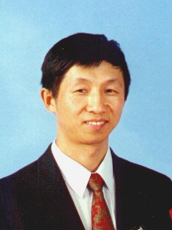 Changming Sun合作论文数The Commonwealth Scientific and Industrial Research Organisation;School of Computer Science and Engineering, The University of New South Wales27
 Dadong Wang合作论文数University of New South Wales;Quantitative Imaging Research Team, CSIRO18
 Luke Domanski合作论文数12
 Tomasz Bednarz合作论文数CSIRO Mathematics, Informatics & Statistics, Sydney, NSW, Australia12
 Karsten Weis合作论文数Institute of Biochemistry, Eidgenössische Technische Hochschule Zürich11Matthew Payne合作论文数8
 David Lovell合作论文数Faculty of Science, School of Computer Science, Queensland University of Technology8
 Ryan Lagerstrom合作论文数Mathematical and Information Sciences, CSIRO8
 Tuan D Pham合作论文数Barts and The London School of Medicine and Dentistry, Queen Mary University of London8
 Bischof Leanne合作论文数6
 William E Hughes合作论文数6
 Horst Vogel合作论文数Shenzhen Institute of Advanced Technology, Chinese Academy of Sciences;University of Chinese Academy of Sciences5
 David James合作论文数Charles Perkins Centre, The University of Sydney;School of Life & Environmental Sciences, Faculty of Science, The University of Sydney;School of Medical Sciences, The University of Sydney5
 Gaudenz Danuser合作论文数Institute of Human Biology;Roche5Gerald Münch合作论文数Psychiatrische Klinik und Poliklinik, Universitäts-Nervenklinik5
 Cynthia Whitchurch合作论文数Quadram Institute;University of Technology Sydney4Christopher F Mugler合作论文数4
 Ruud Hovius合作论文数Institute of Chemical Sciences and Engineering, School of Basic Sciences, École Polytechnique Fédérale de Lausanne4
 Stephanie Heinrich合作论文数Institute for Biochemistry, ETH Zurich4
 Chao Zhang合作论文数Samsung Research China4Evgeny Onischenko合作论文数4
 Ruchika Sachdev合作论文数3Katarina Mele合作论文数3
 Lynne Turnbull合作论文数Ithree Inst, Univ Technol Sydney3
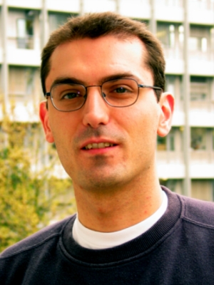 Aaron Ponti合作论文数Department of Biosystems Science and Engineering, Eidgenössische Technische Hochschule Zürich3
 Janet Newman合作论文数3
 Leon Y. Chan合作论文数David H. Koch Institute for Integrative Cancer Research and Howard Hughes Medical Institute, Massachusetts Institute of Technology3
 Ran Su (苏苒)合作论文数College of Intelligence and Computing, Tianjin University3
 Arcot Sowmya合作论文数School of Computer Science and Engineering, Faculty of Engineering, The University of New South Wales3
 Yulia Arzhaeva合作论文数University Medical Center Utrecht3
 Clare Waterman合作论文数Laboratory of Cell and Tissue Morphodynamics, National Heart, Lung, and Blood Institute, National Institutes of Health3Elisa Dultz合作论文数Gene Expression Unit, European Molecular Biology Laboratory, Meyerhofstr. 1, 69117 Heidelberg, Germany3Alex Khassapov合作论文数3
 Mohammadreza Hosseini合作论文数UNSW3
 James Geoffrey Burchfield合作论文数School of Life and Environmental Sciences, The University of Sydney3
 Rhonda June Davey合作论文数3
 Thorsten Wohland合作论文数Department of Chemistry, Faculty of Science, National University of Singapore3
 Maria Hondele合作论文数Dept Biol, Swiss Fed Inst Technol3
 Tim Gureyev合作论文数ARC Centre of Excellence in Advanced Molecular Imaging, School of Physics, The University of Melbourne3Ap Tairi合作论文数2Carina Patrizia Derrer合作论文数2
 Annemarie Kralt合作论文数2Yvonne Ng合作论文数2
 Sasikumar Rajoo合作论文数Swiss Fed Inst Technol, Swiss Fed Inst Technol Zurich2
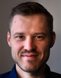 Matthias Wojtynek合作论文数Max Planck Inst Dev Biol, Dept Prot Evolut, Max Planck Ring 5, D-72076 Tubingen, Germany2
 Sabine Schmoelzl合作论文数Institute of Veterinary Medicine, University of Göttingen2
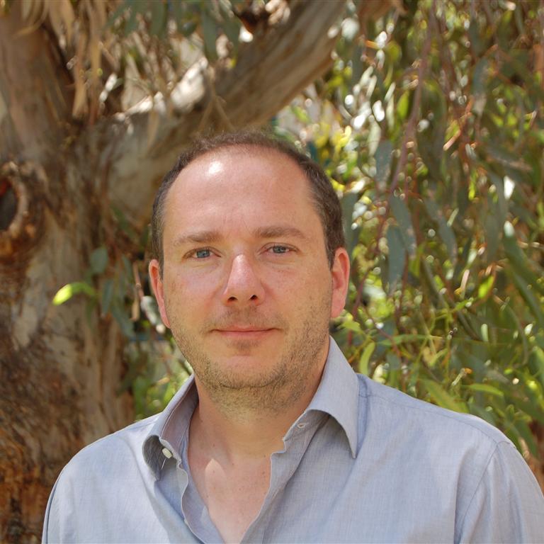 E. Lombi合作论文数Future Industries Institute, University of South Australia2
 Darren Thompson合作论文数Commonwealth Sci. &Ind. Res. Organ.;c;Commonwealth Sci. & Ind. Res. Organ.2
 Prabhakar Ranganathan合作论文数Department of Mechanical and Aerospace Engineering, Faculty of Engineering, Monash University2




