Background:For patients who have patellar instability with patella alta, distalizing the patella via tibial tubercle osteotomy (TTO-d) is one method used to surgically restore stability. Although this procedure is generally associated with favorable outcomes, the effects on maintaining postoperative patellar height remain inadequately understood. Purpose/Hypothesis:This study aimed to assess changes in patellar height after TTO-d in skeletally mature patients. It was hypothesized that the patella would eventually return to a state of alta after TTO-d. Study Design:Case series; Level of evidence, 4. Methods:Skeletally mature patients who underwent a TTO-d procedure with a follow-up lateral knee radiograph at ≥3 months after surgery were included. Caton-Deschamp (CD) and Blackburne-Peele (BP) indices were calculated at various time intervals: preoperative, immediate postoperative, 3-month, 6-month, and ≥1-year follow-up on lateral knee radiographs as available. Paired Student t tests were used to compare patellar height indices at different time points. Result:A total of 35 knees in 28 patients were included in this study, 20 of which had imaging ≥1 year postoperatively. The mean age of the cohort was 22.3 ± 7.95 years (range, 15.5-43.0 years), mean body mass index was 28.0 ± 7.86, mean tibial tubercle distalization was 10.8 ± 4.83 mm at time of surgery, and mean follow-up using the latest available lateral knee radiographs occurred at 2.83 ± 2.96 years postoperatively. Mean preoperative CD and BP indices were 1.46 ± 0.193 and 1.24 ± 0.196, respectively, which decreased to 1.13 ± 0.189 (P < .001) and 0.855 ± 0.209 (P < .001) on immediate postoperative films. In those with imaging ≥1 year out from surgery, mean CD and BP indices increased to 1.34 ± 0.196 (P < .001) and 1.13 ± 0.131, respectively. Additionally, 88% (15/17) of patients with a minimum 1-year radiographic follow-up transitioned from normal CD and BP indices immediately postoperatively to patella alta at latest follow-up. Conclusion:Nearly all patients who underwent TTO-d had progressive increases in both CD and BP indices over time, suggesting a return to a state of patella alta over time. Further studies are needed to compare clinical outcomes of patients who have a confirmed return to alta versus those who do not in order to better understand the clinical significance of this observation.
Background:Multiligament knee injuries (MLKIs) due to high-energy (HE) or polytraumatic (PT) mechanisms result in worse patient outcomes after surgery versus low-energy (LE) or nonpolytraumatic (NPT) mechanisms. There remains a paucity of research in the literature investigating MLKIs resulting from these mechanisms of injury. Purpose:To characterize differences in ligamentous injury/surgery patterns between different mechanism groups: HE versus LE and PT versus NPT. Study Design:Cross-sectional study; Level of evidence, 3. Methods:This is a retrospective chart review of consecutive surgical MLKIs from a single institution. Demographics, clinical/radiographic findings, and intraoperative variables were obtained. HE versus LE mechanisms were stratified. Patients with inadequate data were excluded. PT versus NPT classifications were based on presence of concurrent nonknee injuries. Subgroup comparisons were made based on body mass index, age, sex, number/type of ligaments injured, and surgeries performed. Results:From a total of 226, 176 (78%) patients were included (41 PT [23%] and 135 NPT [77%]; 62 HE [35%] and 114 LE [65%]). PT mean age was 30.7 ± 13.7 years (male, 32 [78%)], while NPT mean age was 27.1 ± 12.3 years [male, 91/135 [67%]). HE mean age was 31.4 ± 13.1 years (male, 49/62 [79%]), and LE mean age was 25.6 ± 11.8 years (male, 73/114 [64%]). Posterior cruciate ligament (PCL) injuries were higher in PT (27/41 [66%]) and HE (42/62 [68%]) versus NPT (58/135 [43%]) and LE (44/114 [39%]), respectively. Lateral collateral ligament (LCL) injuries were higher in PT (29/41 [71%]) and HE (41/62 [66%]) versus NPT (66/135 [49%]) and LE (55/114 [48%]), respectively. The mean number of ligaments injured was higher in PT versus NPT (2.9 vs 2.4) and in HE versus LE (2.8 vs 2.4). LCL surgical interventions were more common in PT (23/41 [55%]) versus NPT (49/135 [36%]) and in HE (33/62 [53%]) versus LE (39/114 [34%]). No other differences in injuries/surgeries existed between PT and NPT or HE and LE, in anterior cruciate, medial collateral, or medial patellofemoral ligaments. Conclusion:PT and HE MLKIs are characterized by increased rates of PCL and LCL injuries compared with LE and NPT. HE and PT patients experience more injured ligaments than LE and NPT and are more likely to require reconstruction/repair of the LCL. These findings suggest injury severity and mechanism may facilitate clinical decision making in MLKIs.
Purpose: To evaluate thigh muscle volume differences in patients undergoing anterior cruciate ligament reconstruction (ACLR) with either allograft tissue or bone−patellar tendon−bone (BPTB) autografts, using 3-dimensional magnetic resonance imaging (MRI) modeling. Methods: This is a retrospective, single-center study of patients undergoing primary ACLR with either ipsilateral BPTB autograft or allograft tissue. Inclusion criteria were age 18-45 years, 5 to 8 years of follow-up, and body mass index <30. Patients were excluded if they had multiligamentous knee injuries, surgical treatment of meniscus tears (repair or reconstruction), and any subsequent knee procedures after the index ACLR. 3D MRI of the bilateral thighs was performed and images were rendered with 3D modeling software and the total thigh, anterior-, posterior-, and medial-thigh compartment musculature were individually segmented and the volumes were calculated. Data were evaluated using a t test, and muscle volumes were standardized as a percentage of the corresponding nonoperative extremity and then tested in comparison with the alternative graft type. Results: Ten allograft and 10 patients with BPTB were included. Patients with allograft were older at surgery (mean age 34.6 vs 23.4 years (P < .001) and at study MRI (mean age 40.9 vs 29.7 years; P < .001). Allografts showed no differences in total thigh or compartmental muscle volumes between the operative and nonoperative limbs. BPTB showed a statistically significant reduction in total thigh volume on the operative side (103 cm3, 97.0%, P = .013), whereas compartment volumes remained similar between limbs. Percentage changes in volume comparing injured with contralateral thigh volume was not significantly different between graft type groups. Conclusions: No significant side-to-side differences were found in individual thigh compartments or total volume after allograft ACLR. BPTB ACLR has a modest effect (∼3%) on long-term total thigh muscle volume. Level of Evidence: Level III, retrospective cohort study.
Background: Minimal clinically important differences (MCIDs) for different patient outcome scores have been reported for various shoulder diseases, including shoulder arthroplasty and the nonoperative treatment of rotator cuff disease. The purpose of this study was to assess the MCID for the American Shoulder and Elbow Surgeons Standardized Shoulder Assessment Form (ASES) score, the Simple Shoulder Test (SST), and a visual analog scale (VAS) measuring pain, after arthroscopic rotator cuff repair. Methods: A total of 202 patients who underwent arthroscopic rotator cuff repair were retrospectively reviewed. ASES, SST, and VAS pain scores were collected preoperatively and at 1 year postoperatively. The MCID was then calculated via a 4-question anchor-based method. Results: The MCID results for the ASES, SST, and VAS pain scores were 27.1, 4.3, and 2.4, respectively. Age at time of surgery, sex, anteroposterior tear size, and worker's compensation status were not associated with MCID values (P > .05). Conclusion: The MCID values determined in the current study are higher than those previously identified for the nonoperative treatment of rotator cuff disease using the same anchor questions. Use of these higher values should be considered when evaluating improvements of individual patients after rotator cuff repair, to determine comparative effectiveness of various rotator cuff repair techniques and to determine sample sizes for prospective comparative trials of rotator cuff repair methods. (C) 2019 Journal of Shoulder and Elbow Surgery Board of Trustees. All rights reserved.
BACKGROUND:Although surgical shoulder stabilization is a substantial cost nationally within the United States, little information exists to analyze this cost. The purpose of this study was to identify factors associated with variation in direct costs with the arthroscopic treatment of glenohumeral instability. METHODS:This was a retrospective study of all patients who underwent arthroscopic treatment of glenohumeral instability between January 12, 2012 and July 11, 2017. Patient and procedure factors were collected. Direct perioperative costs were collected using a validated internal tool. Patient and procedure characteristics significantly associated with costs were identified using multivariate generalized linear models. RESULTS:The study included 302 patients, of whom 12% were undergoing revision and 32% were contact or collision athletes. Anterior instability was present in 73%, whereas 14% had posterior and 10% had multidirectional instability. Of the patients, 67% were recurrent dislocators and 33% were first-time dislocators or subluxators. Remplissage was performed in 13%; biceps tenodesis, 5%; and rotator cuff repair, 3%. An average of 4.0 ± 1.4 anchors were used. Of costs, 39% were operative facility utilization costs and 41% were implant costs. Factors associated with cost increase included an increased number of anchors (P < .0001), posterior vs. anterior instability (P = .001), recurrent instability vs. first-time dislocation (P = .025), remplissage (P = .006), rotator interval closure (P = .021), bicep tenodesis (P = .020), rotator cuff repair (P < .0001), an inpatient stay (P = .003), and repair of humeral avulsion of the glenohumeral ligaments (P = .012). CONCLUSION:Most perioperative costs associated with the arthroscopic treatment of glenohumeral instability are facility utilization and implant costs. Nonmodifiable factors associated with increased cost included posterior direction of instability and recurrent instability. Modifiable factors included additional procedures and inpatient stay.
» Injuries to the posterior root of the lateral meniscus occur frequently in acute knee injuries with concomitant tearing of the anterior cruciate ligament (ACL). » The meniscofemoral ligaments (MFLs), consisting of the anterior MFL (ligament of Humphrey) and the posterior MFL (ligament of Wrisberg), as well as the osseous attachment of the meniscal root, stabilize the lateral meniscus, enabling appropriate load transmission and maintenance of contact forces within the lateral compartment of the knee during loading and range of motion. » In the setting of an ACL injury to the knee with osseous root injury of the posterior root of the lateral meniscus, the MFLs (when present) may stabilize the lateral meniscus against meniscal extrusion, thereby maintaining appropriate contact mechanics within the knee, decreasing the risk of subsequent chondral and meniscal injury and the development of premature osteoarthritis. » Additional study on the indications for posterior meniscal root repair during ACL reconstruction is warranted since the ideal management of lateral root injury in the MFL-intact knee remains unknown.
Background: The purposes of this study were to determine whether acromial morphology (1) could be measured accurately on magnetic resonance images (MRIs) as compared to computed tomographs (CTs) as a gold standard, (2) could be measured reliably on MRIs, (3) differed between patients with rotator cuff tears (RCTs) and those without evidence of RCTs or glenohumeral osteoarthritis, and (4) differed between patients with rotator cuff repairs (RCRs) that healed and those that did not. Methods: This is a retrospective comparative study. We measured coronal, axial, and sagittal acromial tilt; acromial width, acromial anterior and posterior coverage, and glenoid version and inclination on MRI corrected into the plane of the glenoid. We determined accuracy by comparison with CT via intraclass correlation coefficients (ICCs). To determine reliability, these same measurements were made on MRI by 2 observers and ICCs calculated. We compared these measurements between patients with a full-thickness RCT and patients aged > 50 years without evidence of an RCT or glenohumeral osteoarthritis. We then compared these measurements between those patients with healed RCRs and those with a retorn rotator cuff on MRI. In this portion, we only included patients with both a preoperative MRI and a postoperative MRI at least 1 year from RCR. Only those patients without tendon defects on postoperative MRIs were considered to be healed. In these patients, we also radiographically measured the critical shoulder angle. Results: In a validation cohort of 30 patients with MRI and CT, all ICCs were greater than 0.86. In these patients, the inter-rater ICCs of the MRI measurements were >0.53. In our RCT group of 110 patients, there was greater acromial width [mean difference (95% confidence interval) = 0.1 (0, 0.2) mm, P = .012] and significantly less sagittal acromial tilt [9 degrees (5 degrees-12 degrees), P<.001] than in our comparison group of 107 patients. A total of 110 RCRs were included. Postoperative MRI scans were obtained at a mean follow-up of 24.2 +/- 15.8 months, showing 84 patients (76%) had healed RCRs. Aside from acromial width, which was 0.2 mm different and thus did not have clinical significance, there was no association between healing and any of the measured morphologic characteristics. Patients with healed repairs had significantly smaller tears in terms of both width (P<.001) and retraction (P <.001). Conclusion: Although the acromion is wider in RCTs, the difference of 0.1 mm likely has no clinical significance. The acromion is more steeply sloped from posteroinferior to anterosuperior in those with RCTs. These findings call into question subacromial impingement due to native acromial morphology as a cause of rotator cuff tearing. Acromial morphology, critical shoulder angle, and glenoid inclination were not associated with healing after RCR. This study does not support lateral acromioplasty. (C) 2020 Journal of Shoulder and Elbow Surgery Board of Trustees. All rights reserved.
Background: The purpose of this study was to determine the short-term outcomes for patients who underwent revision surgery for shoulder instability, including both revision arthroscopic repair and Latarjet. Methods: This study included patients who underwent revision of a prior arthroscopic labral repair to arthroscopic labral repair or Latarjet at our institution from 2012 to 2017. After collection of preoperative demographic data, preoperative 3-dimensional imaging was reviewed to determine percent glenoid bone loss (%GBL) and to determine whether each shoulder was on-track or off-track. Patients were contacted to obtain postoperative patient-reported outcome metrics including visual analog scale pain, Simple Shoulder Test, American Shoulder and Elbow Surgeons scores, and instability recurrence (full dislocation, subluxation, or subjective apprehension) data at a minimum of 2 years postoperatively. Results: Of 62 patients who met criteria, 45 patients were able to be contacted. Of them, 21 underwent revision arthroscopy and 24 underwent a Latarjet procedure. In the revision arthroscopy group, 5 of 15 had %GBL >20% and 4 of 21 were contact athletes. In the Latarjet group, 11 of 22 had %GBL >20% and 5 of 24 were contact athletes. Of 21 revision arthroscopy patients, 8 underwent concomitant remplissage. Eight of 21 patients in the revision arthroscopy group and 7 of 21 patients in the Latarjet group reported instability postoperatively. Three of 21 patients in the revision arthroscopy group and 2 of 21 patients in the Latarjet group reported full dislocations postoperatively. Zero patients in the revision arthroscopy group and 1 of 21 patients in the Latarjet group underwent reoperation. Conclusion: Our results suggest that both revision Latarjet and arthroscopic stabilization can be of benefit in select circumstances. However, in revision settings, postoperative instability symptoms are common with both procedures.
Background: The optimal surgical treatment of anterior shoulder instability remains controversial. Hypothesis: (1) Implants and facility-related costs are the primary drivers of variation in direct costs between arthroscopic Bankart and Latarjet procedures, and (2) distal tibial allograft (DTA) is more costly than Latarjet as a function of the graft expense. Study Design: Cross-sectional study; Level of evidence, 3. Methods: Intraoperative cost data were derived for all arthroscopic anterior stabilizations and Latarjet and DTA procedures performed at a single academic institution from January 2012 to September 2017. Cost comparisons were made between those undergoing arthroscopic stabilization and Latarjet and between Latarjet and DTA. Multivariate regressions were performed to determine the difference in direct costs accounting for various patient- and surgery-related factors. Results: A total of 87 arthroscopic stabilizations, 44 Latarjet procedures, and 5 DTA procedures were performed during the study period. Arthroscopic Bankart repair was found to be 17% more costly than Latarjet, with suture anchor implant cost being the primary driver of cost. DTA was 2.9-fold more costly than Latarjet, with greater costs across all domains. Multivariate analysis also found the number of prior arthroscopic procedures performed (P = .007) and whether the procedure was performed in an ambulatory or inpatient setting (P < .0001) to be significantly associated with higher direct costs. Conclusion: Latarjet is less costly than arthroscopic Bankart repair, largely because of implant cost. Value-driven strategies to narrow the cost differential could focus on performing these procedures in an outpatient setting in addition to reducing overall implant cost for arthroscopic procedures. Perceived potential benefits of DTA over Latarjet may be outweighed by higher costs.
BACKGROUND:Few studies have investigated the influence of patient-specific variables or procedure-specific factors on the overall cost of anterior cruciate ligament reconstruction (ACLR) in an ambulatory surgery setting. PURPOSE:To determine patient- and procedure-specific factors influencing the overall direct cost of outpatient arthroscopic ACLR utilizing a unique value-driven outcomes (VDO) tool. STUDY DESIGN:Cohort study (economic and decision analysis); Level of evidence, 3. METHODS:All ACLRs performed by 4 surgeons over 2 years were retrospectively reviewed. Cost data were derived from the VDO tool. Patient-specific variables included age, body mass index, comorbidities, American Society of Anesthesiologists (ASA) classification, smoking status, preoperative Patient-Reported Outcomes Measurement Information System (PROMIS) Physical Function Computerized Adaptive Testing (PF-CAT) score, and preoperative Single Assessment Numeric Evaluation (SANE) score. Procedure-specific variables included graft type, revision status, associated injuries and procedures, time from injury to ACLR, surgeon, and operating room (OR) time. Multivariate analysis determined patient- and procedure-related predictors of total direct costs. RESULTS:There were 293 autograft reconstructions, 110 allograft reconstructions, and 31 hybrid reconstructions analyzed. Patient-specific factors did not significantly influence the ACLR cost. The mean OR time was shorter for allograft reconstruction (P < .001). Predictors of an increased direct cost included the use of an allograft or hybrid graft (44.5% and 33.1% increase, respectively; P < .001), increased OR time (0.3% increase per minute; P < .001), surgeon 3 or 4 (9.1% or 5.9% increase, respectively; P < .001 or P = .001, respectively), and concomitant meniscus repair (24.4% increase; P < .001). Within the meniscus repair cohort, all-inside, root, and combined repairs correlated with a 15.5%, 31.4%, and 53.2% increased mean direct cost, respectively, compared with inside-out repairs (P < .001). CONCLUSION:This study failed to identify modifiable patient-specific factors influencing direct costs of ACLR. Allografts and hybrid grafts were associated with an increased total direct cost. Meniscus repair independently predicted an increased direct cost, with all-inside, root, and combined repairs being costlier than inside-out repairs. The time-saving potential of all-inside meniscus repair was not realized in this study, making implant use a significant factor in the overall cost of ACLR with meniscus repair.
customized to the size of the defect.Objective, subjective, and demographic data were prospectively collected and retrospectively reviewed.ASES, SANE, QuickDASH, SF-12 and satisfaction outcome measures were collected pre and post-operatively.Results: 34 patients (13 women, 21 men, mean 52 AE 7 years) were included in this study.16 patients underwent ASCR and 18 patients underwent LDT.Failure of the repair occurred in 1 patient in the ASCR group (6.2%), who suffered a graft tear shown on MRI at 141 days postoperatively, and 2 patients (11%) in the LDT group, both whom progressed to rTSA.Two additional patients (11%) in the LDT group had further surgery around 1-year postoperatively -an arthroscopic cuff repair and hardware removal operation.In those who did not fail, pain significantly decreased postoperatively (p<0.05) in both groups.Only patients who underwent ASCR had a statistically significant functional improvement (p¼0.002 vs. p¼0.161).Mean change in abduction and flexion were -7.3 and 0.6 respectively in the LDT group, compared to 56.0 and 21.7 respectively in the ASCR group.At final follow-up, satisfaction was a median 8/10 points in both groups.Conclusion: Patients who underwent ASCR had significantly improved postoperative scores and range of motion, compared to those who underwent LDT, but longer follow-up is required.
Background: Very limited information exists about factors affecting direct clinical costs of rotator cuff repair surgery. The purpose of this study was to determine the direct cost of outpatient arthroscopic rotator cuff repair surgery using a unique value-driven outcomes tool and to identify patient- and treatment-related variables affecting cost. Methods: Cost data were derived for arthroscopic rotator cuff repairs performed by 3 surgeons from March 2014 to June 2015 using the value-driven outcomes tool. Costs included overall total direct cost, which included facility utilization costs, medication costs, supply costs, and other ancillary costs. Univariate and multivariate regressions were performed to determine the effect of various patient-related and surgical-related factors on costs. Results: There were 170 arthroscopic rotator cuff repairs performed during the study period. Multivariate analysis showed significant correlations between higher total direct cost and the presence of a subscapularis repair being performed (P=.015) and total number of anchors used (P<.0001). Higher body mass index, severe systemic illness, 1 of the 3 surgeons, biceps tenodesis using an anchor, and total sum of anchors were correlated with higher facility utilization costs (P<.04). Severe systemic illness, addition of a subscapularis repair, 1 of the 3 surgeons, and additional subacromial decompression were correlated with higher pharmacy costs (P<.006). The addition of a subscapularis repair, total sum of anchors, and severe muscle changes to the supraspinatus were correlated with higher supply costs (P<.015). Conclusions: From a direct cost perspective, implementation of strategies to reduce overall costs should focus on reducing overall anchor quantity or price. (c) 2017 Journal of Shoulder and Elbow Surgery Board of Trustees. All rights reserved.
BACKGROUND:Recent attention has focused on the optimal surgical treatment for recurrent shoulder instability in young athletes. Collision athletes are at a higher risk for recurrent instability after surgery.PURPOSE:To evaluate variables affecting return-to-play (RTP) rates in Division I intercollegiate football athletes after shoulder instability surgery.STUDY DESIGN:Case series; Level of evidence, 4.METHODS:Invitations to participate were made to select sports medicine programs that care for athletes in Division I football conferences (Pac-12 Conference, Southeastern Conference [SEC], Atlantic Coast Conference [ACC]). After gaining institutional review board approval, 7 programs qualified and participated. Data on direction of instability, type of surgery, time to resume participation, and quality and level of play before and after surgery were collected.RESULTS:There were 168 of 177 procedures that were arthroscopic surgery, with a mean 3.3-year follow-up. Overall, 85.4% of players who underwent arthroscopic surgery without concomitant procedures returned to play. Moreover, 15.6% of athletes who returned to play sustained subsequent shoulder injuries, and 10.3% sustained recurrent instability, resulting in reduction/revision surgery. No differences were noted in RTP rates in athletes who underwent anterior labral repair (82.4%), posterior labral repair (92.9%), combined anterior-posterior repair (84.8%; P = .2945), or open repair (88.9%; P = .9362). Also, 93.3% of starters, 95.4% of utilized players, and 75.7% of rarely used players returned to play. The percentage of games played before the injury was 49.9% and rose to 71.5% after surgery ( P < .0001). Athletes who played in a higher percentage of games before the injury were more likely to return to play; 91% of athletes who were starters before the injury returned as starters after surgery. Scholarship status significantly correlated with RTP after surgery ( P = .0003).CONCLUSION:The majority of surgical interventions were isolated arthroscopic stabilization procedures, with no statistically significant difference in RTP rates when concomitant arthroscopic procedures or open stabilization procedures were performed. Athletes who returned to play often played in a higher percentage of games after surgery than before the injury, and many played at the same or a higher level after surgery.
PURPOSE:The primary purpose of this study was to determine the effect of the preoperative position of the musculotendinous junction (MTJ) on rotator cuff healing after double-row arthroscopic rotator cuff repair. A secondary purpose was to evaluate how tendon length and MTJ position change when the rotator cuff heals. METHODS:Preoperative and postoperative magnetic resonance imaging (MRI) scans of 42 patients undergoing arthroscopic double-row rotator cuff repair were reviewed. Patients undergoing repairs with other constructs or receiving augmented repairs (platelet-rich fibrin matrix) who had postoperative MRI scans were excluded. Preoperative MRI scans were evaluated for anteroposterior tear size, tendon retraction, tendon length, muscle quality, and MTJ position with respect to the glenoid in the coronal plane. The position of the MTJ was referenced off the glenoid face as either lateral or medial. Postoperative MRI scans were evaluated for healing, tendon length, and MTJ position. RESULTS:Of 42 tears, 36 (86%) healed, with 27 of 31 small to medium tears (87%) and 9 of 11 large to massive tears (82%) healing. Healing occurred in 94% of tears that had a preoperative MTJ lateral to the face of the glenoid but only 56% of tears that had a preoperative MTJ medial to the glenoid face (P = .0135). The measured tendon length increased an average of 14.4 mm in patients whose tears healed compared with shortening by 6.4 mm in patients with tears that did not heal (P < .001). The MTJ lateralized an average of 6.1 mm in patients whose tears healed compared with medializing 1.9 mm in patients whose tears did not heal (P = .026). The overall follow-up period of the study was from April 2005 to September 2014 (113 months). CONCLUSIONS:The preoperative MTJ position is predictive of postoperative healing after double-row rotator cuff repair. The position of the MTJ with respect to the glenoid face is a reliable, identifiable marker on MRI scans that can be predictive of healing. LEVEL OF EVIDENCE:Level IV, retrospective review of case series; therapeutic study.
Objectives: The purpose of this study was to evaluate return to play (RTP) rates and variables influencing RTP in division I intercollegiate football athletes following shoulder instability surgery. Methods: Requests for participation in the study were sent to select sports medicine programs caring for athletes participating in football from the PAC-12, SEC, and ACC athletic conferences. After gaining IRB approval, 7 programs were able to participate in the study. Inclusion criteria were restricted to athletes active during the 2004-2013 seasons that required surgical treatment for shoulder instability. Direction of instability, type of surgery, time to resume participation, quality and level of play data both before and after surgery was collected for each athlete. Data was analyzed to determine overall RTP and the influence that scholarship and depth chart position prior to surgery had on RTP. To determine the effect that surgery had on players’ ability to RTP, the percent of games played before and after surgery was determined. Results: 177 shoulder injuries in 153 athletes were identified and met inclusion criteria. Overall, 85.4% of players who underwent arthroscopic surgery without concomitant procedures returned to play. 82.4% of players who underwent anterior labral repair, 88.7% of those that underwent posterior labral repair, and 84.8% who underwent combined anterior-posterior repair returned to sport. Categorized by depth chart position, 93.3% of starters, 95.4% of utilized players, and 75.7% of rarely used players returned to play. The percentage of games played in by athletes prior to injury was 49.9%, and rose to 71.5% following surgery. Athletes who played in a higher percentage of games prior to injury (49.4 +/- 43.4%) were more likely to return to play than athletes who played in a fewer percentage of games (19.6 +/- 39.4%). Of the 42 athletes identified as starters prior to injury that returned to play, 98% continued as starters; 2% became utilized players. Of the 41 players that prior to injury were utilized, 49% became starters, 49% remained utilized, and 2% rarely played following surgery. For the 56 players identified as rarely playing pre-injury that returned to play, 36% became starters, 23% were utilized athletes, and 41% maintained their rarely playing status. Having a scholarship significantly correlated with RTP after surgery. Conclusion: RTP rates in high-level intercollegiate football players undergoing shoulder stabilization surgery was 85%. Posterior labral repair, anterior labral repair, and combined labral repair demonstrated no statistical difference in RTP rates. The majority of surgical interventions were isolated arthroscopic stabilization procedures, and demonstrated no statistically significant difference in RTP rates when concomitant arthroscopic procedures or open stabilization procedures were performed. Finally, athletes who return to play often did so in a higher percentage of games following surgery than they did prior to injury, and many players were utilized at the same or a higher level by their teams following surgery. These findings suggest that the majority of football players at the division 1 level who undergo shoulder stabilization surgery are able to participate fully in their programs, and progress and develop as players following their return to sport.
Reported rates of venous thromboembolism (VTE) after shoulder arthroplasty (SA) range from 0.2% to 13%. Few studies have evaluated the incidence of VTE in a large patient population from a single institution. We conducted a study to determine the incidence of VTE (deep venous thrombosis [DVT] and pulmonary embolism [PE]) in a large series of SAs. Cases of SAs performed at our institution between January 1999 and May 2012 were retrospectively reviewed for development of symptomatic VTE within the first 90 days after surgery. During the study period, 533 SAs (245 anatomical total SAs [TSAs], 112 reverse TSAs, 92 hemiarthroplasties, 84 revision SAs) were performed. Logistic regression analyses were used to evaluate the association of various risk factors with VTE. For the 533 SAs, the symptomatic VTE rate was 2.6% (14 patients), the DVT rate was 0.9% (5), and the PE rate was 2.3% (12). Risk factors significantly correlated with a thrombotic event included raised Charlson Comorbidity Index, preoperative thrombotic event, lower preoperative hemoglobin and hematocrit levels, diabetes, lower postoperative hemoglobin level, use of general endotracheal anesthesia without interscalene nerve block, higher body mass index, and revision SA (P < .05). Our rates of symptomatic VTE events (DVT, PE) after SA are relatively low, though they are higher than the rates in studies that have used large state or national databases. Risk factors associated with thrombosis can be useful in identifying patients at risk for clotting after SA.
Objectives: The Patient Reported Outcomes Measurement Instrument System (PROMIS) Physical Function Computer Adaptive Test (PF-CAT) has been developed by the National Institutes of Health as a means of providing validated outcomes scores. PF-CAT adapts to patient response by applying different validated questions based on prior responses to generate scores using between 4 to 12 questions, and has been validated in a variety of patient populations to assess physical function. The purpose of our study is to compare patient reported outcomes scores in a sports medicine (shoulder and knee injury) patient population by analyzing convergence validity of PF-CAT to IKDC, SST, ASES, and SANE scores. We also wanted to assess and compare time burden placed on patients in completing these outcomes scores. Methods: All patient visits from April through September 2014 presenting with either knee or shoulder complaints were included from a university-based sports medicine clinic, during which both PF-CAT as well as SANE, SST, and ASES legacy outcomes scores for shoulder injury patients, and SANE and IKDC outcomes scores for knee injury patients were obtained via tablet during the initial part of each visit. A total of 415 shoulder and 450 knee clinical evaluations qualified for inclusion in the study. Spearman correlation was used to evaluate pair-wise agreement among outcomes scores. The total scores from each outcome test and the T-scores from the PF-CAT were used in this correlation analysis. For interpretation, results greater than 0.5-0.69 were considered to have fair correlation, while values greater than 0.7 were considered as having good correlation. Significance was assessed at the 0.05 level. Median and interquartile ranges were used to summarize scores. The minimum score (floor effects) and maximum score (ceiling effects) occurrences were tallied and reported as percentages. A Fishers exact test was used to test for a difference in the number of times floor and ceiling values occurred. The Wilcoxon signed rank test was used to test for differences in completion times (measured in seconds). Results: Good correlation was found between IKDC and PF-CAT scores (r=0.7511; p<0.001), and fair correlation was found between SST (r=0.6777; p<0.001) and ASES (r=0.6262; p<0.001). Percent of patients’ scores that hit floor and ceiling values was 15% on SST (p<0.001), 2.5% on ASES (ceiling only, p=0.001), and 0% for PF-CAT shoulder patients. In knee injury patients, 0% of IKDC and 0.44% of PF-CAT scores hit floor or ceiling values (p<0.514). Completion times for PF-CAT averaged 81 seconds versus 359 seconds for IKDC which was significant (p<0.001). Average completions times for shoulder patients completing PF-CAT, SST, and ASES scores was 94, 192, and 157 (349 combined) seconds respectively (p <0.001). Conclusion: PF-CAT holds strong convergence correlation with IKDC outcome scores in a sports medicine knee injury population, and only fair convergence correlation with SST and ASES scores in a sports medicine shoulder injury population. In addition, PF-CAT avoided floor and ceiling effects equal to IKDC outcomes and better than SST and ASES outcomes. Finally, PF-CAT was completed in a shorter time period than IKDC scores as well as compared to SST and ASES times both separately and combined. The PROMIS PF-CAT is an acceptable outcomes tool to evaluate sports medicine knee injuries that places less time burden on patients to complete outcomes scores.
Study objective: The femoral, lateral femoral cutaneous, and obturator nerves (ONs) can reportedly be blocked using a single-injection deep to the fascia iliaca (FI) at the level of the inguinal ligament. Two commonly used methods (the FI compartment and 3-in-1 blocks) have produced inconsistent results with respect to local anesthetic distribution and effect on the ON. To date, no study of either method has been performed using advanced imaging techniques to document both needle placement and local anesthetic distribution. We report the outcome of a series of 3-in-1 and FI blocks performed using ultrasound to guide needle position and magnetic resonance imaging (MRI) to define local anesthetic distribution.Design: Patients were prospectively studied, and images were interpreted using a randomized and blinded protocol.Setting: The study was performed in the perioperative area of an academic orthopedic specialty hospital.Patients: Ten patients (ASA 1-2) having anterior cruciate ligament reconstruction received either 3-in-1 or FI compartment blocks for postoperative analgesia using the surface landmarks described for these techniques.Interventions: Ultrasound was used to position the injecting needle immediately deep to the FI. Local anesthetic distribution was studied using MRI.Measurements: Patients were examined for motor and/or sensory function of the femoral, obturator, and lateral femoral cutaneous nerves. Magnetic resonance imaging was used to document the limits of injectate distribution.Main results: Magnetic resonance imaging showed distribution of injectate over the surface of the iliacus and psoas muscles to the level of the retroperitoneum. No patient showed medial extension of injectate to the ON. At the level of the inguinal ligament, injectate extended laterally toward the anterior superior iliac spine and medially to the femoral vein. All patients had significant weakness with extension of the knee and sensory loss over the anterior, lateral, and medial thigh. No patient demonstrated decreased hip adductor strength.Conclusions: Ultrasound and MRI show consistent superior extension of local anesthetic to the level of the retroperitoneum for both techniques. There was reliable clinical effect on the femoral and lateral femoral cutaneous nerves. However, none of the injections produced evidence of ON block either at the level of the retroperitoneum or the inguinal ligament. (C) 2015 Elsevier Inc. All rights reserved.
Patients with shoulder and rotator cuff pathology who exhibit greater levels of psychological distress report inferior preoperative self-assessments of pain and function. In several other areas of orthopaedics, higher levels of distress correlate with a higher likelihood of persistent pain and disability after recovery from surgery. To our knowledge, the relationship between psychological distress and outcomes after arthroscopic rotator cuff repair has not been similarly investigated.(1) Are higher levels of preoperative psychological distress associated with differences in outcome scores (visual analog scale [VAS] for pain, Simple Shoulder Test, and American Shoulder and Elbow Surgeons score) 1 year after arthroscopic rotator cuff repair? (2) Are higher levels of preoperative psychological distress associated with less improvement in outcome scores (VAS for pain, Simple Shoulder Test, and American Shoulder and Elbow Surgeons score) 1 year after arthroscopic rotator cuff repair? (3) Does the prevalence of psychological distress in a population with full-thickness rotator cuff tears change when assessed preoperatively and 1 year after arthroscopic rotator cuff repair?Eighty-five patients with full-thickness rotator cuff tears were prospectively enrolled; 70 patients (82%) were assessed at 1-year followup. During the study period, the three participating surgeons performed 269 rotator cuff repairs; in large part, the low overall rate of enrollment was related to two surgeons enrolling only two patients total in the initial 14 months of the study. Psychological distress was quantified using the Distress Risk Assessment Method questionnaire, and patients completed self-assessments including the VAS for pain, the Simple Shoulder Test, and the American Shoulder and Elbow Surgeons score preoperatively and 1 year after arthroscopic rotator cuff repair. Fifty of 85 patients (59%) had normal levels of distress, 26 of 85 (31%) had moderate levels of distress, and nine of 85 (11%) had severe levels of distress. Statistical models were used to assess the effect of psychological distress on patient self-assessment of shoulder pain and function at 1 year after surgery.With the numbers available, distressed patients were not different from nondistressed patients in terms of postoperative VAS for pain (1.9 [95% confidence interval {CI}, 1.0-2.8] versus 1.0 [95% CI, 0.5-1.4], p = 0.10), Simple Shoulder Test (9 [95% CI, 8.1-10.4] versus 11 [95% CI, 10.0-11.0], p = 0.06), or American Shoulder and Elbow Surgeons scores (80 [95% CI, 72-88] versus 88 [95% CI, 84-92], p = 0.08) 1 year after arthroscopic rotator cuff repair. With the numbers available, distressed patients also were not different from nondistressed patients in terms of the amount of improvement in scores between preoperative assessment and 1-year followup on the VAS for pain (3 [95% CI, 2.2-4.1] versus 2 [95% CI, 1.4-2.9], p = 0.10), Simple Shoulder Test (5.2 [95% CI, 3.7-6.6] versus 5.0 [95% CI, 4.2-5.8], p = 0.86), or American Shoulder and Elbow Surgeons scale (38 [95% CI, 29-47] versus 30 [95% CI, 25-36], p = 0.16). The prevalence of psychological distress in our patient population was lower at 1 year after surgery 14 of 70 (20%) versus 35 of 85 (41%) preoperatively (odds ratio, 0.36; 95% CI, 0.17-0.74; p = 0.005).Mild to moderate levels of distress did not diminish patient-reported outcomes to a clinically important degree in this small series of patients with rotator cuff tears. This contrasts with reports from other areas of orthopaedic surgery and may be related to a more self-limited course of symptoms in patients with rotator cuff disease or possibly to a beneficial effect of rotator cuff repair on sleep quality or other unrecognized determinants of psychosocial status.Level I, prognostic study.
 Robert T. Burks合作论文数35R T Burks合作论文数15
 Robert Z. Tashjian合作论文数University of Utah Orthopaedic Center, University of Utah Health Sciences Center14
 Stephen K Aoki合作论文数7Jeffrey D. Swenson合作论文数7
 Kent N Bachus合作论文数University of Utah6Amit Lahav合作论文数5
 Julia Crim合作论文数5
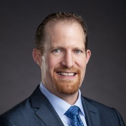 Maak Travis G合作论文数Hospital for Special Surgery, New York, NY 10021, USA. maakt@hss.edu5Davis Jennifer J合作论文数5
 Chalmers Peter N合作论文数Department of Orthopaedic Surgery, Rush University Medical Center5Holmstrom Michael C合作论文数5Gerber J Parry合作论文数4Xerogeanes John W合作论文数Emory Orthopaedic and Spine Center4Daruwalla Jimmy合作论文数4
 Justin J. Ernat合作论文数4John R. West合作论文数4
 Robin L. Marcus合作论文数School of Medicine, University of Utah Health;Department of Physical Therapy and Athletic Training, College of Health, University of Utah4Barbara P. Fink合作论文数4Nathaniel A. Bryan合作论文数3
 Garrett V Christensen合作论文数3Minkyoung Yoo合作论文数3
 Joseph Featherall合作论文数Department of Orthopaedic Surgery, University of Utah Health3
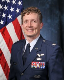 Richard J Robins合作论文数Dept Orthopaed, US Air Force Acad3
 Man Hung合作论文数3
 David Bardana合作论文数3Granger Erin K合作论文数3M. G. Scuderi合作论文数3Paul C. Lastayo合作论文数3
 Robert E. Burks合作论文数3
 Nicholas Brown合作论文数3
 Leland E. Dibble合作论文数3Michael Q. Potter合作论文数2Mark G. Luker合作论文数2Ryan E. Dobbs合作论文数2Brian J Snow合作论文数2Evelyn Loose合作论文数2
 Jun Kawakami合作论文数2Michael Dienst合作论文数2
 Angela Presson合作论文数Division of Epidemiology, Department of Internal Medicine, School of Medicine, University of Utah;Department of Pediatrics, School of Medicine, University of Utah2
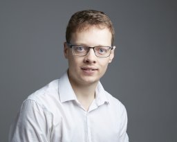 Wylie James D合作论文数Department of Orthopaedic Surgery, University of Utah2
 Yue Zhang合作论文数2Karns Michael R合作论文数2
 Richard Nelson合作论文数Department of Medicine and Epidemiology, School of Veterinary Medicine, University of California2Friederichs Matthew G合作论文数2
 Eric C. Mccarty合作论文数University of Colorado;School of Medicine;Department of Orthopaedics;Rehabilitation Services, University of Colorado Hospital2Byron Bankhead合作论文数2Timothy C. Beals合作论文数2




