Molecular neuroimaging with positron emission tomography (PET) and single-photon emission computed tomography (SPECT) enables quantification of specific molecular targets in the living brain. Despite its scientific impact, molecular neuroimaging research has historically faced challenges due to high costs, small sample sizes, laboratory-specific analysis pipelines, and limited large-scale data sharing. These factors have hindered reproducibility and the broader reuse of valuable PET datasets. The OpenNeuroPET initiative was established to address these barriers by developing standards, infrastructure, and open-source tools for organizing, sharing, and analyzing molecular neuroimaging data. Through collaborations across Europe and North America, OpenNeuroPET has supported the PET extension of the Brain Imaging Data Structure (PET-BIDS), providing a standardized framework for PET datasets and metadata. Building on PET-BIDS, tools such as PET2BIDS, ezBIDS, and BIDSCoin facilitate data conversion and curation. In parallel, OpenNeuro now hosts PET-BIDS datasets for open sharing, while complementary platforms such as PublicnEUro enable GDPR-compliant controlled access. Emerging open-source workflows and BIDS applications further support automated, reproducible PET preprocessing and quantitative analysis, promoting harmonized processing across centers. Together, these developments mark an important step toward an open molecular neuroimaging ecosystem in which datasets, software, and workflows can be transparently shared, reused, and scaled for collaborative research.
Borderline personality disorder (BPD) is a severe psychiatric condition associated with high rates of suicide and poor interpersonal functioning. There are no FDA-approved medications, and evidence-based psychotherapies such as dialectical behavior therapy (DBT) are difficult to access and vulnerable to patient dropout. Novel treatment directions are urgently needed. Mindfulness is the core skill in DBT. Our team has developed a mindfulness-based real-time neurofeedback (mbNF) paradigm where individuals learn to reduce default mode network (DMN) versus control network activation in order to increase present-moment awareness. Here, we describe the study protocol for the MIndfulness-based Neurofeedback to Augment DBT Psychotherapy for Borderline Personality Disorder (MIND-BPD) trial. Participants with BPD (N = 52) enrolled in the study will be randomly assigned (1:1 ratio) to receive one session of either real or sham mbNF. Following neurofeedback, all participants will be enrolled in a 6-month DBT psychotherapy group. Supported by a R61/R33 grant from the NIH, the primary outcome of the R61 phase of this trial is change in DMN connectivity between pre- and post-NF resting-state scans, as defined by increased within-network connectivity between the medial prefrontal cortex (mPFC) and posterior cingulate cortex (PCC), and increased connectivity between the mPFC and dorsolateral prefrontal coftex (dlPFC). Change in self-reported mindfulness between pre- and post-NF is a key secondary outcome. This study is registered in the US Clinical Trials Registry (NCT06446765).
The clinical high risk (CHR) phase of psychosis, characterized by attenuated psychotic symptoms and cognitive impairments, represents a critical window for intervention. Neurobiological alterations observed in schizophrenia (e.g., default mode network (DMN) hyperconnectivity and weakened DMN anticorrelations with the central executive network (CEN)), have been shown to extend to CHR. While neurochemical alterations, specifically glutamate (Glu) and gamma-aminobutyric acid (GABA) activity, are implicated in schizophrenia, their involvement in CHR remains less clear, as does their influence on the large-scale network alterations (e.g., DMN). Previous studies suggest that real-time neurofeedback (rtNFB) using functional magnetic resonance imaging (fMRI) modulates DMN connectivity, offering a potential therapeutic intervention for CHR. However, the efficacy of this approach in modulating neurobiological mechanisms in CHR remains unexplored. This protocol describes a randomized controlled trial evaluating the efficacy of modulating atypical DMN connectivity in CHR individuals. Forty CHR participants (aged 18–30) will be randomized to either a real-rtNFB (N = 20) or sham-rtNFB (N = 20) condition. In the real-rtNFB group, neurofeedback will be derived from DMN-CEN activity, while the sham group will receive feedback from the somatomotor (SMOT) network. We will measure network connectivity pre- and post-intervention, testing two hypotheses: (1) real-rtNFB will reduce DMN hyperconnectivity and strengthen DMN-CEN anticorrelations in CHR, with no such changes occurring in the sham condition, and (2) post-intervention connectivity in the real-rtNFB group will more closely resemble that of neurotypical (NT; N = 20) individuals. A secondary aim will explore the relationship between network connectivity and neurotransmitter concentrations (GABA and Glu) across all participants, and whether they are altered pre- and post-rtNFB. This study will contribute to our understanding of the neurobiological mechanisms of CHR and assess whether rtNFB can alter atypical connectivity in CHR. Ultimately, we aim to inform intervention strategies that could improve both clinical and cognitive outcomes in CHR individuals. The study has been registered with ClinicalTrials.gov (ID: NCT06492343, https://clinicaltrials.gov/study/NCT06492343, Last Update Posted: July 9th, 2024).
Our laboratory recently developed [11C]PS13 as a PET radioligand to selectively measure cyclooxygenase-1 (COX-1). The cyclooxygenase enzyme family converts arachidonic acid into prostaglandins and thromboxanes, which mediate inflammation. The total brain uptake of [11C]PS13, which is composed of both specific binding and background uptake, can be accurately quantified with gold standard methods of compartmental modeling. This study sought to quantify the specific binding of [11C]PS13 to COX-1 in healthy human brain using scans performed with arterial input function at baseline and after blockade by the COX-1-selective inhibitor ketoprofen. Methods: Eight healthy volunteers underwent two 90-min [11C]PS13 PET scans with radiometabolite-corrected arterial input function, at baseline and about 2 h after oral administration of ketoprofen (75 mg). Results: Two-tissue compartment modeling effectively identified the total uptake of radioactivity in the brain (as distribution volume), showing the highest densities in the hippocampus, the occipital cortex, and the banks of the central sulcus. All brain regions exhibited displaceable and specific binding, and thus none could be used as a reference region. Ketoprofen blocked approximately 84% of the binding sites on COX-1 in the whole brain. After full occupancy was extrapolated, the average whole-brain values of [11C]PS13 were 1.6 ± 0.8 mL·cm-3 for specific uptake, 1.7 ± 0.6 mL·cm-3 for background uptake, and 1.1 ± 0.5 for the specific-to-background ratio. The hippocampus had the highest specific-to-background ratio value of 2.7 ± 0.9. Conclusion: [11C]PS13 exhibited high specific binding to COX-1 in the human brain, but its quantification requires arterial blood sampling.
BACKGROUND AND HYPOTHESIS:Auditory hallucinations (AHs) affect 60-80 % of schizophrenia patients and often resist antipsychotic treatment. AHs involve superior temporal gyrus (STG) hyperactivity and disrupted auditory-cognitive control connectivity. Real-time fMRI neurofeedback (NFB) enables voluntary modulation of targeted brain regions. We previously showed STG-targeted NFB with mindfulness meditation reduced STG activation and AHs in one session. However, whether effects are specific to hallucination-related regions versus placebo, and whether NFB modulates broader networks, remained unclear. STUDY DESIGN:This randomized, sham-controlled trial examined NFB specificity and network effects. Twenty-three adults with schizophrenia/schizoaffective disorder and medication-resistant hallucinations practiced mindfulness meditation while receiving neurofeedback from either STG (n = 10, Real-NFB) or motor cortex (n = 13, Sham-NFB control). Sham participants subsequently received Real-NFB, providing within-subject comparison. STUDY RESULTS:Both groups showed reduced AHs post-NFB without group differences. However, compared to Sham-NFB, Real-NFB produced greater reductions in secondary auditory cortex activation and connectivity between auditory cortex and cognitive control regions (dorsolateral prefrontal cortex and anterior cingulate). These connectivity reductions persisted in the Real-after-Sham condition. Both groups showed reduced primary auditory cortex activation, suggesting mindfulness meditation independently regulates bottom-up hallucination processes. CONCLUSIONS:Region-specific NFB targeting produces distinct neural changes beyond symptom reduction. STG-targeted NFB differentially modulates auditory-cognitive control networks, potentially restoring the disrupted balance between bottom-up sensory processing and top-down control in AHs. These findings highlight the importance of anatomically-informed NFB targets and provide mechanistic insights for developing precision interventions for treatment-resistant psychiatric symptoms.
Cyclooxygenase enzymes (COX-1 and COX-2) synthesize pro-inflammatory cytokines that may contribute to Alzheimer's Disease and Major Depression Disorder pathogenesis. Our lab recently developed a potent PET radioligand, 11C-PS13, to target COX-1, which is primarily located in microglia. We have previously shown that 11C-PS13 uptake in the monkey brain is selective for COX-1 as it can be displaced by COX-1 but not by COX-2 selective agents. This study aimed to assess the specificity of 11C-PS13 to COX-1 by blockade studies using ketoprofen, a highly selective COX-1 inhibitor.
Motivation: Many cutting-edge MR neuroimaging paradigms require real-time decision making and precise FOV positioning. We present two software tools to support such paradigms. Goal(s): Develop two modules. 1) vSend: opens a socket and sends imaging data to another computer in a vendor-agnostic format, enabling real-time analysis. 2) AAhijack: reads a matrix from a socket and overwrites the Siemens AutoAlign matrix, enabling online slice prescription. Approach: Modules are implemented as Siemens image reconstruction modules (ICE functors) in C++ and two slice prescription systems utilizing the modules are demonstrated. Results: The slice prescription systems have comparable performance and various advantages and disadvantages. Impact: The software tools presented have enabled a variety of cutting-edge MR neuroimaging paradigms including real-time fMRI, motion tracker calibration, real-time shimming, fetal head-pose detection and automated FOV prescription, reacquisition planning and single-slice BOLD imaging FOV prescription.
Motivation: Very high quality of MR spectroscopic imaging (MRSI) data is needed for robust and reproducible metabolite quantification. This critically depends on the B0 shimming and scan stability. Integrated RF-receive/B0-shim arrays significantly improve spectral quality. Goal(s): Real-time motion correction and multicoil shimming update with an integrated RF-receive/B0-shim array for robust whole-brain MRSI. Approach: We developed a rapid navigator for head tracking and B0 fieldmapping in combination with rapid processing for real-time update of multicoil shim currents and MRSI localization. Results: Real-time motion correction and multicoil shimming provides significantly narrower linewidth, higher signal-to-noise, reduced quantification errors and reproducible metabolic imaging. Impact: Whole-brain MRSI is a unique method for non-invasive mapping of brain neurochemistry, and in combination with real-time motion correction and multicoil shim array update provides robust and reproducible quantitative metabolic imaging for clinical use.
Background Adolescence is characterized by a heightened vulnerability for Major Depressive Disorder (MDD) onset, and currently, treatments are only effective for roughly half of adolescents with MDD. Accordingly, novel interventions are urgently needed. This study aims to establish mindfulness-based real-time fMRI neurofeedback (mbNF) as a non-invasive approach to downregulate the default mode network (DMN) in order to decrease ruminatory processes and depressive symptoms. Methods Adolescents ( N = 90) with a current diagnosis of MDD ages 13–18-years-old will be randomized in a parallel group, two-arm, superiority trial to receive either 15 or 30 min of mbNF with a 1:1 allocation ratio. Real-time neurofeedback based on activation of the frontoparietal network (FPN) relative to the DMN will be displayed to participants via the movement of a ball on a computer screen while participants practice mindfulness in the scanner. We hypothesize that within-DMN (medial prefrontal cortex [mPFC] with posterior cingulate cortex [PCC]) functional connectivity will be reduced following mbNF (Aim 1: Target Engagement). Additionally, we hypothesize that participants in the 30-min mbNF condition will show greater reductions in within-DMN functional connectivity (Aim 2: Dosing Impact on Target Engagement). Aim 1 will analyze data from all participants as a single-group, and Aim 2 will leverage the randomized assignment to analyze data as a parallel-group trial. Secondary analyses will probe changes in depressive symptoms and rumination. Discussion Results of this study will determine whether mbNF reduces functional connectivity within the DMN among adolescents with MDD, and critically, will identify the optimal dosing with respect to DMN modulation as well as reduction in depressive symptoms and rumination. Trial Registration This study has been registered with clinicaltrials.gov, most recently updated on July 6, 2023 (trial identifier: NCT05617495).
In multi-inversion EPI (MI-EPI), each slice samples a distinct inversion time during each inversion recovery, providing an efficient method for estimating T 1 . MI-EPI is vulnerable to through-plane motion, which results in slices sampling a subset of the desired inversion times and wrong TIs will be attributed to the slices. This cannot be corrected retrospectively. We introduce prospective motion correction in MI-EPI using volumetric navigators ( vNavs ). vNavs are acquired at the beginning of the inversion recovery thus the effects of their excitation pulses must be modeled for T 1 estimation. This provides improved T 1 estimation accuracy in the presence of subject motion.
Subject motion results in intra-scan ∆B0 changes which are uncompensated in conventional ∆B0 shimming methods. Rapidly switchable shim currents and ∆B0 vNavigators together enable motion-compensated shimming. We have demonstrated successful measurement, calculation, and application of motion-compensated slice-by-slice shims using and AC/DC coil and vNav ∆B0 maps. Motion-compensated slice-by-slice homogeneity shimming improves shim robustness to subject motion and enables compatibility with changing slice prescriptions of prospective motion correction.
Purpose To compare prospective motion correction (PMC) and retrospective motion correction (RMC) in Cartesian 3D-encoded MPRAGE scans and to investigate the effects of correction frequency and parallel imaging on the performance of RMC. Methods Head motion was estimated using a markerless tracking system and sent to a modified MPRAGE sequence, which can continuously update the imaging FOV to perform PMC. The prospective correction was applied either before each echo train (before-ET) or at every sixth readout within the ET (within-ET). RMC was applied during image reconstruction by adjusting k-space trajectories according to the measured motion. The motion correction frequency was retrospectively increased with RMC or decreased with reverse RMC. Phantom and in vivo experiments were used to compare PMC and RMC, as well as to compare within-ET and before-ET correction frequency during continuous motion. The correction quality was quantitatively evaluated using the structural similarity index measure with a reference image without motion correction and without intentional motion. Results PMC resulted in superior image quality compared to RMC both visually and quantitatively. Increasing the correction frequency from before-ET to within-ET reduced the motion artifacts in RMC. A hybrid PMC and RMC correction, that is, retrospectively increasing the correction frequency of before-ET PMC to within-ET, also reduced motion artifacts. Inferior performance of RMC compared to PMC was shown with GRAPPA calibration data without intentional motion and without any GRAPPA acceleration. Conclusion Reductions in local Nyquist violations with PMC resulted in superior image quality compared to RMC. Increasing the motion correction frequency to within-ET reduced the motion artifacts in both RMC and PMC.
Changes in subject position move susceptibility interfaces and therefore ∆B 0 field patterns in the brain. We apply a prospective real time (TR-to-TR) shim updating scheme using dual echo EPI volume navigators to correct motion-induced changes in ∆B 0 fields to reduce distortion in 2D EPI. Shim fields were produced by a 32 channel AC/DC head shim array. TR-to-TR shimming reduced EPI distortion in all head positions in in vivo experiments.
Objective: Head motion is one of the most common sources of artefacts in brain MRI. When imaging young children, general anaesthesia is common, which is a limited resource. We evaluate the performance of markerless prospective motion correction (PMC) and selective reacquisition in a complete clinical protocol for brain MRI, comparing acquisitions with and without instructed intentional head motion.Materials and Methods: Image quality metrics and ratings were analysed for scans with and without PMC - acquired with six 2D- and 3D-encoded sequences in twenty-two healthy adults. The influence of PMC on motion-artefact-related changes in cortical thickness estimates was quantified using a general linear model.Results: For the motion-degraded 3D-encoded MPR and FLAIR sequences, image quality increased with PMC and reacquisition (p<0.001, corrected). Motionless scans with PMC showed slightly reduced (p < 0.05, corrected), but still diagnostic image quality. Cortical thickness estimates were widely correlated with motion level in the uncorrected scans (p<0.05), which was not apparent to the same extent in PMC scans. For the motion-degraded 2D-encoded TSE, STIR and DWI sequences we observed higher image quality with PMC and reacquisition (p<0.001, corrected), though the effect size varied. We did not observe an improvement in the T2* sequence (p>0.05, corrected), which is known to be sensitive to motion-related changes in B0. Discussion: Using PMC and selective reacquisition in sequences for standard clinical brain MRI improves diagnostic image quality when there is head motion.
In fetal‐brain MRI, head‐pose changes between prescription and acquisition present a challenge to obtaining the standard sagittal, coronal and axial views essential to clinical assessment. As motion limits acquisitions to thick slices that preclude retrospective resampling, technologists repeat ~55‐second stack‐of‐slices scans (HASTE) with incrementally reoriented field of view numerous times, deducing the head pose from previous stacks. To address this inefficient workflow, we propose a robust head‐pose detection algorithm using full‐uterus scout scans (EPI) which take ~5 seconds to acquire. Our ~2‐second procedure automatically locates the fetal brain and eyes, which we derive from maximally stable extremal regions (MSERs). The success rate of the method exceeds 94% in the third trimester, outperforming a trained technologist by up to 20%. The pipeline may be used to automatically orient the anatomical sequence, removing the need to estimate the head pose from 2D views and reducing delays during which motion can occur.
Purpose Fetal brain Magnetic Resonance Imaging suffers from unpredictable and unconstrained fetal motion that causes severe image artifacts even with half-Fourier single-shot fast spin echo (HASTE) readouts. This work presents the implementation of a closed-loop pipeline that automatically detects and reacquires HASTE images that were degraded by fetal motion without any human interaction. Methods A convolutional neural network that performs automatic image quality assessment (IQA) was run on an external GPU-equipped computer that was connected to the internal network of the MRI scanner. The modified HASTE pulse sequence sent each image to the external computer, where the IQA convolutional neural network evaluated it, and then the IQA score was sent back to the sequence. At the end of the HASTE stack, the IQA scores from all the slices were sorted, and only slices with the lowest scores (corresponding to the slices with worst image quality) were reacquired. Results The closed-loop HASTE acquisition framework was tested on 10 pregnant mothers, for a total of 73 acquisitions of our modified HASTE sequence. The IQA convolutional neural network, which was successfully employed by our modified sequence in real time, achieved an accuracy of 85.2% and area under the receiver operator characteristic of 0.899. Conclusion The proposed acquisition/reconstruction pipeline was shown to successfully identify and automatically reacquire only the motion degraded fetal brain HASTE slices in the prescribed stack. This minimizes the overall time spent on HASTE acquisitions by avoiding the need to repeat the entire stack if only few slices in the stack are motion-degraded.
 Andre Van Der Kouwe合作论文数Laboratory for Computational Neuroimaging, Athinoula A. Martinos Center for Biomedical Imaging, Massachusetts General Hospital, Harvard Medical School;Department of Human Biology, University of Cape Town19
 Matthew Dylan Tisdall合作论文数Department of Radiology, Perelman School of Medicine, University of Pennsylvania12
 M. S. Atkins合作论文数Computing Science;Simon Fraser University11
 Tim Lee合作论文数BC Cancer Research Centre;The University of British Columbia10
 Frost Robert合作论文数FMRIB Centre, University of Oxford9
 David I. Mclean合作论文数Washington State Transportation Center;Washington State University;Department of Civil & Environmental Engineering Pullman;Washington State Transportation Center, Washington State University7
 Oliver Hinds合作论文数Department of Cognitive and Neural Systems;Boston;University;McGovern Institute for Brain Research;Massachusetts Institute of Technology;Department of Radiology;Massachusetts General Hospital;Athinoula A. Martinos Center;Medical School, Harvard;Harvard Medical School, Boston ; University7
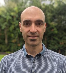 Borjan Gagoski合作论文数Boston Children's Hospital7
 Susan Whitfield-Gabrieli合作论文数Massachusetts Institute of Technology7Harvey Lui合作论文数7
 Hans Breiter合作论文数Warren Wright Adolescent Center, Feinberg School of Medicine, Northwestern University;Mass General Research Institute, Massachusetts General Hospital;Laboratory of Neuroimaging and Genetics, Massachusetts General Hospital;Toggle AI, Inc.;Sprk.AI, Inc.5Hoffmann Malte合作论文数5
 Clemens C.C. Bauer合作论文数Instituto de Neurobiología, Universidad Nacional Autónoma de México5
 Patricia Ellen Grant合作论文数Harvard Medical School;Maternal Fetal Care Center, Boston Children’s Hospital;Brain Injury Center, Boston Children’s Hospital5
 Elfar Adalsteinsson合作论文数Research Laboratory of Electronics, Massachusetts Institute of Technology;Institute for Medical Engineering & Science, Massachusetts Institute of Technology5Paolo Zanotti Fregonara合作论文数4Jose Montero Santamaria合作论文数4Raven Cureton合作论文数4Anthony Galassi合作论文数4Nafiseh Ghazanfari合作论文数University Medical Center Groningen, University of Groningen4
 Michelle Hampson合作论文数Neuroscience, Biological & Biomedical Sciences, Yale University4
 Robert Innis合作论文数National Institute of Mental Health, National Institutes of Health4Sami S. Zoghbi合作论文数National Institute of Mental Health, National Institutes of Health4
 Victor Pike合作论文数Section on PET Radiopharmaceutical Sciences, National Institute of Mental Health, National Institutes of Health4
 A. Eden Evins合作论文数Center for Addiction Medicine, Massachusetts General Hospital4
 Jeih-San Liow合作论文数National Institute of Mental Health, National Institutes of Health4
 Jinsoo Hong合作论文数Molecular Imaging Branch, National Institute of Mental Health, National Institutes of Health;Section on PET Radiopharmaceutical Sciences, National Institute of Mental Health, National Institutes of Health4
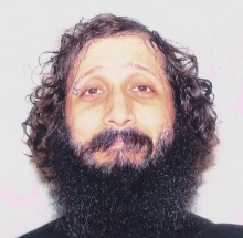 Jonathan Polimeni合作论文数Harvard Medical School;Athinoula A. Martinos Center for Biomedical Imaging, Massachusetts General Hospital;Division of Health Sciences and Technology, Harvard-MIT4
 Douglas Greve合作论文数Massachusetts General Hospital;Laboratory for Computational Neuroimaging, Athinoula A. Martinos Center for Biomedical Imaging, Mass General Research Institute;Harvard Medical School4H. Umesha Shetty合作论文数4
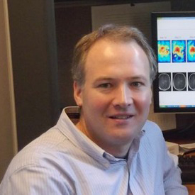 Ovidiu Andronesi合作论文数Harvard Medical School, Harvard University;Athinoula A. Martinos Center for Biomedical Imaging, Mass General Research Institute, Massachusetts General Hospital, Mass General Brigham4Stefan L Glimberg合作论文数3Madeline Jenkins合作论文数3Carson Knoer合作论文数3Nicolas Arango合作论文数3Jakob M Slipsager合作论文数3
 Stephanie M. Greer合作论文数Hopelab, 100 Calif St,Suite 1150, San Francisco, CA 94111 USA3
 Jiahe Zhang合作论文数bNortheastern University3V Calderon合作论文数3
 Martin Nørgaard合作论文数Rigshosp, Neurobiol Res Unit, Copenhagen, Denmark3
 Dustin Scheinost合作论文数Multi-Modal Imaging, Neuroinformatics, & Data Science Laboratory, Department of Radiology & Biomedical Imaging, Yale School of Medicine, Yale University;Child Study Center, Yale School of Medicine, Yale University3
 Judson A Brewer合作论文数Department of Behavioral and Social Sciences,School of Public Health, Brown University/Mindfulness Center at Brown,School of Public Health, Brown University3
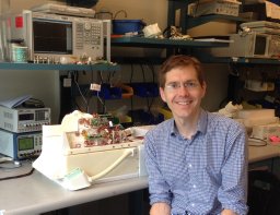 Stockmann Jason P合作论文数Massachusetts General Hospital Martinos Center for Imaging3
 John Gabrieli合作论文数Athinoula A. Martinos Imaging Center, McGovern Institute for Brain Research, Massachusetts Institute of Technology;Department of Brain and Cognitive Sciences, Massachusetts Institute of Technology3
 Reuter Martin合作论文数3
 Stephen Laconte合作论文数Biomedical Imaging Technology Center, Emory University & Georgia Institute of Technology3
 Divya Varadarajan合作论文数3
 Laurence M. Hirshberg合作论文数Development Center, Neuro3
 Satrajit Ghosh合作论文数McGovern Institute for Brain Research, Massachusetts Institute of Technology;Department of Otolaryngology Head and Neck Surgery, Medical School, Harvard University;Harvard Division of Medical Sciences3




