The human calcium-sensing receptor (CaSR) detects fluctuations in the extracellular Ca2+ concentration and maintains Ca2+ homeostasis1,2. It also mediates diverse cellular processes not associated with Ca2+ balance3-5. The functional pleiotropy of CaSR arises in part from its ability to signal through several G-protein subtypes6. We determined structures of CaSR in complex with G proteins from three different subfamilies: Gq, Gi and Gs. We found that the homodimeric CaSR of each complex couples to a single G protein through a common mode. This involves the C-terminal helix of each G alpha subunit binding to a shallow pocket that is formed in one CaSR subunit by all three intracellular loops (ICL1-ICL3), an extended transmembrane helix 3 and an ordered C-terminal region. G-protein binding expands the transmembrane dimer interface, which is further stabilized by phospholipid. The restraint imposed by the receptor dimer, in combination with ICL2, enables G-protein activation by facilitating conformational transition of G alpha. We identified a single G alpha residue that determines Gq and Gs versus Gi selectivity. The length and flexibility of ICL2 allows CaSR to bind all three G alpha subtypes, thereby conferring capacity for promiscuous G-protein coupling. Structures of the human calcium-sensing receptor can be bound into complex with G proteins from three different G alpha subtypes while maintaining G-protein-binding specificity.
The genomes of most mesophilic organisms encode multiple A TP- B inding C assette F (ABCF) proteins. EttA, one of four E. coli paralogs, regulates synthesis of the first peptide bond on the ribosome dependent on ATP/ADP ratio, while A ntibiotic Re sistance factors (AREs), paralogs in other organisms, both regulate and directly mediate resistance to ribosome-targeted antibiotics. However, the physiological functions remain unclear for most paralogs, and the mechanism-of-action has yet to be rigorously established for any paralog. We herein present single particle cryogenic electron microscopy structures of ribosome complexes of all four E. coli ABCF paralogs (EttA, Uup, YbiT, and YheS), which, together with previously determined ARE structures, show that ABCFs control the binding geometry of the tRNA in the peptidyl-tRNA-binding (P) site on the ribosome. They modulate the position of its acceptor stem relative to the peptidyl transferase center (PTC) in a manner that can either promote (EttA and Uup) or disrupt (YbiT, YheS, and the AREs) proper catalytic geometry. The YbiT/70S reconstructions include a conformation with no density for ribosomal protein bL33, and structural analyses support the exchange of this sub-stoichiometric ribosomal protein being functionally related to conformational changes in YbiT controlled by sequence variations in the strongly non-canonical Signature Sequence in its first ABC domain. Our studies establish general structural/enzymological principles by which the ATPase activity of ABCF proteins controls translation elongation coupled to modulation of conformation and stereochemistry in the catalytic core of the ribosome.
ABSTRACT Cystic fibrosis (CF) is caused by mutations in a chloride channel called the human Cystic Fibrosis Transmembrane Conductance Regulator (hCFTR). We used cryo-EM global conformational ensemble reconstruction to characterize the mechanism by which the breakthrough drug VX445 (Elexacaftor) simultaneously corrects both protein-folding and channel-gating defects caused by CF mutations. VX445 drives hCFTR molecules harboring the gating-defective G551D mutation towards the open-channel conformation by binding to a site in the first transmembrane domain. This binding interaction reverses the usual pathway of allosteric structural communication by which ATP binding activates channel conductance, which is blocked by the G551D mutation. Our ensemble reconstructions include a 3.4 Å non-native structure demonstrating that detachment of the first nucleotide-binding domain of hCFTR is directly coupled to local unfolding of the VX445 binding site. Reversal of this unfolding transition likely contributes to its corrector activity by cooperatively stabilizing NBD1 and the transmembrane domains of hCFTR during biogenesis. Summary Cryo-EM global conformational ensemble reconstruction has been used to characterize the mechanism-of-action of a breakthrough pharmaceutical that corrects fatal protein-folding and channel-gating defects in the human cystic fibrosis transmembrane conductance regulator (CFTR).
The human extracellular calcium-sensing (CaS) receptor controls plasma Ca2+ levels and contributes to nutrient-dependent maintenance and metabolism of diverse organs. Allosteric modulation of the CaS receptor corrects disorders of calcium homeostasis. Here, we report the cryogenic-electron microscopy reconstructions of a near-full-length CaS receptor in the absence and presence of allosteric modulators. Activation of the homodimeric CaS receptor requires a break in the transmembrane 6 (TM6) helix of each subunit, which facilitates the formation of a TM6-mediated homodimer interface and expansion of homodimer interactions. This transformation in TM6 occurs without a positive allosteric modulator. Two modulators with opposite functional roles bind to overlapping sites within the transmembrane domain through common interactions, acting to stabilize distinct rotamer conformations of key residues on the TM6 helix. The positive modulator reinforces TM6 distortion and maximizes subunit contact to enhance receptor activity, while the negative modulator strengthens an intact TM6 to dampen receptor function. In both active and inactive states, the receptor displays symmetrical transmembrane conformations that are consistent with its homodimeric assembly.
An amendment to this paper has been published and can be accessed via a link at the top of the paper.
The endoplasmic reticulum (ER) is a highly dynamic network of membranes. Here, we combine live-cell microscopy with in situ cryo-electron tomography to directly visualize ER dynamics in several secretory cell types including pancreatic β-cells and neurons under near-native conditions. Using these imaging approaches, we identify a novel, mobile form of ER, ribosome-associated vesicles (RAVs), found primarily in the cell periphery, which is conserved across different cell types and species. We show that RAVs exist as distinct, highly dynamic structures separate from the intact ER reticular architecture that interact with mitochondria via direct intermembrane contacts. These findings describe a new ER subcompartment within cells.
Cyclic nucleotide–gated (CNG) channels convert cyclic nucleotide (CN) binding and unbinding into electrical signals in sensory receptors and neurons. The molecular conformational changes underpinning ligand activation are largely undefined. We report both closed- and open-state atomic cryo-EM structures of a full-length Caenorhabditis elegans cyclic GMP−activated channel TAX-4, reconstituted in lipid nanodiscs. These structures, together with computational and functional analyses and a mutant channel structure, reveal a double-barrier hydrophobic gate formed by two S6 amino acids in the central cavity. cGMP binding produces global conformational changes that open the cavity gate located ~52 Å away but do not alter the structure of the selectivity filter—the commonly presumed activation gate. Our work provides mechanistic insights into the allosteric gating and regulation of CN-gated and nucleotide-modulated channels and CNG channel−related channelopathies. Cryo-EM structures of a C. elegans cGMP-activated channel TAX-4 in lipid nanodiscs reveal a hydrophobic gate in the central cavity and, together with electrophysiology, provide mechanistic insights into the gating and regulation of CNG channels.
The human GABAB receptor—a member of the class C family of G-protein-coupled receptors (GPCRs)—mediates inhibitory neurotransmission and has been implicated in epilepsy, pain and addiction1. A unique GPCR that is known to require heterodimerization for function2–6, the GABAB receptor has two subunits, GABAB1 and GABAB2, that are structurally homologous but perform distinct and complementary functions. GABAB1 recognizes orthosteric ligands7,8, while GABAB2 couples with G proteins9–14. Each subunit is characterized by an extracellular Venus flytrap (VFT) module, a descending peptide linker, a seven-helix transmembrane domain and a cytoplasmic tail15. Although the VFT heterodimer structure has been resolved16, the structure of the full-length receptor and its transmembrane signalling mechanism remain unknown. Here we present a near full-length structure of the GABAB receptor, captured in an inactive state by cryo-electron microscopy. Our structure reveals several ligands that preassociate with the receptor, including two large endogenous phospholipids that are embedded within the transmembrane domains to maintain receptor integrity and modulate receptor function. We also identify a previously unknown heterodimer interface between transmembrane helices 3 and 5 of both subunits, which serves as a signature of the inactive conformation. A unique ‘intersubunit latch’ within this transmembrane interface maintains the inactive state, and its disruption leads to constitutive receptor activity. The structure of the GABAB receptor in an inactive state reveals, amongst other features, a latch between the two subunits that locks the transmembrane domain interface, and the presence of large phospholipids that may modulate receptor function.
When the ribosome encounters a stop codon, it recruits a release factor (RF) to hydrolyze the ester bond between the peptide chain and tRNA. RFs have structural motifs that recognize stop codons in the decoding center and a GGQ motif for induction of hydrolysis in the peptidyl transfer center 70 Å away. Surprisingly, free RF2 is compact, with only 20 Å between its codon-reading and GGQ motifs. Cryo-EM showed that ribosome-bound RFs have extended structures, suggesting that RFs are compact when entering the ribosome and then extend their structures upon stop codon recognition. Here we use time-resolved cryo-EM to visualize transient compact forms of RF1 and RF2 at 3.5 and 4 Å resolution, respectively, in the codon-recognizing ribosome complex on the native pathway. About 25% of complexes have RFs in the compact state at 24 ms reaction time, and within 60 ms virtually all ribosome-bound RFs are transformed to their extended forms.
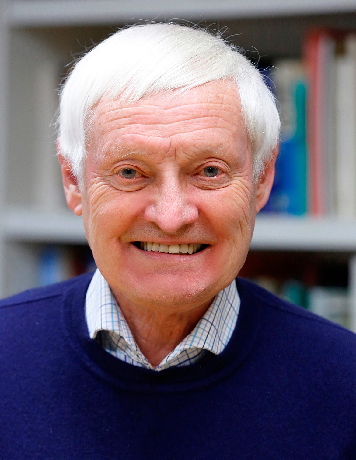 Joachim Frank (阿希姆·弗兰克)合作论文数Howard Hughes Medical Institute, Department of Biochemistry and Molecular Biophysics, College of Physicians and Surgeons, Columbia University;Department of Biological Sciences, Columbia University116
 Pawel Penczek合作论文数Department of Biochemistry and Molecular Biology, McGovern Medical School, University of Texas Health Science Center at Houston46
 Rajendra K. Agrawal合作论文数Department of Biomedical Sciences, State University of New York at Albany44
 Terence Wagenknecht合作论文数26
 Yelshanskaya Maria V合作论文数Department of Biochemistry and Molecular Biophysics, Columbia University17
 Knud H. Nierhaus合作论文数Institute of Medical Physics and Biophysics, Charité Universitätsmedizin Berlin;Max-Planck-Institut fur Molekulare Genetik17
 Sidney Fleischer合作论文数Department of Molecular Biology, Vanderbilt University17
 Irene S. Gabashvili合作论文数17
 Amedee des Georges合作论文数Chemistry & Biochemistry, the City College of New York15E.C. Twomey合作论文数13A.I. Sobolevsky合作论文数13cmt spahn合作论文数13M Radermacher合作论文数13
 Ziao Fu合作论文数12Sengupta Jayati合作论文数11
 Wayne A. Hendrickson合作论文数Department of Physiology and Cellular Biophysics, Columbia University Medical Center;Department of Biochemistry & Molecular Biophysics, Columbia University;Photon Sciences Directorate, Brookhaven National Laboratory10
 Oliver Biggs Clarke合作论文数Department of Medical Biology, The University of Melbourne10Nils Burkhardt合作论文数10Hstau Y. Liao合作论文数9
 Christian M. T. Spahn合作论文数Institut für Medizinische Physik und Biophysik, Charité – Universitätsmedizin Berlin9Mans Ehrenberg合作论文数9
 Robert Langlois合作论文数Department of Bioengineering, University of Illinois at Chicago Bioinformatics Program9J Berkowitz合作论文数8
 Poul Nissen合作论文数Department of Molecular Biology and Genetics, Aarhus University;Faculty of Natural Sciences, Aarhus University8
 Tatyana Pestova合作论文数Department of Cell Biology, SUNY Downstate Health Sciences University8
 Satish Kumar Srivastava合作论文数University of Texas Medical Branch7
 Manjuli R. Sharma合作论文数NY State Department of Health, Wadsworth Center7S. kaledhonkar合作论文数6G. Indrisiunaite合作论文数6J. Frank合作论文数6M. Sun合作论文数6Hb Xin合作论文数6
 Christopher U.T. Hellen合作论文数Department of Cell Biology, SUNY Downstate Health Sciences University6Adriana Verschoor合作论文数6Roland Beckmann合作论文数Department of Biochemistry, Ludwig-Maximilians-University of Munich6Tanvir R. Shaikh合作论文数Wadsworth Center, New York State Department of Health6
 Yaser Hashem合作论文数IBMC, Université Louis Pasteur de Strasbourg6Zheng Liu合作论文数5Ab Heagle合作论文数5
 Susan Madison-Antenucci合作论文数Parasitol Lab, New York State Dept Hlth5
 Andrew R. Marks合作论文数Department of Physiology & Cellular Biophysics, Vagelos College of Physicians and Surgeons, Columbia University;Department of Biomedical Engineering, The Fu Foundation School of Engineering and Applied Science, Columbia University5Albert E. Dahlberg合作论文数5
 Maria G Gomezlorenzo合作论文数5
 Ralf Junemann合作论文数5Mikel Valle合作论文数5Z. Fu合作论文数4M. Ehrenberg合作论文数4B. Shah合作论文数4Amy B. Heagle合作论文数4




