BACKGROUND:The Fassier-Duval (FD) telescopic rod is the standard of care for treating long bone fractures and deformities in children with osteogenesis imperfecta (OI). Despite its advantages, revision surgery remains common. This study aimed to analyze the medium-to long-term outcomes of FD rodding and identify risk factors for revision surgery. METHODS:A retrospective review was conducted on all OI patients who received lower limb FD rods between 2009 and 2020. Data on patient demographics, OI type, surgical details, and follow-up were collected. The primary outcome was revision surgery. We used Kaplan-Meier survival analysis and Cox proportional hazards modeling to identify risk factors associated with earlier revision. RESULTS:The study included 169 operative limbs in 80 patients with a median follow-up of 64 months. The overall revision rate was 36.69%. The most common reasons for revision were refracture (41.94%), recurrence of deformity (29.03%), and rod bending (27.42%). Survival analysis identified the cut point of the age at surgery of ≤30 months ( P =0.0003) and OI type IV ( P =0.034) as significant risk factors for earlier revision. For each additional month of age at surgery, the risk of revision decreased by 1.4% (HR=0.986, 95% CI: 0.979-0.994, P <0.001). CONCLUSIONS:In our 12-year experience, FD rods demonstrated a medium-term revision rate of ∼37%. Younger age at surgery (≤30 mo) and a diagnosis of OI type IV are critical predictors of earlier implant failure. These findings highlight the unique surgical challenges in the youngest and the more ambulatory OI populations. Further study on surgical technique and implant modification for these very young OI patients is warranted to improve surgical outcomes and implant survival. LEVELS OF EVIDENCE:Level III-prognostic studies.
Neurofibromatosis type 1 (NF-1) leads to cutaneous, neurological, and musculoskeletal manifestations. Congenital pseudarthrosis of the tibia treatment is considered aggressive in achieving bone union. Most of those patients get a short limb at the end of treatment because of pathological bone resection and malalignment. One of the manifestations that can appear in patients is localized hemihypertrophy of the limb. In our case, we present the first time a coexistence of congenital pseudarthrosis of the tibia and hemihypertrophy in the same leg was reported.
Arthrogryposis multiplex congenita (AMC) represents a large, rare group of congenital conditions. This study addressed major challenges in AMC research posed by the lack of systematic frameworks for data collection and the use of inconsistent terminologies and text descriptions. We aimed to systematically review the Human Phenotype Ontology (HPO) terms, encode AMC phenotypic traits as HPO terms, and pilot test the encoding process in a cohort of children with AMC. An international consensus-based dataset for AMC was used to extract phenotypic traits from the fetal period to adulthood. The encoding process was developed by an international expert panel to expand and revise HPO ontology for joint contractures, as the main characterizing traits in AMC. Using a pre-tested mapping algorithm, the HPO mapping process resulted in a 62% complete match, a 12% incomplete match, and a 26% no match. The encoding process included 37 new terms and annotations and 13 re-structures across 10 different joints. The implemented annotations significantly increased the number of available HPO terms for joint contractures in a cohort of children with AMC (p-value = 0.04). Our encoding and annotation approach may be used as a blueprint for systematic HPO (re)annotations for musculoskeletal and non-musculoskeletal phenotypic traits of AMC.
Going to the hospital can be scary for children, especially when they must go through a painful procedure. Doctors, nurses, and other health professionals may use special distraction techniques to help take children’s minds off the pain. There is a cool new way to help distract children from painful procedures. It is called virtual reality (VR). A study was done at a children’s hospital that specializes in bone care. The researchers wanted to know if VR was easy to use at the hospital to help the children deal with pain. The study included 44 children who had different kinds of procedures done, like having a needle put in their vein, removing stitches, having blood taken, and more. The researchers concluded that VR can help. VR is fast and easy to use, almost everyone liked it, and it works. To help with pain relief, virtual reality has all the ingredients to be a recipe for success!
Objective To investigate the extent of extraskeletal manifestations along with inpatient outcomes and complications associated with osteogenesis imperfecta (OI). Study design This cross-sectional study utilized the Kids' Inpatient Database as a part of the Healthcare Cost and Utilization Project to investigate inpatient hospital outcomes and management in patients with OI from 1997 through 2016. Data regarding hospital characteristics, cost of treatment, inpatient outcomes, and procedures were collected and analyzed. Results There were 7291 admissions that listed OI as a diagnosis in the Kids' Inpatient Database from 1997 through 2016. Unexpectedly, more than one-third of all admissions in these children with OI presented with an extraskeletal manifestation. The rate of major complications was 3.85%. The rate of minor complications was 19.4%, most commonly respiratory problems. The mortality rate was 18.2% in the neonatal period and 1.0% in all other admissions. Total charges of hospital stay increased over the years. Conclusions We identified a striking prevalence of extraskeletal manifestations in OI along with inpatient outcomes and complications associated with OI, of which respiratory complications were predominant. We observed a significant financial burden for patients with OI and identified additional risks for financial crisis, in addition to disparities in care identified among socioeconomic groups. These data contribute to a more holistic understanding of OI from diagnosis to management.
The Wnt signaling pathway is a key molecular process during fracture repair. Although much of what we now know about the role of this pathway in bone is derived from in vitro and animal studies, the same cannot be said about humans. As such, we hypothesized that Wnt signaling will also be a key process in humans during physiological fracture healing as well as in the development of a nonunion (hypertrophic and oligotrophic). We further hypothesized that the expression of Wnt-signaling pathway genes/proteins would exhibit a differential expression pattern between physiological fracture callus and the pathological nonunion tissues. We tested these two hypotheses by examining the mRNA levels of key Wnt-signaling related genes: ligands (WNT4, WNT10a), receptors (FZD4, LRP5, LRP6), inhibitors (DKK1, SOST) and modulators (CTNNB1 and PORCN). RNA sequencing from calluses as well as from the two nonunion tissue types, revealed that all of these genes were expressed at about the same level in these three tissue types. Further, spatial expression experiments identified the cells responsible of producing these proteins. Robust expression was detected in osteoblasts for the majority of these genes except SOST which displayed low expression, but in contrast, was mostly detected in osteocytes. Many of these genes were also expressed by callus chondrocytes as well. Taken together, these results confirm that Wnt signaling is indeed active during both human physiological fracture healing as well as in pathological nonunions.
 Noémi Dahan-Oliel合作论文数School of Physical & Occupational Therapy, Faculty of Medicine and Health Sciences, McGill University25
 Frank Rauch合作论文数23
 François Fassier合作论文数Division of Orthopaedic Surgery, Department of Surgery, Faculty of Medicine and Health Sciences, McGill University20
 Maryam Tabrizian合作论文数Department of Biomedical Engineering, McGill University;Biomat’X Research Laboratories, McGill University13Ashby Elizabeth合作论文数12
 Yousef Marwan合作论文数11
 Ziyad S Haidar合作论文数Center for Biorecognition and Biosensors, McGill University11
 Mitchell Bernstein合作论文数Department of Pediatric Surgery, Faculty of Medicine and Health Sciences, McGill University9N. Alam合作论文数Shriners Hosp Children8
 Makhdom Asim M合作论文数Faculty of Medicine and Health Sciences, King Abdul Aziz University8Kathleen Montpetit合作论文数Shriners Hospital for Children8Sofia Addab合作论文数7
 Mohammad Alzahrani合作论文数Montreal Children Hospital, McGill University;Department of Orthopaedic Surgery, University of Dammam;McGill University, University of Dammam7
 Bettina M Willie合作论文数Department of Bioengineering, University of Utah7
 Argerie Tsimicalis合作论文数Ingram School of Nursing, Faculty of Medicine and Health Sciences, McGill University6
 Louis-Nicolas Veilleux合作论文数Shriners Hospital for Children-Canada and Department of Pediatrics (L.-N.V., A.P.-L., McGill University;Centre de Réadaptation Marie Enfant (L.-N.V., A.P.-L.),, Sainte-Justine University Hospital;and Département de Kinanthropologie (M.L.),, Université du Québec à Montréal;Sainte-Justine University Hospital, Université du Québec à Montréal6
 Harold J P van Bosse合作论文数6Kyle Kavaseri合作论文数5Dominique Lauzier合作论文数5Kelly Thorstad合作论文数5
 Khaled Abu Dalu合作论文数5Caroline Elfassy合作论文数5
 Chantal Janelle合作论文数Department of Pediatric Surgery, Faculty of Medicine and Health Sciences, McGill University5Haque T合作论文数5
 Tanya Bedard合作论文数5
 Francis H. Glorieux合作论文数Shriners Hospitals for Children;McGill University5
 Isabelle Villemure合作论文数Research Center Sainte-Justine Hospital5A Izadpanah合作论文数4Vasiliki Betty Darsaklis合作论文数4Jessica Collins合作论文数4
 Marianne Gagnon合作论文数4
 Mohammed A. Alotaibi合作论文数4Saran Neil合作论文数4
 Mekhail Mina合作论文数Department of Biomedical Engineering, McGill University4
 Audrey Parent合作论文数4
 Annie Pouliot-Laforte合作论文数4
 Laurent Ballaz合作论文数4
 Judith G Hall合作论文数BC Children’s Hospital Research Institute;Department of Pediatrics, Faculty of Medicine, University of British Columbia4Francois Fassier合作论文数4Peter Kloen合作论文数afdeling Orthopedie, AMC Amsterdam4Mehdi Aarabi合作论文数4
 Rene St-Arnaud合作论文数Department of Human Genetics, School of Biomedical Sciences, Faculty of Medicine and Health Sciences, McGill University;Shriners Hospitals for Children Canada4Mathushan Subasri合作论文数3V Rosen合作论文数3David Bertrand合作论文数3
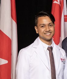 Nehal Islam合作论文数Dept Epidemiol Biostat & Occupat Hlth, McGill Univ3M DUHAIME合作论文数3
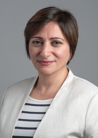 Hadil Al-Jallad合作论文数Division of Orthopaedic Surgery, Department of Surgery, Faculty of Medicine and Health Sciences, McGill University3




