Background The optimal ventilation strategy during cardiopulmonary resuscitation (CPR) remains undetermined. Synchronizing ventilation to chest compressions has been proposed to enhance end-organ perfusion and oxygenation. This study evaluates the pulmonary function and injury in a synchronized ventilation (SV) strategy using lower peak pressure compared to chest compression synchronized ventilation (CCSV) with higher peak pressures and intermittent positive pressure ventilation (IPPV). Materials/methods 35 pigs underwent cardiac arrest, followed by basic and advanced life support with mechanical chest compressions and ventilation according to randomized groups: IPPV (peak pressure (Ppeak) 40 mbar), SV (Ppeak 20 mbar), and CCSV (Ppeak 40 mbar). Arterial blood gases, ventilation-perfusion (V/Q) ratios, and hemodynamics were assessed during CPR and after return of spontaneous circulation (ROSC). Pulmonary histopathology and inflammatory markers (IL-6, TNF-α) were analyzed post-mortem. Results During CPR, CCSV demonstrated superior oxygenation compared to SV (paO2: CCSV 190.61 mmHg, SV 96.02 mmHg; p = 0.006), while CCSV and IPPV utilized significant higher airway pressures. SV showed the highest mean arterial pressure. Lactate levels were non-significantly highest in CCSV during CPR. ROSC rates were lower in CCSV (4/10) than in IPPV and SV (both 9/10); all non-ROSC CCSV cases exhibited pneumothoraces. Post-CPR, increased low V/Q and shunt fractions were observed in CCSV and SV exhibited reduced IL-6 expression. Conclusions SV resulted in lower oxygenation but utilized lower airway pressures compared with CCSV and IPPV, produced less lactate, and achieved ROSC rates comparable to IPPV with fewer complications compared to CCSV. These findings suggest that SV may represent a viable alternative ventilation strategy during CPR. Further studies are needed to confirm these results.
Lung injury caused by respiratory infection is a major cause of hospitalization and mortality and a leading origin of sepsis. Sepsis-associated encephalopathy and delirium are frequent complications in patients with severe lung injury, yet the pathogenetic mechanisms remain unclear. Here, 70 female C57BL/6 mice were subjected to a single full-body-exposure with nebulized lipopolysaccharide (LPS). Neuromotor impairment was assessed repeatedly and brain, blood, and lung samples were analyzed at survival points of 24 h, 48 h, 72 h, and 96 h after exposure. qRT-PCR revealed increased mRNA-expression of TNFα and IL-1β 24 h and 48 h after LPS-exposure in the lung, concomitantly with increased amounts of proteins in bronchoalveolar lavage and interstitial lung edema. In the cerebral cortex, at 72 h and/or 96 h after LPS exposure, the inflammation- and activity-associated markers TLR4, GFAP, Gadd45b, c-Fos, and Arc were increased. Therefore, single exposure to nebulized LPS not only triggers an early inflammatory reaction in the lung but also induces a delayed neuroinflammatory response. The identified mechanisms provide new insights into the pathogenesis of sepsis-associated encephalopathy and might serve as targets for future therapeutic approaches.
Background Sepsis is a common disease in intensive care units worldwide, which is associated with high morbidity and mortality. This process is often associated with multiple organ failure including acute lung injury. Although massive research efforts have been made for decades, there is no specific therapy for sepsis to date. Early and best treatment is crucial. Lidocaine is a common local anesthetic and used worldwide. It blocks the fast voltage-gated sodium (Na+) channels in the neuronal cell membrane responsible for signal propagation. Recent studies show that lidocaine administered intravenously improves pulmonary function and protects pulmonary tissue in pigs under hemorrhagic shock, sepsis and under pulmonary surgery. The aim of this study is to show that lidocaine inhalative induces equivalent effects as lidocaine intravenously in pigs in a lipopolysaccharide (LPS)-induced sepsis with acute lung injury. Methods After approval of the local State and Institutional Animal Care Committee, to induce the septic inflammatory response a continuous infusion of lipopolysaccharide (LPS) was administered to the pigs in deep anesthesia. Following induction and stabilisation of sepsis, the study medication was randomly assigned to one of three groups: (1) lidocaine intravenously, (2) lidocaine per inhalation and (3) sham group. All animals were monitored for 8 h using advanced and extended cardiorespiratory monitoring. Postmortem assessment included pulmonary mRNA expression of mediators of early inflammatory response (IL-6 & TNF-alpha), wet-to-dry ratio and lung histology. Results Acute respiratory distress syndrome (ARDS) was successfully induced after sepsis-induction with LPS in all three groups measured by a significant decrease in the PaO2/FiO2 ratio. Further, septic hemodynamic alterations were seen in all three groups. Leucocytes and platelets dropped statistically over time due to septic alterations in all groups. The wet-to-dry ratio and the lung histology showed no differences between the groups. Additionally, the pulmonary mRNA expression of the inflammatory mediators IL-6 and TNF-alpha showed no significant changes between the groups. The proposed anti-inflammatory and lung protective effects of lidocaine in sepsis-induced acute lung injury could not be proven in this study.
Aims: Influencing the inflammatory response represents an important branch in ARDS research. The naturally occurring polyphenol derivative resveratrol has already been confirmed to have strong anti-inflammatory effects on the cardiac and metabolic system. In the present study, we investigated the propagated anti-inflammatory effects of intravenous resveratrol in a porcine ARDS model.Main methods: 20 domestic pigs (30 +/- 2 kg; approval G20-1-135), divided into three groups: 1. resveratrol high dose (HD; n = 8), single bolus of 20 mg/kg over 15 min. 2. resveratrol low dose (LD; n = 8), single bolus of 10 mg/kg over 15 min. 3. Vehicle (n = 4), with the carrier solution DMSO over 15 min administered after ARDS induction. ARDS induction: using BAL/oleic acid and a subsequent test period of 8 h. Measurement parameters: Hemodynamics/spirometry data were collected continuously, BGA/laboratory parameters repetitively. Post-mortem: analysis of pulmonary inflammatory markers.Statistics: Two-way analysis of variance (repeated measurement) and Student-Newman-Keuls method.Key findings: Resveratrol HD significantly reduced the expression of TNF-alpha in lung tissue compared to the LD group (p < 0.05). A significantly increased functional residual capacity (FRC) could be demonstrated for the HD group at the end of the test (p < 0.05 for HD vs. LD/vehicle). Further, resveratrol HD reduced statistically the EVLWI compared to LD/vehicle (p < 0.05 at T4/T8).Significance: In this study, resveratrol HD ameliorated pulmonary mechanics as reported for the FRC and EVLWI. Further, the proposed anti-inflammatory effects of resveratrol, a significant reduction in the expression of TNF-alpha was observed in the HD group.
Sepsis and septic shock are frequently encountered in patients treated in intensive care units (ICUs) and are among the leading causes of death in these patients. It is caused by a dysregulated immune response to an infection. Even with optimized treatment, mortality rates remain high, which makes further insights into the pathophysiology and new treatment options necessary. Lipopolysaccharide (LPS) is a component of the cell membrane of gram-negative bacteria, which are often responsible for infections causing sepsis and septic shock. The severity and high mortality of sepsis and septic shock make standardized experimental studies in humans impossible. Thus, an animal model is needed for further studies. The pig is especially well suited for this purpose as it closely resembles humans in anatomy, physiology, and size. This protocol provides an experimental model for endotoxemic shock in pigs by LPS infusion. We were able to reliably induce changes frequently observed in septic shock patients, including hemodynamic instability, respiratory failure, and acidosis. This will allow researchers to gain valuable insight into this highly relevant condition and evaluate new therapeutic approaches in an experimental setting.
Sepsis and septic shock are frequently encountered in patients treated in intensive care units (ICUs) and are among the leading causes of death in these patients. It is caused by a dysregulated immune response to an infection. Even with optimized treatment, mortality rates remain high, which makes further insights into the pathophysiology and new treatment options necessary. Lipopolysaccharide (LPS) is a component of the cell membrane of gram-negative bacteria, which are often responsible for infections causing sepsis and septic shock. The severity and high mortality of sepsis and septic shock make standardized experimental studies in humans impossible. Thus, an animal model is needed for further studies. The pig is especially well suited for this purpose as it closely resembles humans in anatomy, physiology, and size. This protocol provides an experimental model for endotoxemic shock in pigs by LPS infusion. We were able to reliably induce changes frequently observed in septic shock patients, including hemodynamic instability, respiratory failure, and acidosis. This will allow researchers to gain valuable insight into this highly relevant condition and evaluate new therapeutic approaches in an experimental setting.
Abstract Objective This study aimed to determine whether ultra-low tidal volume ventilation (ULTVV) applied during cardiopulmonary resuscitation (CPR) compared with standard ventilation (intermittent positive pressure ventilation, IPPV) can reduce pulmonary end-organ damage in the post-resuscitation period. Methods A prospective, randomized trial was conducted using a porcine model (n = 45). The animals were divided into three groups: IPPV, ULTVV, and a sham control group. Juvenile male pigs underwent CPR after inducing ventricular fibrillation and received the designated ventilation intervention [IPPV: tidal volume 6–8 ml per kilogram body weight (ml/kg BW), respiratory rate 10/min, FiO2 1.0; ULTVV: tidal volume 2–3 ml/kg BW, respiratory rate 50/min, FiO2 1.0]. A 20-h observation period followed if return of spontaneous circulation was achieved. Histopathological examination using the diffuse alveolar damage scoring system was performed on postmortem lung tissue samples. Arterial and venous blood gas analyses and ventilation/perfusion measurements via multiple inert gas elimination technique (MIGET) were repeatedly recorded during the experiment. Results Out of the 45 experiments conducted, 28 animals were excluded based on predefined criteria. Histopathological analysis showed no significant differences in lung damage between the ULTVV and IPPV groups. ULTVV demonstrated adequate oxygenation and decarboxylation. MIGET measurements during and after resuscitation revealed no significant differences between the intervention groups. Conclusion In the short-term follow-up phase, ULTVV demonstrated similar histopathological changes and functional pulmonary parameters compared to standard ventilation. Further research is needed to investigate the long-term effects and clinical implications of ULTVV in resuscitation settings.
Background: Tracheal intubation in critical care is a high-risk procedure requiring significant expertise and airway strategy modification. We hypothesise that awake tracheal intubation is associated with a lower incidence of severe adverse events compared to standard tracheal intubation in critical care patients. Methods: Records were acquired for all tracheal intubations performed from 2020 to 2022 for critical care patients at a tertiary hospital. Each awake tracheal intubation case, using a videolaryngoscope with a hyperangulated blade (McGrath® MAC X-Blade), was propensity matched with two controls (1:2 ratio; standard intubation videolaryngoscopy (VL) and direct laryngoscopy (DL) undergoing general anaesthesia). The primary endpoint was the incidence of adverse events, defined as a mean arterial pressure of <55 mmHg (hypotension), SpO2 < 80% (desaturation) after sufficient preoxygenation, or peri-interventional cardiac arrest. Results: Of the 135 tracheal intubations included for analysis, 45 involved the use of an awake tracheal intubation. At least one adverse event occurred after tracheal intubation in 36/135 (27%) of patients, including awake 1/45 (2.2%; 1/1 hypotension), VL 10/45 (22%; 6/10 hypotension and 4/10 desaturation), and DL 25/45 (47%; 10/25 hypotension, 12/25 desaturation, and 3/25 cardiac arrest; p < 0.0001). Conclusions: In this retrospective observational study of intubation practices in critical care patients, awake tracheal intubation was associated with a lower incidence of severe adverse events than anaesthetised tracheal intubation.
The optimal ventilation strategy during cardiopulmonary resuscitation (CPR) has eluded scientists for years. This porcine study aims to validate the hypothesis that ultra-low tidal volume ventilation (tidal volume 2–3 mL kg−1; ULTVV) minimizes renal and hepatic end-organ damage when compared to standard intermittent positive pressure ventilation (tidal volume 8–10 mL kg−1; IPPV) during CPR. After induced ventricular fibrillation, the animals were ventilated using an established CPR protocol. Upon return of spontaneous circulation (ROSC), the follow-up was 20 h. After sacrifice, kidney and liver samples were harvested and analyzed histopathologically using an Endothelial, Glomerular, Tubular, and Interstitial (EGTI) scoring system for the kidney and a newly developed scoring system for the liver. Of 69 animals, 5 in the IPPV group and 6 in the ULTVV group achieved sustained ROSC and were enlisted, while 4 served as the sham group. Creatinine clearance was significantly lower in the IPPV-group than in the sham group (p < 0.001). The total EGTI score was significantly higher for ULTVV than for the sham group (p = 0.038). Aminotransferase levels and liver score showed no significant difference between the intervention groups. ULTVV may be advantageous when compared to standard ventilation during CPR in the short-term ROSC follow-up period.
The optimal ventilation method for patients suffering from cardiac arrest and receiving cardiopulmonary resuscitation (CPR) remains unclear, and the recent guidelines do not provide detailed information.[1-3] During CPR, changing thoracic pressures due to chest compressions and ventilation pressure influence venous return and cardiac output, respectively.[4-6] Novel mechanical ventilation modes that are synchronized to chest compressions may improve blood flow and oxygenation.[4] In this porcine trial, we investigated the feasibility of a specially designed chest compression synchronized ventilation (CCSV) mode with peak pressures limited to 40 mbar (1 mbar=0.1 kPa) and an experimental synchronized ventilation (SV) limited to 20 mbar, respectively, intended to achieve a more lung-protective ventilation pattern during resuscitation. We evaluated whether the different synchronized pressure levels ameliorate hemodynamics, gas exchange and pulmonary function and compared it to intermittent positive pressure ventilation (IPPV).
The calcium sensitiser levosimendan, which is used as an inodilator to treat decompensated heart failure, may also exhibit anti-inflammatory properties. We examined whether treatment with levosimendan improves cardiopulmonary function and is substantially beneficial to the inflammatory response in acute respiratory response syndrome (ARDS). Levosimendan was administered intravenously in a new experimental porcine model of ARDS. For comparison, we used milrinone, another well-known inotropic agent. Our results demonstrated that levosimendan intravenously improved hemodynamics and lung function in a porcine ARDS model. Significant beneficial alterations in the inflammatory response and lung injury were not detected.
Introduction Perioperative red blood cell (RBC) transfusions have been associated with increased morbidity and worse oncological outcome in some solid neoplasms. In order to elucidate whether RBC transfusions themselves, the preoperative anemia of cancer (AOC), or the impaired global health status might explain this impact on patients with endometrial cancer (EC) or ovarian cancer (OC), we performed a retrospective, single-institution cohort study. Materials and methods Women older than 60 years with EC or OC were included. The influence of RBC transfusions, AOC, and frailty status determined by the G8 geriatric screening tool (G8 score), as well as the clinical-pathological cancer characteristics on progression-free survival (PFS) and overall survival (OS), was determined by using the Kaplan-Meier method and the Cox regression analyses. Results In total, 263 patients with EC (n = 152) and OC (n = 111) were included in the study. Patients with EC receiving RBC transfusions were faced with a significantly shorter 5-year PFS (79.8% vs. 26.0%; p < 0.001) and 5-year OS (82.6% vs. 25.7%; p < 0.001). In multivariable analyses, besides established clinical-pathological cancer characteristics, the RBC transfusions remained the only significant prognostic parameter for PFS (HR: 1.76; 95%-CI [1.01–3.07]) and OS (HR: 2.38; 95%-CI [1.50–3.78]). In OC, the G8 score stratified the cohort in terms of PFS rates (G8-non-frail 53.4% vs. G8-frail 16.7%; p = 0.010) and AOC stratified the cohort for 5-year OS estimates (non-anemic: 36.7% vs. anemic: 10.6%; p = 0.008). Multivariable Cox regression analyses determined the G8 score and FIGO stage as independent prognostic factors in terms of PFS (HR: 2.23; 95%-CI [1.16–4.32] and HR: 6.52; 95%-CI [1.51–28.07], respectively). For OS, only the TNM tumor stage retained independent significance (HR: 3.75; 95%-CI [1.87–7.53]). Discussion The results of this trial demonstrate the negative impact of RBC transfusions on the prognosis of patients with EC. Contrastingly, the prognosis of OC is altered by the preoperative global health status rather than AOC or RBC transfusions. In summary, we suggested a cumulatively restrictive transfusion management in G8-non-frail EC patients and postulated a more moderate transfusion management based on the treatment of symptomatic anemia without survival deficits in OC patients.
Background Interorgan cross-talk describes the phenomenon in which a primarily injured organ causes secondary damage to a distant organ. This cross-talk is well known between the lung and brain. One theory suggests that the release and systemic distribution of cytokines via the bloodstream from the primarily affected organ sets in motion proinflammatory cascades in distant organs. In this study, we analysed the role of the systemic distribution of cytokines via the bloodstream in a porcine ARDS model for organ cross-talk and possible inflammatory changes in the brain. Methods After approval of the State and Institutional Animal Care Committee, acute respiratory distress syndrome (ARDS) induction with oleic acid injection was performed in seven animals. Eight hours after ARDS induction, blood (35–40 ml kg –1 ) was taken from these seven ‘ARDS donor’ pigs. The collected ‘ARDS donor’ blood was transfused into seven healthy ‘ARDS-recipient’ pigs. Three animals served as a control group, and blood from these animals was transfused into three healthy pigs after an appropriate ventilation period. All animals were monitored for 8 h using advanced cardiorespiratory monitoring. Postmortem assessment included cerebral (hippocampal and cortex) mediators of early inflammatory response (IL-6, TNF-alpha, iNOS, sLCN-2), wet-to-dry ratio and lung histology. TNF-alpha serum concentration was measured in all groups. Results ARDS was successfully induced in the ‘ARDS donor’ group, and serum TNF-alpha levels were elevated compared with the ‘ARDS-recipient’ group. In the ‘ARDS-recipient’ group, neither significant ARDS alterations nor upregulation of inflammatory mediators in the brain tissue were detected after high-volume random allogenic ‘ARDS-blood’ transfusion. The role of the systemic distribution of inflammatory cytokines from one affected organ to another could not be confirmed in this study.
Background: Invasive ventilation during cardiopulmonary resuscitation (CPR) is very complex due to unique thoracic pressure conditions. Current guidelines do not provide specific recommendations for ventilation during ongoing chest compressions regarding positive end-expiratory pressure (PEEP). This trial examines the cardiopulmonary effects of PEEP application during CPR. Methods: Forty-two German landrace pigs were anaesthetised, instrumented, and randomised into six intervention groups. Three PEEP levels (0, 8, and 16 mbar) were compared in high standard and ultralow tidal volume ventilation. After the induction of ventricular fibrillation, mechanical chest compressions and ventilation were initiated and maintained for thirty minutes. Blood gases, ventilation/perfusion ratio, and electrical impedance tomography loops were taken repeatedly. Ventilation pressures and haemodynamic parameters were measured continuously. Postmortem lung tissue damage was assessed using the diffuse alveolar damage (DAD) score. Statistical analyses were performed using SPSS, and p values <0.05 were considered significant. Results: The driving pressure (Pdrive) showed significantly lower values when using PEEP 16 mbar than when using PEEP 8 mbar (p = 0.045) or PEEP 0 mbar (p < 0.001) when adjusted for the ventilation mode. Substantially increased overall lung damage was detected in the PEEP 0 mbar group (vs. PEEP 8 mbar, p = 0.038; vs. PEEP 16 mbar, p = 0.009). No significant differences in mean arterial pressure could be detected. Conclusion: The use of PEEP during CPR seems beneficial because it optimises ventilation pressures and reduces lung damage without significantly compromising blood pressure. Further studies are needed to examine long-term effects in resuscitated animals.
The use of extracorporeal life support (ECLS) as part of cardio-circulatory support has increased rapidly in recent years. Severe hyperlactatemia is not uncommon in this group of patients. Lactate peak concentrations and lactate clearance have already been identified as independent marker for mortality in critical ill patients without mechanical device support. The aim of this study was to determine a supposed correlation between the variables lactate peak concentration and clearance in the blood and mortality in the ECLS context. Therefore, a total of 51 cardiac surgery ICU patients with ECLS therapy were included in this retrospective, clinical observational study (survivors n = 23; non-survivors n = 28). Lactate measurement was performed before, during and after ECLS therapy. Further, common ICU scores (SAPSII, SOFA, TISS28), the rates of transfusion and the different vasopressor therapies will be compared. Significant elevated peak lactate levels and poor lactate clearance were associated with higher mortality during ECLS therapy (p < 0.001). Deceased patients had higher SAPSII scores (p < 0.001), received more transfusions (p < 0.001) and presented with higher rates of epinephrine (p < 0.001). In conclusion, hyperlactatemia during ECLS therapy is a time sensitive emergency. Lactate cannot be cleared in all patients. Reversible causes should be explored and treated. In cases where the cause is irreversible, the prognosis of elevated lactate concentrations and reduced clearance is very poor.
Background: Shedding of the endothelial glycocalyx can be observed regularly during sepsis.Moreover, sepsis may be associated with acute respiratory distress syndrome (ARDS), which requires lung protective ventilation with the two cornerstones of application of low tidal volume and positive endexpiratory pressure.This study investigated the effect of a lung protective ventilation on the integrity of the endothelial glycocalyx in comparison to a high tidal volume ventilation mode in a porcine model of sepsis-induced ARDS.Methods: After approval by the State and Institutional Animal Care Committee, 20 male pigs were anesthetized and received a continuous infusion of lipopolysaccharide to induce septic shock.The animals were randomly assigned to either low tidal volume ventilation, high tidal volume ventilation, or sham ventilation groups and observed for 6 hours.In addition to the gas exchange parameters and hematologic analyses, the serum hyaluronic acid concentrations were determined from central venous blood and from pre-and postpulmonary and pre-and postcerebral circulation.Post-mortem analysis included histopathological evaluation and determination of the pulmonary and cerebral wet-to-dry ratios.Results: Both sepsis groups developed ARDS within 6 hours of the experiment and showed significantly increased serum levels of hyaluronic acid in comparison to the sham group.No significant differences in the hyaluronic acid concentrations were detected before and after pulmonary and cerebral circulation.There was also no significant difference in the serum hyaluronic acid concentrations between the two sepsis groups.Post-mortem analysis showed no significant difference between the two sepsis groups. Conclusion:In a porcine model of septic shock and ARDS, the serum hyaluronic acid levels were significantly elevated in both sepsis groups in comparison to the sham group.Intergroup comparison between lung protective ventilated and high tidal ventilated animals revealed no significant differences in the serum hyaluronic acid levels.
Endotracheal intubation is often a basic requirement for translational research in porcine models for various interventions that require a secured airway or high ventilation pressures. Endotracheal intubation is a challenging skill, requiring a minimum number of successful endotracheal intubations to achieve a high success rate under optimal conditions, which is often unachievable for non-anaesthesiology researchers. Due to the specific porcine airway anatomy, a difficult airway can usually be assumed. The impossibility of establishing a secure airway can result in injury, adverse events, or death of the laboratory animal. Using a prospective, randomized, controlled evaluation approach, it has been shown that fiberopticassisted endotracheal intubation takes longer but has a higher first-pass success rate than conventional intubation without causing clinically relevant drops in oxygen saturation. This model presents a standardized regimen for endoscopically guided endotracheal intubation, providing a secured airway, especially for researchers who are inexperienced in the technique of endotracheal intubation via direct laryngoscopy. This procedure is expected to minimize animal suffering and unnecessary animal losses.
Abstract Background Acute respiratory distress syndrome (ARDS) is a common disease in intensive care medicine. Despite intensive research, mortality rates are high, not even in COVID-19 ARDS. Thereby, pigs offer some advantages to study the characteristics of ARDS. Many different ARDS models exist. Most of the articles published focused on histopathological and microscopic lung alterations to identify the most suitable animal ARDS model. “Macroscopic” observations and descriptions are often missing. Therefore, we performed a post-hoc comparison of two common ARDS models for pigs: lipopolysaccharide (LPS) vs. a double-hit model (bronchoalveolar lavage + oleic acid infusion). We investigated hemodynamic, spirometric and laboratory changes as another main clinical part of ARDS. Results The groups were compared by two-way analysis of variance (ANOVA) with a post-hoc Student–Newman–Keuls test. A p value lower than 0.05 was accepted as significant. All animals (n = 8 double-hit ARDS; n = 8 LPS ARDS) survived the observation period of 8 h. ARDS induction with reduced oxygen indices was successful performed in both models (76 ± 35/225 ± 54/212 ± 79 vs. 367 ± 64; T0/T4/T8 vs. BLH for double-hit; 238 ± 57/144 ± 59 vs. 509 ± 41; T4/T8 vs. BLH for LPS; p < 0.05). ARDS induced with LPS leads to more hemodynamic (mean arterial pulmonary pressure 35 ± 3/30 ± 3 vs. 28 ± 4/23 ± 4; T4/T8 LPS vs. double-hit; p < 0.05; doses of norepinephrine 1.18 ± 1.05 vs. 0.11 ± 0.16; LPS vs. double-hit for T8; p < 0.05) and inflammatory (pulmonary IL-6 expression: 2.41e−04 ± 1.08e−04 vs. 1.45e−05 ± 7.26e−06; LPS vs. double-hit; p < 0.05) alterations. ARDS induced by double-hit requires a more invasive ventilator strategy to maintain a sufficient oxygenation (PEEP at T4: 8 ± 3 vs. 6 ± 2; double-hit vs. LPS; p < 0.05). Conclusions Both animal ARDS models are feasible and are similar to human presentation of ARDS. If your respiratory research focus on hemodynamic/inflammation variables, the LPS-induced ARDS is a feasible model. Studying different ventilator strategies, the double-hit ARDS model offers a suitable approach.
Background: Shedding of the endothelial glycocalyx can be observed regularly during sepsis. Moreover, sepsis may be associated with acute respiratory distress syndrome (ARDS), which requires lung protective ventilation with the two cornerstones of application of low tidal volume and positive end-expiratory pressure. This study investigated the effect of a lung protective ventilation on the integrity of the endothelial glycocalyx in comparison to a high tidal volume ventilation mode in a porcine model of sepsis-induced ARDS. Methods: After approval by the State and Institutional Animal Care Committee, 20 male pigs were anesthetized and received a continuous infusion of lipopolysaccharide to induce septic shock. The animals were randomly assigned to either low tidal volume ventilation, high tidal volume ventilation, or no-LPS-group groups and observed for 6 h. In addition to the gas exchange parameters and hematologic analyses, the serum hyaluronic acid concentrations were determined from central venous blood and from pre- and postpulmonary and pre- and postcerebral circulation. Post-mortem analysis included histopathological evaluation and determination of the pulmonary and cerebral wet-to-dry ratios. Results: Both sepsis groups developed ARDS within 6 h of the experiment and showed significantly increased serum levels of hyaluronic acid in comparison to the no-LPS-group. No significant differences in the hyaluronic acid concentrations were detected before and after pulmonary and cerebral circulation. There was also no significant difference in the serum hyaluronic acid concentrations between the two sepsis groups. Post-mortem analysis showed no significant difference between the two sepsis groups. Conclusion: In a porcine model of septic shock and ARDS, the serum hyaluronic acid levels were significantly elevated in both sepsis groups in comparison to the no-LPS-group. Intergroup comparison between lung protective ventilated and high tidal ventilated animals revealed no significant differences in the serum hyaluronic acid levels.
Background A laryngeal tube is often used as an alternative to intubation with direct laryngoscopy during cardiopulmonary resuscitation. However, in a study with piglets undergoing simulated cardiac arrest, reduced carotid artery blood flow was associated with the insertion of different supraglottic airway devices, such as a laryngeal tube. Limited by its construction, secondary tracheal intubation through a laryngeal tube was difficult or impossible in contrast to a second generation laryngeal mask. The new disposable intubating laryngeal tube with suction (iLTS-D ® ) seems to facilitate tracheal intubation. We hypothesized that iLTS-D, when inflated to the recommended air volume, does not reduce cerebral oxygenation in patients with cardiovascular diseases undergoing elective cardiac surgery. Methods This single-center prospective, controlled observational study was approved by the local ethics committee (Ethical Committee No. 2018-13716). Forty adult patients undergoing elective cardiac surgery requiring tracheal intubation were included in this study. The exclusion criteria were age <18 years and a high risk of aspiration, inability to consent, height <155 cm, or pregnancy. Prior to insufflation and deflation of the cuffs, we performed cerebral oximetry via near-infrared spectroscopy. The primary outcome was a significant reduction in NIRS in the context of the preinduction baseline value after inflation of the cuffs with the recommended air volume, defined as a ≥25% decrease from baseline or an absolute value ≤ 50%. The secondary endpoints were differences in time points, insertion success rates, and complications. Results There was no significant reduction in cerebral oximetry after inflation with the recommended cuff volume and an initially measured cuff pressure of >120 cmH2O. Overall, tracheal intubation was achieved in a median of 20 s [interquartile range 15-23 s] and enabled sufficient ventilation and tracheal intubation through the iLTS-D in all patients. Traces of blood on the cuffs (after removing the iLTS-D) and a sore throat (evaluated 2 h after removing the tracheal tube) were observed in one patient. Conclusion Our results showed no association between the insertion of the iLTS-D and reduced cerebral oxygenation in patients undergoing elective cardiac surgery. Trial registration ClinicalTrials.gov , NCT03942809.
 Ziebart Alexander合作论文数18Ruemmler Robert合作论文数17Hartmann Erik K合作论文数15Miriam Renz合作论文数13Kamuf Jens合作论文数11Katja Mohnke合作论文数10Julian Riedel合作论文数10Moritz Gosling合作论文数4Duenges Bastian合作论文数4Andrea Urmann合作论文数3Marc Kriege合作论文数3
 Thomas Ott合作论文数3Katharina Ritter合作论文数2
 Eric Hotz合作论文数2
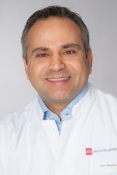 Hazem El Beyrouti合作论文数2
 Matthias David合作论文数2Liu Tanghua合作论文数2Thomas Rainer合作论文数2Arno Schad合作论文数2Joerg Albers合作论文数1Moritz Schaefer合作论文数1
 Michael K.E. Schafer合作论文数1Christiane Westphalen合作论文数1Andreas Garcia Bardon合作论文数1Valerie Catherine Linz合作论文数1Mehmet Oezkur合作论文数1Amelie Löwe合作论文数1Thomas Baller合作论文数1Victoria Buschmann合作论文数1Elisabeth Britten合作论文数1Leah Müllejans合作论文数1Wolfgang Weikel合作论文数1
 Mona Wanda Schmidt合作论文数1
 Katharina Anic合作论文数1
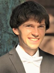 Markus Schepers合作论文数Univ Med Ctr, Johannes Gutenberg Univ Mainz1Christian Möllmann合作论文数1
 Roxana Schwab合作论文数1Slavomir Krajnak合作论文数1Conradi Roland合作论文数1
 Marco Battista合作论文数1Dohle Daniel-Sebastian合作论文数West German Heart Center, University of Duisburg-Essen1
 Marcus Schmidt合作论文数für Molekulare Onkologie, Universitätsmedizin Mainz1
 Marc Bodenstein合作论文数1Annette Hasenburg合作论文数1
 Florian Heid合作论文数1
 Walburgis Brenner合作论文数1




