Sleep spindles are rhythmic electroencephalographic signatures of non-rapid-eye-movement sleep. Their dysregulation has been implicated in several neuropsychiatric illnesses. Spindles have a characteristic waxing and waning shape, but the cellular and circuit mechanisms controlling their shape are not well understood. Recent but sparse research has implied that sleep spindle shape becomes abnormal in post-traumatic stress disorder (PTSD). PTSD patients have dysfunctional GABA A receptors in midline thalamic regions, areas involved in the orchestration of sleep spindles. We modelled this GABA A dysfunction within thalamocortical (TC) neurons using localized CRISPR-Cas9 technology to test the hypothesis that GABA dysfunction would dysregulate sleep spindle shape and cause symptoms of PTSD, in mouse model behavioral evaluations. We found sleep spindles were shorter and abnormally shaped, having lost their characteristic waxing and waning shape, in mice with GABA A receptor knock-down in TC neurons (TC-α1KD). TC-α1KD mice failed to recover from learned fearful reactions following an aversive stimulus. We tested this with a contextual fear conditioning paradigm using electric foot shocks. A control group with intact GABA A receptors successfully habituated to the fear conditioned location in subsequent visits to that context without foot shocks. In contrast, TC-α1KD mice never habituated, suggesting abnormally extended fearful memories. The number of inhibitory post synaptic currents in TC neurons were significantly decreased in vitro , confirming an effective knock-down. Our results imply that abnormally shaped sleep spindles may serve as a biomarker of GABA A receptor dysfunction in TC neurons which may be involved in abnormal fear processing in PTSD. We postulate GABA A receptor dysfunction in TC neurons may be underlying pathophysiology of PTSD and our findings here may inspire the development of screens, diagnostics and objective characteristics of stress related disorders, including PTSD.
Sleep abnormalities and dysfunction of gamma band (30-80 Hz) activity generated by parvalbumin (PV) interneurons are early characteristics of Alzheimer's disease (AD) which correlate with the severity of amyloid-β deposition (Aβ) and cognitive impairment. However, the timing of these alterations in vivo with respect to disease progression is unclear. Here, in longitudinal recordings from APP/PS1/PV-cre (AD mice) from 3-6 months, we found reduced sleep slow-wave power (0.5-4 Hz) in hippocampus and medial prefrontal cortex in AD mice as young as 3 months old, compared to non-AD (PV-cre) mice, well before overt pathology. This finding was primarily due to reductions in the NREM delta range (1.5-4 Hz), a hallmark of restorative functions of sleep. In contrast, beta (15-30 Hz) power linked to insomnia was significantly higher across all sleep-wake states. Loss of deep NREM sleep was not compensated by an increase in NREM sleep time, instead NREM sleep during the dark (active) phase was slightly but significantly lower in AD mice. 40-Hz auditory steady-state responses and associated evoked calcium responses of hippocampal PV neurons recorded using fiber photometry were also impaired by 3 months old. However, Y-maze performance in 3- and 6-month-old AD mice was not significantly different from non-AD mice. These results reveal reduced deep sleep and PV-associated 40-Hz activity as very early changes amenable to early intervention occurring prior to cognitive deficits. Furthermore, they establish APP/PS1 mice as a good model to causally test the relationship between sleep, PV neuronal activity and amyloid-mediated pathology.
Sleep spindles are cortical electrical oscillations considered critical for memory consolidation and sleep stability. The timing and pattern of sleep spindles are likely to be important in driving synaptic plasticity during sleep as well as preventing disruption of sleep by sensory and internal stimuli. However, the relative importance of factors such as sleep depth, cortical up/down-state, and temporal clustering in governing sleep spindle dynamics remains poorly understood. Here, we analyze sleep data from 1,025 participants, statistically modeling the simultaneous influences of multiple factors on moment-to-moment spindle production using a point process-generalized linear model framework. Results reveal fingerprint-like timing patterns, characterized by a refractory period followed by a period of increased spindle activity, which are highly individualized yet consistent night-to-night, with increased variability with age. Strikingly, short-term (<15 s) temporal patterns of past spindle history are the main determinant of spindle timing, accounting for over 70% of the statistical deviance—surpassing the contribution of factors such as cortical up/down-state (slow oscillation phase), sleep depth, and long-term history (15 to 90 s, including ~50 s infraslow activity). Short-term history has a statistically significant influence in over 98% of the population, suggesting it is a near-universal feature of spindle activity. Short-term history and slow oscillation phase exert independent effects on spindle timing. Our results establish a robust statistical framework to examine abnormalities in sleep spindle timing observed in neurological disorders and aging, as well as the relationship between individualized sleep spindle timing, cognition, and sleep stability.
Prolonged wakefulness induces a homeostatic sleep response (HSR) with increases in non-REM sleep time and delta power. The quality of wakefulness impacts the magnitude and form of the HSR but little is known about the neural mechanisms. Opto-stimulation of basal forebrain (BF) glutamatergic neurons (stim) leads to arousal and avoidance behavior. Here we compared the HSR following 4 hr of prolonged wakefulness induced by opto-stimulation of BF glutamate neurons and compared to sleep deprivation (SD) induced by gentle handling (GH). Finally, we compared the c-Fos activation pattern of the whole brain after 4hr of opto-stimulation compared to 4hr SD induced by GH. Mice (vGlut2-Cre or C57BL6) were implanted with microdialysis/optodialysis probes targeting the BF and EEG/EMG electrodes. Unilateral opto-stimulations of BF vGluT2 (20Hz, 5s On-55s Off) neurons and SD were performed during ZT3-ZT7 with or without the administration of ionotropic glutamate receptor antagonists (DNQX + D-AP5) and allowed 3h of recovery sleep. For cFos mapping whole brains were collected after 4hr of stim or SD with time-matched undisturbed controls, and analyzed for activated cFos (Lifecanvas, Inc). BF vGluT2 stimulation caused rapid arousal and, compared to baseline day, mice stayed awake (93.3 ±1.2%. N=10) during the 4 h of stimulation, comparable to that noted for SD (~95%) by GH. Stim conditions showed a significant hourly decrease in wakefulness from 2-4h which was further decreased by optodialysis of the ionotropic glutamate receptors antagonists, effects not seen in SD condition. Unlike SD group, the recovery NREM sleep remained unaltered following BF vGluT2 stim. SD increased NREM delta (0.5-4Hz) but the stim group showed a selective increase in 2.5-3.5Hz range. The whole brain c-Fos mapping revealed different activation patterns between stimulation and SD groups for multiple brain regions including the habenula and hypothalamus. Our data suggests a differential pattern of arousal and HSR following BF vGluT2 stimulation, and resulting activation of aversive brain circuits, when compared to that of GH-induced SD. R01 NS119227 (R.B.); VA Biomedical Laboratory Research and Development Service Merit Awards I01 BX001404, I01 BX006105 (R.B.), I01 BX002774 (J.T.M.), I01 BX004673 (R.E.B.). I01 BX006550 (DSU); IK2 BX 004905 (DSU)
Sleep-wake scoring in vivo electrophysiologic signals is necessary in many basic and translational studies. Performing this manually is a burden and bottleneck. Thus, many attempts have been made to automate it. Recently, with increased access to machine learning (ML) technologies, a new wave of attempts is occurring. The overwhelmingly common strategy deployed involves leveraging large datasets to train completely novel, yet relatively simple, ML-models. However, evidence suggests this does little to help with a common problem that is ML-models can perform poorly with new or unfamiliar data. Instead, transfer-learning – re-training highly sophisticated ML models – is known to be flexible and dependable, robustly dealing with novel data. We demonstrate transfer-learning of GoogLeNet provides highly dependable sleep-wake scoring across several diverse mouse electrophysiologic datasets, and matches the scoring of the person who trained it. We term our freely-available code “Sleep-Deep-Learner”. Transfer-learning was used to retrain GoogLeNet – accessed first unmodified, from the pretrained deep-neural-nets available via MATLAB. High-level final layers were replaced to classify wavelet transforms of epochs as wakefulness, NREM sleep or REM sleep. We used F1 scores to test how closely Sleep-Deep-Learner mimics two independent expert scorers. We validated performance in wild-type EEG, EEG altered by the hypnotic agent zolpidem, LFP data, data from an Alzheimer’s disease model and even sub-cortical data (hippocampus). We also reproduced findings of a CRISPR-based study previously completed with manual scoring. We ensure accurate fine-grain sleep architecture with hypnograms and bout analyses. Automated scores were very similar to either expert scorer regardless of dataset. We estimate this reduces labor burden of scoring to one twelfth. We provide a transfer-learning based approach to automating sleep-wake scoring. This has the advantage that automated sleep-wake scores agree with those of the expert scorer using the application. This, unbiased, flexible approach of retraining before each scoring session means there is no dependence on familiarity with novel data to perform accurately. VA Biomedical Laboratory Research and Development Service CDA-2 IK2 BX004905 (D.S.U.) and Merit Awards I01BX006550 (D.S.U.) I01 BX001404 and I01 BX006105(R.B.); I01 BX004673 (R.E.B.) and NIH K01 AG068366 (FK), R01 NS119227 (R.B.)
Sleep spindles have been identified as key potential biomarkers for numerous neurological and psychiatric disorders, as well as for aging. Thus, studying spindle mechanisms provides valuable insight into the pathophysiology of various disorders. In addition to translatable human studies, animal model studies can more directly and invasively probe and systematically alter spindle networks. While various animal studies have shown distinct morphological differences between human and animal model spindles, the assumptions underlying approaches to spindle detection remain relatively unchanged across species. To assess the generalizability and validity of existing studies, it is crucial to understand how spindle activity and detection assumptions vary across species. Recently, new quantitative approaches have identified broader classes of human spindle-like transient events that are more informative than traditionally detected spindles. In this study, we extend these approaches to non-human primates and rodents to characterize spindle activity across species, validate signal processing assumptions, and understand implications for past and future studies. We analyzed sleep period electroencephalogram (EEG) recordings of central electrodes from adult humans and mature macaques (N=5), rats (N=8), and mice (N=4) from baseline or control data from previously published studies. For each record, we used the DYNAM-O toolbox to quantify and visualize the dynamics of thousands of spindle-like transient oscillatory peaks across the night. We find distinct morphological and distributional differences in spindles between species. With increasingly lower-order species, spindles become markedly less morphologically distinct and more variable in their dynamics. In humans, fast spindles (12-16Hz) appear on spectrograms as distinct events in clear, narrow-band frequency. In primates, spindles are less separable and fall within a wider frequency range (8-14Hz). In rats, we observe one diffuse mode (6-16Hz) with little separability in events. Mice (6-20Hz) possess the least separable events and the most variability. Given the diffuse morphology and high variability of spindles in animal models relative to humans, traditionally detected spindles likely represent a very small fraction of the underlying activity. Thus, caution should be taken, particularly in rodents, in the interpretation of studies focusing on individual spindles and their relationship to other waveforms. MJP: 1RF1AG079917-01A1, REB: I01 BX004673, FK: NIH K01 AG068366
American Servicemembers are routinely exposed to prolonged periods of intense stress during deployment and combat exposure. These stressors are critical risk factors for the pathogenesis and severity of psychiatric disease—a leading cause of permanent disability within VA that significantly increases risk of suicide. However, the identity of the regions, cell types, and mechanisms integrating the effect of stress on sleep disruption remain poorly characterized, thus hindering the development of effective treatments. I have previously found that BF Npas1+ neurons project to numerous brain regions implicated in arousal, reward, and the stress response, and disrupt normal sleep behavior and oscillatory activity when chemogenetically activated, and are therefore potential mediators for stress-induced sleep disruption and psychiatric disease. I explored this using a chronic multimodal stress paradigm (10 days, 4h/day) in mice. Mice were implanted with EEG and EMG electrodes to monitor sleep behavior and oscillations before and during the 10 days of stress. Stress-sensitive mice were identified by a reduction in their sucrose preference. Immunohistochemistry was performed in basal forebrain tissue for the transcription factor NPAS1 and the immediate early gene cFOS. Preliminary findings indicate that Npas1+ neurons in the basal forebrain (BF) are potently activated by stress (30-40+% cFOS-expressing), correlating with disruptions in sucrose preference. These stress-sensitive mice tended to spend less time awake (n=8, p=0.13) and in NREM sleep (p=0.09) during the light-(active) period, with more dramatic shifts seen in the dark-(inactive) period with wake (p< 0.05) and NREM sleep (p< 0.01). Individual wake and NREM bouts tended to be less frequent after stress. Furthermore, slow and delta wave decreased following stress, correlating to the degree of increased NREM sleep. Sleep-spindle density also significantly decreased following chronic stress. Notably, the proportion of cFOS-expressing Npas1+ neurons correlates with the post-stress change in wake (p=0.07), and with NREM (p=0.04) and REM sleep (p=0.01). I conclude Npas1+ basal forebrain neurons are potently activated by stress and may play a key role in stress-induced sleep and psychiatric illness. This work was supported by US VA Merit Awards I01 BX004673, I01 BX002774, and the HMS DSM T32 Training Program in Sleep, Circadian and Respiratory Neurobiology.
Sleep-wake scoring is a time-consuming, tedious but essential component of clinical and preclinical sleep research. Sleep scoring is even more laborious and challenging in rodents due to the smaller EEG amplitude differences between states and the rapid state transitions which necessitate scoring in shorter epochs. Although many automated rodent sleep scoring methods exist, they do not perform as well when scoring new datasets, especially those which involve changes in the EEG/EMG profile. Thus, manual scoring by expert scorers remains the gold standard. Here we take a different approach to this problem by using a neural network to accelerate the scoring of expert scorers. Sleep-Deep-Learner creates a bespoke deep convolution neural network model for individual electroencephalographic or local-field-potential (LFP) records via transfer learning of GoogLeNet, by learning from a small subset of manual scores of each EEG/LFP record as provided by the end-user. Sleep-Deep-Learner then automates scoring of the remainder of the EEG/LFP record. A novel REM sleep scoring correction procedure further enhanced accuracy. Sleep-Deep-Learner reliably scores EEG and LFP data and retains sleep-wake architecture in wild-type mice, in sleep induced by the hypnotic zolpidem, in a mouse model of Alzheimer's disease and in a genetic knock-down study, when compared to manual scoring. Sleep-Deep-Learner reduced manual scoring time to 1/12. Since Sleep-Deep-Learner uses transfer learning on each independent recording, it is not biased by previously scored existing datasets. Thus, we find Sleep-Deep-Learner performs well when used on signals altered by a drug, disease model, or genetic modification.
Summary Attention is impaired in many neuropsychiatric disorders, as well as by sleep disruption, leading to decreased workplace productivity and increased risk of accidents. Thus, understanding the neural substrates is important. Here we test the hypothesis that basal forebrain neurons that contain the calcium‐binding protein parvalbumin modulate vigilant attention in mice. Furthermore, we test whether increasing the activity of basal forebrain parvalbumin neurons can rescue the deleterious effects of sleep deprivation on vigilance. A lever release version of the rodent psychomotor vigilance test was used to assess vigilant attention. Brief and continuous low‐power optogenetic excitation (1 s, 473 nm @ 5 mW) or inhibition (1 s, 530 nm @ 10 mW) of basal forebrain parvalbumin neurons was used to test the effect on attention, as measured by reaction time, under control conditions and following 8 hr of sleep deprivation by gentle handling. Optogenetic excitation of basal forebrain parvalbumin neurons that preceded the cue light signal by 0.5 s improved vigilant attention as indicated by quicker reaction times. By contrast, both sleep deprivation and optogenetic inhibition slowed reaction times. Importantly, basal forebrain parvalbumin excitation rescued the reaction time deficits in sleep‐deprived mice. Control experiments using a progressive ratio operant task confirmed that optogenetic manipulation of basal forebrain parvalbumin neurons did not alter motivation. These findings reveal for the first time a role for basal forebrain parvalbumin neurons in attention, and show that increasing their activity can compensate for disruptive effects of sleep deprivation.
Sleep occurs in all animals but its amount, form, and timing vary considerably between species and between individuals. Currently, little is known about the basis for these differences, in part, because we lack a complete understanding of the brain circuitry controlling sleep-wake states and markers for the cell types which can identify similar circuits across phylogeny. Here, I explain the utility of an "Evo-devo" approach for comparative studies of sleep regulation and function as well as for sleep medicine. This approach focuses on the regulation of evolutionary ancient transcription factors which act as master controllers of cell-type specification. Studying these developmental transcription factor cascades can identify novel cell clusters which control sleep and wakefulness, reveal the mechanisms which control differences in sleep timing, amount, and expression, and identify the timepoint in evolution when different sleep-wake control neurons appeared. Spatial transcriptomic studies, which identify cell clusters based on transcription factor expression, will greatly aid this approach. Conserved developmental pathways regulate sleep in mice, Drosophila, and C. elegans. Members of the LIM Homeobox (Lhx) gene family control the specification of sleep and circadian neurons in the forebrain and hypothalamus. Increased Lhx9 activity may account for increased orexin/hypocretin neurons and reduced sleep in Mexican cavefish. Other transcription factor families specify sleep-wake circuits in the brainstem, hypothalamus, and basal forebrain. The expression of transcription factors allows the generation of specific cell types for transplantation approaches. Furthermore, mutations in developmental transcription factors are linked to variation in sleep duration in humans, risk for restless legs syndrome, and sleep-disordered breathing. This paper is part of the "Genetic and other molecular underpinnings of sleep, sleep disorders, and circadian rhythms including translational approaches" collection.
ABSTRACT Sleep spindles are critical for memory consolidation and strongly linked to neurological disease and aging. Despite their significance, the relative influences of factors like sleep depth, cortical up/down states, and spindle temporal patterns on individual spindle production remain poorly understood. Moreover, spindle temporal patterns are typically ignored in favor of an average spindle rate. Here, we analyze spindle dynamics in 1008 participants from the Multi-Ethnic Study of Atherosclerosis using a point process framework. Results reveal fingerprint-like temporal patterns, characterized by a refractory period followed by a period of increased spindle activity, which are highly individualized yet consistent night-to-night. We observe increased timing variability with age and distinct gender/age differences. Strikingly, and in contrast to the prevailing notion, individualized spindle patterns are the dominant determinant of spindle timing, accounting for over 70% of the statistical deviance explained by all of the factors we assessed, surpassing the contribution of slow oscillation (SO) phase (∼14%) and sleep depth (∼16%). Furthermore, we show spindle/SO coupling dynamics with sleep depth are preserved across age, with a global negative shift towards the SO rising slope. These findings offer novel mechanistic insights into spindle dynamics with direct experimental implications and applications to individualized electroencephalography biomarker identification.
COPYRIGHT © 2023 Brown and de Lecea. This is an open-access article distributed under the terms of the Creative Commons Attribution License (CC BY). The use, distribution or reproduction in other forums is permitted, provided the original author(s) and the copyright owner(s) are credited and that the original publication in this journal is cited, in accordance with accepted academic practice. No use, distribution or reproduction is permitted which does not comply with these terms. Editorial: Insights in sleep and circadian rhythms: 2021
EDITORIAL article Front. Neurosci., 17 January 2023Sec. Sleep and Circadian Rhythms Volume 17 - 2023 | https://doi.org/10.3389/fnins.2023.1133907
EDITORIAL article Front. Neurosci., 01 August 2023Sec. Sleep and Circadian Rhythms Volume 17 - 2023 | https://doi.org/10.3389/fnins.2023.1254248
ABSTRACTSleep-wake scoring is a time-consuming, tedious but essential component of clinical and pre-clinical sleep research. Sleep scoring is even more laborious and challenging in rodents due to the smaller EEG amplitude differences between states and the rapid state transitions which necessitate scoring in shorter epochs. Although many automated rodent sleep scoring methods exist, they do not perform as well when scoring new data sets, especially those which involve changes in the EEG/EMG profile. Thus, manual scoring by expert scorers remains the gold-standard. Here we take a different approach to this problem by using a neural network to accelerate the scoring of expert scorers. Sleep-Deep-Net (SDN) creates a bespoke deep convolution neural network model for individual electroencephalographic or local-field-potential records via transfer learning of GoogleNet, by learning from a small subset of manual scores of each EEG/LFP record as provided by the end-user. SDN then automates scoring of the remainder of the EEG/LFP record. A novel REM scoring correction procedure further enhanced accuracy. SDN reliably scores EEG and LFP data and retains sleep-wake architecture in wild-type mice, in sleep induced by the hypnotic zolpidem, in a mouse model of Alzheimer’s disease and in a genetic knock-down study, when compared to manual scoring. SDN reduced manual scoring time to 1/12. Since SDN uses transfer learning on each independent recording, it is not biased by previously scored existing data sets. Thus, we find SDN performs well when used on signals altered by a drug, disease model or genetic modification.STATEMENT OF SIGNIFICANCESleep medicine is often critically advanced by translational research based onin vivoelectrophysiologic mouse data. A necessary but time-consuming step in this field is scoring epochs of recordings into wakefulness, non-rapid-eye-movement sleep and non-rapid-eye-movement sleep. Despite efforts to automate this, manual scoring remains the gold-standard since automatic methods poorly handle data that is not similar enough to data used during development. Here, we describe a novel automated sleep scoring method that involves retraining a deep-convolution-neural-net capable of computer vision to score sleep-wake patterns after learning from a small set of manual scores within a record. This avoids biasing the model to expect data to be the same as its training set from previous records.
Here we describe a novel group of basal forebrain (BF) neurons expressing neuronal PAS domain 1 (Npas1), a developmental transcription factor linked to neuropsychiatric disorders. Immunohistochemical staining in Npas1-cre-2A-TdTomato mice revealed BF Npas1 + neurons are distinct from well-studied parvalbumin or cholinergic neurons. Npas1 staining in GAD67-GFP knock-in mice confirmed that the vast majority of Npas1 + neurons are GABAergic, with minimal colocalization with glutamatergic neurons in vGlut1-cre-tdTomato or vGlut2-cre-tdTomato mice. The density of Npas1 + neurons was high, 5-6 times that of neighboring cholinergic, parvalbumin or glutamatergic neurons. Anterograde tracing identified prominent projections of BF Npas1 + neurons to brain regions involved in sleep-wake control, motivated behaviors and olfaction such as the lateral hypothalamus, lateral habenula, nucleus accumbens shell, ventral tegmental area and olfactory bulb. Chemogenetic activation of BF Npas1 + neurons in the light (inactive) period increased the amount of wakefulness and the latency to sleep for 2-3 hr, due to an increase in long wake bouts and short NREM sleep bouts. Non-REM slow-wave (0-1.5 Hz) and sigma (9-15 Hz) power, as well as sleep spindle density, amplitude and duration, were reduced, reminiscent of findings in several neuropsychiatric disorders. Together with previous findings implicating BF Npas1 + neurons in stress responsiveness, the anatomical projections of BF Npas1 + neurons and the effect of activating them suggest a possible role for BF Npas1 + neurons in motivationally-driven wakefulness and stress-induced insomnia. Identification of this major subpopulation of BF GABAergic neurons will facilitate studies of their role in sleep disorders, dementia and other neuropsychiatric conditions involving BF. SIGNIFICANCE STATEMENT:We characterize a group of basal forebrain (BF) neurons in the mouse expressing neuronal PAS domain 1 (Npas1), a developmental transcription factor linked to neuropsychiatric disorders. BF Npas1 + neurons are a major subset of GABAergic neurons distinct and more numerous than cholinergic, parvalbumin or glutamate neurons. BF Npas1 + neurons target brain areas involved in arousal, motivation and olfaction. Activation of BF Npas1 + neurons in the light (inactive) period increased wakefulness and the latency to sleep due to increased long wake bouts. Non-REM sleep slow waves and spindles were reduced reminiscent of findings in several neuropsychiatric disorders. Identification of this major subpopulation of BF GABAergic wake-promoting neurons will allow studies of their role in insomnia, dementia and other conditions involving BF.
Sleep disorders are widespread in society and are prevalent in military personnel and in Veterans. Disturbances of sleep and arousal mechanisms are common in neuropsychiatric disorders such as schizophrenia, post-traumatic stress disorder, anxiety and affective disorders, traumatic brain injury, dementia, and substance use disorders. Sleep disturbances exacerbate suicidal ideation, a major concern for Veterans and in the general population. These disturbances impair quality of life, affect interpersonal relationships, reduce work productivity, exacerbate clinical features of other disorders, and impair recovery. Thus, approaches to improve sleep and modulate arousal are needed. Basic science research on the brain circuitry controlling sleep and arousal led to the recent approval of new drugs targeting the orexin/hypocretin and histamine systems, complementing existing drugs which affect GABAA receptors and monoaminergic systems. Non-invasive brain stimulation techniques to modulate sleep and arousal are safe and show potential but require further development to be widely applicable. Invasive viral vector and deep brain stimulation approaches are also in their infancy but may be used to modulate sleep and arousal in severe neurological and psychiatric conditions. Behavioral, pharmacological, non-invasive brain stimulation and cell-specific invasive approaches covered here suggest the potential to selectively influence arousal, sleep initiation, sleep maintenance or sleep-stage specific phenomena such as sleep spindles or slow wave activity. These manipulations can positively impact the treatment of a wide range of neurological and psychiatric disorders by promoting the restorative effects of sleep on memory consolidation, clearance of toxic metabolites, metabolism, and immune function and by decreasing hyperarousal.
The perinatal environment interacts with the genotype of the developing organism resulting in a unique phenotype through a developmental or perinatal programming phenomenon. However, it remains unclear how this phenomenon differentially affects particular targets expressing specific drinking responses depending on the perinatal conditions. The main goal of the present study was to compare the dipsogenic responses induced by different thirst models as a function of two perinatal manipulation models, defined by the maternal free access to hypertonic sodium solution and a partial aortic ligation (PAL-W/Na) or a sham-ligation (Sham-W/Na). The programmed adult offspring of both perinatal manipulated models responded similarly when was challenged by overnight water dehydration or after a sodium depletion showing a reduced water intake in comparison to the non-programmed animals. However, when animals were evaluated after a body sodium overload, only adult Sham-W/Na offspring showed drinking differences compared to PAL and control offspring. By analyzing the central neurobiological substrates involved, a significant increase in the number of Fos + cells was found after sodium depletion in the subfornical organ of both programmed groups and an increase in the number of Fos + cells in the dorsal raphe nucleus was only observed in adult depleted PAL-W/Na. Our results suggest that perinatal programming is a phenomenon that differentially affects particular targets which induce specific dipsogenic responses depending on matching between perinatal programming conditions and the osmotic challenge in the latter environment. Probably, each programmed-drinking phenotype has a particular set point to elicit specific repertoires of mechanisms to reestablish fluid balance.
Identification of mechanisms which increase deep sleep could lead to novel treatments which promote the restorative effects of sleep. Here, we show that knockdown of the α3 GABA A -receptor subunit from parvalbumin neurons in the thalamic reticular nucleus using CRISPR-Cas9 gene editing increased the thalamocortical delta (1.5–4 Hz) oscillations which are implicated in many health-promoting effects of sleep. Inhibitory synaptic currents in thalamic reticular parvalbumin neurons were strongly reduced in vitro. Further analysis revealed that delta power in long NREM bouts prior to NREM-REM transitions was preferentially affected by deletion of α3 subunits. Our results identify a role for GABA A receptors on thalamic reticular nucleus neurons and suggest antagonism of α3 subunits as a strategy to enhance delta activity during sleep.
Sleep abnormalities are widely reported in patients with Alzheimer's disease (AD) and are linked to cognitive impairments. Sleep abnormalities could be potential biomarkers to detect AD since they are often observed at the preclinical stage. Moreover, sleep could be a target for early intervention to prevent or slow AD progression. Thus, here we review changes in brain oscillations observed during sleep, their connection to AD pathophysiology and the role of specific brain circuits. Slow oscillations (0.1–1 Hz), sleep spindles (8–15 Hz) and their coupling during non-REM sleep are consistently reduced in studies of patients and in AD mouse models although the timing and magnitude of these alterations depends on the pathophysiological changes and the animal model studied. Changes in delta (1–4 Hz) activity are more variable. Animal studies suggest that hippocampal sharp-wave ripples (100–250 Hz) are also affected. Reductions in REM sleep amount and slower oscillations during REM are seen in patients but less consistently in animal models. Thus, changes in a variety of sleep oscillations could impact sleep-dependent memory consolidation or restorative functions of sleep. Recent mechanistic studies suggest that alterations in the activity of GABAergic neurons in the cortex, hippocampus and thalamic reticular nucleus mediate sleep oscillatory changes in AD and represent a potential target for intervention. Longitudinal studies of the timing of AD-related sleep abnormalities with respect to pathology and dysfunction of specific neural networks are needed to identify translationally relevant biomarkers and guide early intervention strategies to prevent or delay AD progression.
 Robert W. McCarley合作论文数Harvard Medical School55
 James J. Mckenna合作论文数31
 Radhika Basheer合作论文数Division of Sleep Medicine, Harvard Medical School, Harvard Univeristy;The Baker Center for Children and Families, Harvard Medical School, Harvard Univeristy27
 Robert E. Strecker合作论文数Division of Sleep Medicine, Harvard Medical School;Department of Psychology, Stonehill College, Harvard Medical School;Veterans Affairs Boston Healthcare System;Faculty of Psychiatry, Harvard Medical School22Fumi Katsuki合作论文数Department of Neurobiology and Anatomy, Wake Forest School of Medicine;Department of Neurobiology and Anatomy, Wake Forest School of Medicine19
 James G. McNally合作论文数Department of X-Ray Microscopy, Helmholtz Zentrum Berlin17
 James T. Mckenna合作论文数Laboratory of Neuroscience, VA Boston Healthcare System and Harvard Medical School16R. Basheer合作论文数15Y. Yanagawa合作论文数15David S Uygun合作论文数Dept. of Psychiatry, VA Boston Healthcare System and Harvard Medical School15
 Chun Yang合作论文数Division of Sleep Medicine, Harvard Medical School;Boston VA Research Institute, Inc.15James M. McNally合作论文数Department of Psychiatry, VA Boston Healthcare System;Department of Psychiatry, Harvard Medical School;Department of Psychiatry, Harvard Medical School15R. E. Strecker合作论文数14S. Winston合作论文数13
 Stephen Thankachan合作论文数Jawaharlal Nehru University School of Life Sciences13C. Yang合作论文数9
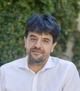 Karl Deisseroth合作论文数Department of Bioengineering, Stanford University8Bernat Kocsis合作论文数7
 Anna Kalinchuk合作论文数University of Helsinki Institute of Biomedicine7
 Tae Kim合作论文数6
 Lichao Chen合作论文数6Stuart Winston合作论文数6
 Thakkar Mahesh M合作论文数Harry S. Truman Memorial Veterans' Hospital and Department of Neurology, University of Missouri5
 Charu Shukla合作论文数5Felipe L. Schiffino合作论文数The University of Delaware5w robert合作论文数4
 Jee Hyun Choi合作论文数Korea Institute of Science &Department of Neuroscience, University of Science &Technology, Daejon 305-333 Korea. jeechoi@kist.re.kr;Korea Institute of Science & Technology, University of Science & Technology4Yunren Bolortuya合作论文数4Michael J. Prerau合作论文数Critical Care, and Pain Medicine, Department of Anesthesia4S. Franciosi合作论文数3
 L. Chen合作论文数3
 Mackenzie Gamble合作论文数School of Medicine, Boston University3Eunjin Hwang合作论文数Ctr Neurosci, Korea Inst Sci & Technol3Spencer Kevin M合作论文数3
 Yuchio Yanagawa合作论文数Gunma University3
 Jaime Tartar合作论文数3
 Christopher P Ward合作论文数2Janneke C Zant合作论文数2Deepak Shrivastava合作论文数2
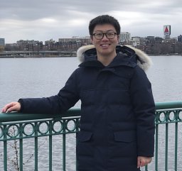 Shuqiang Chen合作论文数Graduate Program for Neuroscience, Boston University2
 Hio-Been Han合作论文数Program Brain & Cognit Engn, Korea Adv Inst Sci & Technol2
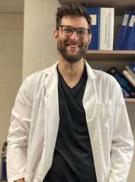 Michael Leonard合作论文数Department of Pharmacology, Vanderbilt University2
 Mingjian He合作论文数2
 John McCoy合作论文数Marketing Department, The Wharton School, University of Pennsylvania2
 Tatiana Korotkova合作论文数2Pan Jen Q合作论文数Stanley Center for Psychiatric Research, Broad Institute of Harvard and MIT2
 Sato Honma合作论文数Center for Sleep and Circadian Rhythm Disorders, Sapporo Hanazono Hospital2
 Seiji Nishino合作论文数SCN lab, Departmrnt of Psychiatry and Behavioral Sciences, Stanford University School of Medicine2




