Background Anoxic brain injury is the leading cause of death following successful resuscitation after out-of-hospital cardiac arrest. No experimental neuroprotective therapy has translated into clinical benefit. Collaborative efforts to harmonize methods may be essential for translational success. Neurofilament light chain (NfL) is a biomarker of neuroaxonal injury with emerging use in clinical practice, but data on its robustness and association with outcomes in porcine models is limited. Our aim was to use NfL as a quantitative marker to compare brain injury severity in different porcine cardiac arrest models. Methods Forty-four pigs (Sus scrofa domestica) were resuscitated after cardiac arrest in different experimental models in four laboratories. Plasma samples were collected prior to the arrest and at 4 and 24 hours after return of spontaneous circulation. NfL concentration was measured using a Single molecule array (Simoa) immunoassay. Results NfL levels changed significantly over time (p <0.001), with similar temporal patterns observed across all laboratories. Inter-laboratory comparisons indicated essentially consistent NfL levels, with modest differences at 24 hours. NfL levels at 24 hours showed an association with no-flow duration (n = 43) in both correlation and univariate regression analyses (p = 0.034, p = 0.041), while no significant associations were found with functional outcomes (n = 26) (p = 0.32, p = 0.20). Conclusion NfL increased consistently across four porcine cardiac arrest models, and higher 24-hour NfL levels were associated with longer no-flow duration, supporting its robustness as a biomarker of neuroaxonal injury. Larger studies are needed to clarify its relationship with functional outcome, as the exploratory analyses were underpowered.
Background:The current landscape of emergency care (EC) is marked by high demand, leading to issues such as emergency department boarding, overcrowding, and subsequent delays that impact the quality and safety of patient care. Integrating data science into EC can enhance decision-making with predictive, preventative, personalized, and participatory approaches. However, gaps in adherence to fairness, accountability, interpretability, and responsibility are evident, particularly due to barriers to data-sharing, which often result in a lack of transparency and robust oversight in these applications. Objective:The FAIR-EC (Fair, Accountable, Interpretable, and Responsible-Emergency Care) collaboration adapts the existing Fair, Accountable, Interpretable, and Responsible principles to address emerging challenges as data science integrates with EC. This initiative aims to transform EC by establishing ethical artificial intelligence standards specifically tailored for this integration. By bridging the gap between EC professionals, data scientists, and other stakeholders, the collaboration promotes international cooperation that leverages advanced data science techniques to enhance EC outcomes across different care settings. Methods:We propose a federated research design to analyze extensive datasets from various global institutions without compromising patient privacy. This approach transforms epidemiological research with advanced data science techniques, emphasizing the harmonization of data for comprehensive analyses across different health care systems. Results:The FAIR-EC initiative has facilitated the identification and harmonization of datasets from diverse geographical regions, enabling the examination of regional variations in EC practices. As of paper submission, participating sites have identified retrospective EC datasets totaling >2 million records (eg, Duke Health >400,000 and Singapore General Hospital >1.7 million records). Initial projects have demonstrated feasibility and operational readiness, including implementation of federated workflows and ongoing development of a federated scoring system, cross-site evaluation, and adaptation of association studies and predictive models across various regions. Cross-site harmonization and pilot analyses are underway (with local ethics approvals in progress), and first multisite results are expected to be submitted in mid-late 2026, with additional project-level publications anticipated in 2027. These efforts highlight the feasibility of leveraging advanced data science techniques to address the complexities of EC while preserving patient privacy without centralizing individual-level data. This project was funded from September 1, 2022, to August 31, 2023. Conclusions:FAIR-EC integrates data science ethically and effectively into EC, addressing challenges such as fragmented data, real-time handoffs, and public health crises. Its federated design harmonizes diverse data streams while preserving privacy, and its emphasis on ethical artificial intelligence aligns with the dynamic nature of EC. Despite challenges in data variability and system complexity, FAIR-EC establishes a strong foundation for innovation in global EC.
OBJECTIVES:The 2024 Society for Academic Emergency Medicine Consensus Conference focused on developing a pathway to build and support a diverse and sustainable emergency medicine (EM) clinician-scientist workforce. The underlying premise is that the specialty of EM needs a robust clinician-scientist workforce to fulfill its research mission of creating new knowledge to improve patient care and outcomes. METHODS:Preconference workgroups assessed existing pathways to develop and support EM clinician-scientists and generated unranked lists of strategies to holistically and comprehensively grow the clinician-scientist workforce. These strategies were refined and prioritized during a one-day, in-person conference, which was followed by a virtual conference to reach consensus on metrics, goals, and timelines for implementation. RESULTS:Overarching strategies included fostering a departmental culture that values research, addressing barriers to recruiting and retaining a diverse research work force, and enhancing the national reputation of EM research. At the undergraduate and medical school stage, creating a portfolio of medium- and long-term research training opportunities with EM faculty mentors was the highest priority. At the resident and fellow stage, top priorities were dedicated research training built into EM residencies and clinical fellowships. Early-career faculty strategies prioritized departmental support for federally funded K awards. Mid-career faculty strategies prioritized securing federal support for research mentoring, leading institutional training grants, and building research teams that include PhD scientists. At all stages, we addressed recruitment and retention of trainees and faculty from disadvantaged and underserved groups. CONCLUSIONS:These prioritized strategies with respective metrics, goals, timelines, and responsible parties provide a roadmap for EM to build a broadly inclusive and sustainable clinician-scientist workforce, capable of creating the new knowledge needed to advance emergency medical care. Successful implementation will require substantial commitment and investment from national EM organizations and academic department chairs. The result will be improved care and outcomes for the patients and communities we serve.
Introduction: During cardiopulmonary resuscitation (CPR), chest compressions induce oscillations in capnography. The relative magnitude of these oscillations can be quantified by the Airway Opening Index (AOI). AOI is hypothesized to be reflective of airway patency and effective ventilation, and may be associated with higher incidence of return of spontaneous circulation (ROSC). Aim: We sought to determine whether AOI is measurable and associated with ROSC using retrospective analyses of human out-of-hospital cardiac arrest (OHCA) and swine resuscitation. Methods: Human OHCA cases received attempted resuscitation from a single metropolitan EMS system between 2015-2021. Yorkshire swine received either 5 or 10 minutes of induced ventricular fibrillation followed by mechanical CPR. Cases were excluded if capnography was missing or artifacted. AOI was calculated using 4 established methods (Fig. 1). Median AOI for each AOI method was compared between humans and swine and according to ROSC status using Wilcoxon rank-sum test. Results: Among the 2095 eligible human cases, 50% (n=1046) achieved ROSC. Among the 37 eligible swine, 70% (n=27) achieved ROSC. Median AOI [Methods 1-4] was higher in humans ([14.9-22.2%]) than swine ([4.1-7.5%], p<0.001). AOI by all 4 methods was higher among those who achieved ROSC compared to No-ROSC for humans (Fig. 2) and for swine (Fig. 3). The difference in AOI according to ROSC was greater in swine than humans ([4.0-8.2%] vs [2.1-3.5%] respectively, p<0.05). Conclusion: AOI was measured in human OHCA and an experimental swine model. AOI was associated with ROSC in both animals and humans. AOI magnitude was significantly smaller in swine, though swine had a greater AOI difference between ROSC and No-ROSC cases. Further investigation will explore whether these differences are due to different thoracic anatomy and/or experimental conditions between humans and swine. Overall, the results validate the experimental swine model as a platform to investigate whether AOI is a modifiable target to increase the likelihood of ROSC, with a goal to improve outcomes for cardiac arrest.
Introduction: The primary goals of cardiopulmonary resuscitation (CPR) are to generate adequate myocardial blood flow to enable restoration of mechanical function and adequate brain blood flow to minimize ischemic injury. While the correlation between myocardial blood flow and coronary perfusion pressure during CPR is well established, the correlation between cerebral blood flow and cerebral perfusion pressure is less clear. Common carotid artery (CCA) blood flow is an accessible metric of brain blood flow during CPR in large animal studies. Here, we evaluate the relationships between CCA and several pressure values during CPR. Methods: Analysis was performed on 28 swine. Animals were instrumented to monitor arterial blood pressure (ABP), central venous blood pressure (CVP), intracranial pressure (ICP) and common carotid artery flow. Cerebral perfusion pressure (CePP) was calculated as mean arterial pressure (MAP) minus ICP. Following baseline measurements, ventricular fibrillation cardiac arrest was initiated and CPR started after 8 minutes. Pressure waveforms from the first 8 minutes of CPR (prior to epinephrine administration) were separated into 5 second segments used to calculate all parameters ( A ). Datapoints from the same individual were averaged together and relationships between variables were evaluated using ordinary least-squares regression. Models were compared using R 2 and Root Mean Squared Error (RMSE). Results: Figure panel A illustrates pressures and flow during the chest compression cycle. Regression between ABP value at the systolic peak and % baseline CCA flow ( B ) was weakest (R 2 = 0.31, p=0.003, RMSE=7.1%). Regression between MAP and % baseline CCA flow ( C ) resulted in a moderate positive relationship (R 2 = 0.46, p<0.001, RMSE = 6.2%), while regression between CePP and % baseline CCA flow ( D ) was slightly weaker (R 2 = 0.34, p=0.001, RMSE = 6.9%). Conclusion: While all three parameters showed positive, significant relationships with percent prearrest CCA flow, they may not be adequately reliable surrogates for achieving specific brain blood flow goals during CPR. While all three parameters showed positive, significant relationships with percent prearrest CCA flow, they may not be adequately reliable surrogates for achieving specific brain blood flow goals during CPR. These results emphasize the importance of developing new techniques to quantitatively monitor brain blood flow during physiology-guided CPR.
OBJECTIVE:To assess the cumulative effect of multiple interventions on time interval to first chest compression and survival outcomes of out-of-hospital cardiac arrest (OHCA). METHODS:We conducted a secondary analysis of a prospective national cohort study on adult, non-traumatic OHCA in Singapore. Six nationwide interventions were implemented sequentially, including the introduction of fire-bikers, dispatch-assisted cardiopulmonary resuscitation, a first responder public cardiopulmonary resuscitation training program, the myResponder phone application, the Save-A-Life public access defibrillation program and first responder high-performance cardiopulmonary resuscitation training, dividing the study period into seven distinct time periods. The outcomes were system-level estimate of time interval to first chest compression and survival outcomes. RESULTS:The time interval to first chest compression for OHCA patients was 10.6 (8.5, 13.4), 10.9 (8.8, 13.9), 7.5 (0.0, 11.9), 5.0 (0.0, 12.1), 5.0 (2.4, 11.9), 4.3 (2.0, 10.5) and 4.5 (2.1, 11.3) minutes from period 1 to 7, respectively. Interventions were significantly associated with reduced time interval to first chest compression (β-estimate -4.09, 95 % confidence interval (CI): -4.81, -3.37), and increased likelihood of survival to hospital discharge (odds ratio (OR) 2.09; 95 % CI, 1.39-3.14) and survival with favorable neurological outcomes (OR 3.06; 95 % CI, 1.79-5.25) after implementation of the six nationwide interventions, compared with pre-intervention. The time interval to first chest compression significantly explained 21.17 % and 22.67 % of the relationship between interventions and survival to discharge and favorable neurological outcomes, respectively. CONCLUSION:The implementation of cumulative multiple interventions was significantly associated with reduced time to first chest compression and improved OHCA survival outcomes.
Background: Brain injury is a major cause of death and disability after cardiac arrest (CA). Quantitative histology enables the assessment of neuronal damage in preclinical models and CA patients. However, manual quantification of labeled neurons is time-consuming and variable, posing a significant challenge to the comprehensive assessment of the brain injury severity and neuroprotective therapy's efficacy. Hypothesis: Machine learning (ML) approach can identify and quantify neuronal damage from brain histological images of swine CA models with comparable accuracy to human raters. Methods: We developed a swine CA model to simulate out-of-hospital CA with 5 or 10 minutes of untreated ventricular fibrillation. Following 24 hours of standardized post-CA care, the animals were euthanized by transcardial perfusion with 4% paraformaldehyde. The brains were post-fixed, cryoprotected, and cryosectioned. Coronal sections (20 μm) containing the caudate putamen were stained with Fluoro-Jade C to label injured neurons. Three blinded human raters quantified Fluoro-Jade C-positive neurons in 136 images from 15 animals. These images were split into training (n=54), validation (n=27), and testing (n=55) sets for ML model development. We compared transfer learning models including VGG16, MobileNetV2, DeepLabv3+, and SegFormer. Model performance was evaluated on individual cells via precision, recall, and F1-score, then by comparing cell counts to the human raters for the best performing model. Results: Human raters showed strong reliability in image-wise counts of Fluoro-Jade C neurons with an average pairwise correlation coefficient of R=0.936. The SegFormer model demonstrated the best performance, with a test-set R=0.989 compared to neurons identified by 2 of 3 human raters, or an average R=0.967 when compared to each rater individually (Figures 1 and 2). On an individual cell level, the model yielded a precision of 0.789, a recall of 0.709, and an F1-score of 0.747 (Table 1). Conclusions: We developed and validated a reliable automated ML approach to quantify neuronal damage after CA in a swine model. Future studies will focus on validating the ML models for other brain regions, other stainings, and application in quantitative histology for CA patients.
Introduction:In the United States, only 8.2% of people treated for out-of-hospital cardiac arrest (OHCA) in 2023 survived with good neurological function. The interval from the onset of cardiac arrest to the start of CPR and defibrillation is strongly associated with survival and neurologic recovery. We present our process of conducting stakeholder engagement sessions to engage an OHCA Learning Community to develop an intervention to decrease time to first treatment (CPR and AED) and improve survival from OHCA in Michigan's Washtenaw and Livingston Counties. Methods:We conducted a CPR survey, a Community Engagement Studio, and three stakeholder engagement sessions with the OHCA Learning Community in Washtenaw and Livingston Counties in Michigan to achieve three goals: (1) increasing public awareness of OHCA, (2) engaging diverse and underserved communities, and (3) developing an intervention. Results:As a result of these sessions, we identified improving in-home OHCA response, addressing disparities in underserved and minority communities, and increasing capacity among families and friends as the key targets for intervention. Conclusion:Based on these sessions, we developed a HeartSafe Home intervention that aims to prepare household members to respond to a cardiac arrest at home.
Introduction: Our understanding of the epidemiology of out-of-hospital cardiac arrest (OHCA) has increased with the establishment of national registries. However, the use of EMS-treated OHCA as the indicator of incidence and the denominator for outcomes underestimates the burden of disease, limits the ability to benchmark EMS system performance regionally and over time, and does not inform the overall population health impact of OHCA care. Goals: The goal of this analysis is to create a methodology to quantify the public health impact of OHCA care in the United States. Methods: Publicly available national and state data from the Cardiac Arrest Registry to Enhance Survival (CARES) and Center for Disease Control and Prevention (CDC) WONDER database for the years 2020 to 2024 were used. CARES data included population covered, total population, incidence of EMS treated OHCA, and survival rate. CDC data included the annual number of all-cause deaths. These data were used to calculate the percent of total deaths that were EMS treated OHCA, and the percent reduction in all-cause mortality attributable to EMS OHCA treatment (Tables 1-3). Results: Between 2020 and 2024, and estimated 7.9% of all-cause deaths in the United States were EMS-treated OHCAs. EMS-treated OHCA survival rate averaged 9.6% resulting in an 0.8% reduction in all-cause mortality attributable to OHCA treatment or an average of 27,312 annual deaths prevented. State level variability in all-cause deaths treated as OHCA ranged from 5.5% to 10.5%. EMS treated OHCA survival rates ranged from 6.6% to 15.2%. The reduction in all-cause mortality attributable to OHCA treatment ranged from 0.4% to 1.3%. Notably, variability in both survival rate and the percent of all-cause deaths treated as OHCA contributed to the state level variability in all-cause mortality reduction attributable to OHCA care. At the national level, the reduction in all cause mortality attributable to OHCA care increased from 0.8% in 2020 to 0.9% in 2024. The greatest improvement occurred in Alaska (1.1% to 1.4%) Delaware (1.1% to 1.5%), Hawaii (1.0 to 1.5%) and Utah (0.6% to 1.3%). These improvements were predominantly driven by improved survival rates rather than changes in the percent of all-cause deaths treated as OHCA. Conclusion: These results provide a novel and informative methodology to benchmark the public health impact of OHCA treatment, compare systems of care, and monitor trends over time.
BACKGROUND:We aimed to investigate the association between the time taken to start dispatcher-assisted cardiopulmonary resuscitation (DA-CPR) and survival outcomes for OHCA. METHODS:This was a retrospective analysis using the Singapore Pan-Asian Resuscitation Outcomes Study data between 2012 and 2021. We included all adult, witnessed, non-traumatic OHCA patients who received DA-CPR. The exposure of interest was time interval from emergency call to start of DA-CPR. Patients were divided into three groups based on previous studies. The outcome was defined as survival to 30-days with favorable neurological outcomes. Multivariable logistic regression analysis was performed. Restricted cubic spline curves were used to explore non-linear relationships. RESULTS:3,861 OHCA patients were included in this analysis. Patients were grouped as follows: short (0-179 s), medium (180-239 s), and long (≥240 s) to start DA-CPR. Adjusted odds ratios [95% CI] for survival to 30-days with favorable neurological outcomes were: medium 0.82 [0.52-1.28], long 0.63 [0.40-0.98]. The restricted cubic spline curve showed a monotonic decrease in the odds ratio for survival to 30-days with favorable neurological outcomes. CONCLUSIONS:This study found that among non-traumatic, witnessed OHCA patients who received DA-CPR, a shorter time to start DA-CPR was associated with better 30-day survival with favorable neurological outcomes.
BACKGROUND:Anoxic brain injury is a common mode of death following out-of-hospital cardiac arrest (OHCA). We assessed the course of regional cerebral oxygen saturation (rSO2) during resuscitation to understand its association with return of spontaneous circulation (ROSC) and functional survival. METHODS:We conducted a prospective observational investigation of OHCA patients treated by Emergency Medical Services (EMS) in a suburban community. Real-time rSO2 was characterized overall and according to ROSC and favorable survival defined by Cerebral Performance Category (CPC) 1-2. We also calculated ΔrSO2, defined as the change in rSO2 from pre- to post-ROSC among those who achieved ROSC, and compared to a time-matched rSO2 difference among those receiving CPR who did not achieve ROSC. RESULTS:Of 140 eligible cases, 93 were enrolled. Of these, 55 % (n = 51) achieved ROSC and 10 % (n = 9) survived with CPC 1-2. Upon cerebral oximeter application, the median rSO2 was 33 % (interquartile range = 22.45 %). Initial rSO2 did not predict subsequent ROSC (38 % ROSC vs 27 % no ROSC, AUC = 0.61, p = 0.13) or subsequent favorable survival (45 % CPC 1-2 vs 32 % no survival with CPC 1-2, AUC = 0.77, p = 0.17). However, real-time rSO2 and ΔrSO2 were greater upon ROSC versus time-matched ongoing pulselessness (rSO2 = 60 % vs. 33 %, AUC = 0.84, p < 0.001; ΔrSO2 = 11 % vs. 1 %, AUC = 0.85, p < 0.001). Among those who achieved ROSC, rSO2 and ΔrSO2 during the peri-ROSC period was greater among those with subsequent favorable survival (rSO2 = 63 % vs. 46 %, AUC = 0.74, p = 0.06; ΔrSO2 = 29 % vs. 10 %, AUC = 0.77, p = 0.04) CONCLUSION: Greater values of rSO2 and ΔrSO2 identified instantaneous ROSC and predicted favorable neurological survival among those who achieved ROSC.
Introduction: Bilateral absent cortical N20 somatosensory evoked potentials (SSEPs) at 12 and 24 hours after return of spontaneous circulation (ROSC) can predict poor neurologic outcome in cardiac arrest patients. However, it remains unknown if early SSEPs can be used to assess the efficacy of neuroprotective therapies after cardiac arrest. We hypothesized that the rate of SSEP recovery during the first 24 hours after ROSC can differentiate brain injury severity. Methods: Eighteen Yorkshire swine (50-60 kg) were subjected to 5 (VF-5) or 10 minutes (VF-10) of ventricular fibrillation followed by advanced cardiac life support until ROSC. Sham animals were included to control for the effects of sedatives and instrumentation. Bilateral median nerves were stimulated with 35 mA at the forepaws. Cortical signal amplitudes were collected for 1 hour before cardiac arrest (baseline), for the first 4 hours after ROSC, and at 6, 12, 18, and 24 hours after ROSC for 15 minutes at each time point. Two-way ANOVAs were performed to determine inter- and intra-group differences using GraphPad Prism. Results: VF-5 (n= 9) and VF-10 (n=9) animals showed similar and significant N20 suppression from baseline during the first 6 hours after ROSC compared to sham animals (n = 4; Table 1 , Figure 1 ), but both recovered during the first 24 hours after ROSC. VF-10 animals showed trends toward slower N20 recovery compared to VF-5 animals. VF-5 animals demonstrated mean (SD) N20 recovery to 97.1 (33.5)% of baseline by 24 hours after ROSC, whereas VF-10 animals (n= 9) had mean (SD) N20 recovery of 63.9 (28.7)% of baseline by 24 hours after ROSC. Sham animals (n=4) demonstrated the known attenuating effect of Propofol on N20 amplitude to a mean (SD) of 68.9 (15.4)% of baseline by 24 hours after ROSC. Conclusions: Our study demonstrates the feasibility of using serial N20 cortical SSEPs to assess brain function recovery and potentially quantify brain injury severity during the first 24 hours after ROSC in swine cardiac arrest models. Future studies are needed to better understand the impact of prolonged sedation on N20 amplitude and correlate the rate of N20 recovery with blood-based biomarkers and histological findings to confirm the construct validity of early SSEPs in our swine cardiac arrest models.
IntroductionFewer than 10% of individuals who suffer out-of-hospital cardiac arrest (OHCA) survive with good neurologic function. Bystander CPR more than doubles the chance of survival, and telecommunicator-CPR (T-CPR) during a 9-1-1 call substantially improves the frequency of bystander CPR.ObjectiveWe examined the barriers to initiation of T-CPR.MethodsWe analyzed the 9-1-1 call audio from 65 EMS-treated OHCAs from a single US 9-1-1 dispatch center. We initially conducted a thematic analysis aimed at identifying barriers to the initiation of T-CPR. We then conducted a conversation analysis that examined the interactions between telecommunicators and bystanders during the recognition phase (i.e., consciousness and normal breathing).ResultsWe identified six process themes related to barriers, including incomplete or delayed recognition assessment, delayed repositioning, communication gaps, caller emotional distress, nonessential questions and assessments, and caller refusal, hesitation, or inability to act. We identified three suboptimal outcomes related to arrest recognition and delivery of chest compressions, which are missed OHCA identification, delayed OHCA identification and treatment, and compression instructions not provided following OHCA identification. A primary theme observed during missed OHCA calls was incomplete or delayed recognition assessment and included failure to recognize descriptors indicative of agonal breathing (e.g., "snoring", "slow") or to confirm that breathing was effective in an unconscious victim.ConclusionsWe observed that modifiable barriers identified during 9-1-1 calls where OHCA was missed, or treatment was delayed, were often related to incomplete or delayed recognition assessment. Repositioning delays were a common barrier to the initiation of chest compressions.
Cardiac arrest (CA) is one of the leading causes of death worldwide. Due to hypoxic ischemic brain injury, CA survivors may experience variable degrees of neurological dysfunction. This study, for the first time, describes the progression of CA-induced neuropathology in the rat. CA rats displayed neurological and exploratory deficits. Brain MRI revealed cortical and striatal edema at 3 days (d), white matter (WM) damage in corpus callosum (CC), external capsule (EC), internal capsule (IC) at d7 and d14. At d3 a brain edema significantly correlated with neurological score. Parallel neuropathological studies showed neurodegeneration, reduced neuronal density in CA1 and hilus of hippocampus at d7 and d14, with cells dying at d3 in hilus. Microgliosis increased in cortex (Cx), caudate putamen (Cpu), CA1, CC, and EC up to d14. Astrogliosis increased earlier (d3 to d7) in Cx, Cpu, CC and EC compared to CA1 (d7 to d14). Plasma levels of neurofilament light (NfL) increased at d3 and remained elevated up to d14. NfL levels at d7 correlated with WM damage. The study shows the consequences up to 14d after CA in rats, introducing clinically relevant parameters such as advanced neuroimaging and blood biomarker useful to test therapeutic interventions in this model.
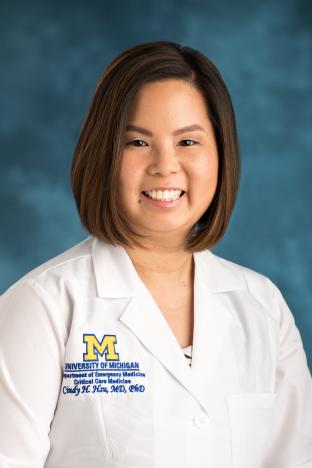 Cindy H. Hsu合作论文数Department of Emergency Medicine, Medical School, University of Michigan;Department of Surgery, Division of Acute Care Surgery, Medical School, University of Michigan43
 Clifton Callaway合作论文数Department of Emergency Medicine, School of Medicine, University of Pittsburgh;UPMC Health Plan32
 Jerry Nolan合作论文数School of Physiology, Pharmacology & Neuroscience, University of Bristol;Warwick Medical School, University of Warwick31
 Laurie Morrison合作论文数Sunnybrook Health Science Centre, Institute of Medical Science, Department of Emergency, Temerty Faculty of Medicine, University of Toronto27
 Peter Morley合作论文数Consultant in Anaesthesia and Intensive Care Medicine, Royal United Hospital27
 Gavin Perkins合作论文数Warwick Medical School, The University of Warwick26
 Mohamad H. Tiba合作论文数24
 Brian J. O’Neil合作论文数22
 Peter J. Kudenchuk合作论文数Division of Cardiology, Arrhythmia Services, Department of Medicine, University of Washington;University of Washington Medical Center21
 Jasmeet Soar合作论文数North Bristol NHS Trust21
 Vinay M. Nadkarni合作论文数Children's Hospital of Philadelphia19de Caen Allan合作论文数19
 Benjamin S. Abella合作论文数Department of Emergency Medicine, Perelman School of Medicine, University of Pennsylvania;Center for Clinical Epidemiology and Biostatistics, University of Pennsylvania;Center for Resuscitation Science, University of Pennsylvania;Penn Acute Research Collaboration, University of Pennsylvania19
 Tzong-Luen Wang合作论文数School of Medicine, Fu Jen Catholic University19Bernd W. Bottiger合作论文数19
 McCracken Brendan M合作论文数18
 Thomas Sanderson合作论文数Medical School, University of Michigan-Ann Arbor17
 Raina M. Merchant合作论文数Department of Medical Ethics and Health Policy, Perelman School of Medicine, University of Pennsylvania;Center for Digital Health, Penn Medicine17
 Farhan Bhanji合作论文数Institute of Health Sciences Education, Faculty of Medicine and Health Sciences, McGill University;Steinberg Centre for Simulation and Interactive Learning, McGill University17
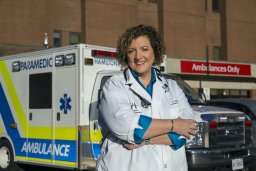 Michelle Welsford合作论文数Department of Emergency Medicine, Hamilton Paramedic Base Hospital Program17
 David F. Gaieski合作论文数Department of Emergency Medicine, Home of Sidney Kimmel Medical College, Thomas Jefferson University17Katherine Berg合作论文数Ctr Resuscitat Sci, Beth Israel Deaconess Med Ctr16Michael M. Parr合作论文数16
 Alvaro Rojas-Pena合作论文数Department of Surgery, University of Michigan Medical School16
 Claudio Sandroni合作论文数16
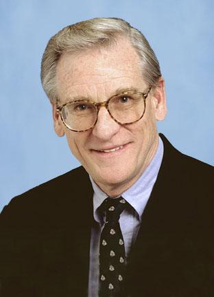 Robert H. Bartlett合作论文数Division of Acute Care Surgery, Department of Surgery, University of Michigan16Eunice M. Singletary合作论文数16
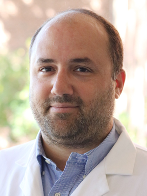 Demetris Yannopoulos合作论文数Departments of Neurology and Medicine—Cardiovascular Division, University of Minnesota Medical Cente16Xiaodan Ren合作论文数15
 Theresa Mariero Olasveengen合作论文数Department of Anesthesia and Intensive Care Medicine, Division of Critical Care, Institute of Clinical Medicine, Faculty of Medicine, University of Oslo;Oslo University Hospital15Mary Fran Hazinski合作论文数15Tonia C. Nicholson合作论文数14
 Brahmajee K. Nallamothu合作论文数Department of Internal Medicine, Medical School, University of Michigan14
 Eric Lavonas合作论文数Denver Hlth & Hosp Author, Rocky Mt Poison & Drug Ctr14
 Ian R Drennan合作论文数14
 Charles Deakin合作论文数Faculty of Medicine, University of Southampton14
 Lance Becker合作论文数Zucker School of Medicine, Hofstra University;Feinstein Institutes for Medical Research;Zucker School of Medicine at Hofstra/Northwell;Department of Emergency, Long Island Jewish Medical Center;Department of Emergency, North Shore University Hospital14
 Maaret Castrén合作论文数Department of Emergency Medicine and Services, Helsinki University Hospital14
 Monica E Kleinman合作论文数14
 James Cranford合作论文数Injury Prevention Center, Medical School, University of Michigan-Ann Arbor13Ian Maconochie合作论文数Department of Infectious Disease, Faculty of Medicine, Imperial College London13
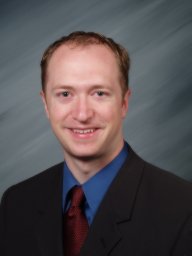 Jason A Bartos合作论文数Dept Med, Div Cardiol, Univ Minnesota13
 Tom P. Aufderheide合作论文数Department of Emergency Medicine, Medical College of Wisconsin13
 Michael W. Donnino合作论文数Emergency Department, Center for Resuscitation Science, Beth Israel Deaconess Medical Center, Harvard Medical School13
 Robert Greif合作论文数University Hospital Bern, University of Bern13Mathias Johan Holmberg合作论文数12Riess Matthias L合作论文数12
 Nikolaos Nikolaou合作论文数12
 Giuseppe Ristagno合作论文数12




