Human blood leukocytes continually flux between blood and tissues. The first step in extravasation of blood-borne cells involves shear-resistant binding to the endothelial surface, a process mediated by adhesive interactions between an endothelial lectin, E-selectin, and a tetrasaccharide known as sialyl Lewis X (sLeX) displayed on circulating cells. Human neutrophils and monocytes express high levels of "E-selectin ligands" (glycoconjugates that contain sLeX) and are thereby operationally primed to extravasate at inflammatory loci. However, the capacity of human blood dendritic cells (DCs) to similarly engage E-selectin is unknown. To address this question, we performed multiparameter flow cytometry, biochemical analyses, and measurements of transcripts encoding key glycosyltransferases programming sLeX biosynthesis. Our results reveal that a unifying feature of all subsets of human blood DCs is the display of carbohydrates that endow robust E-selectin binding. This feature is also characteristic of their ontogenic precursors. Notably, while bulk RNA sequencing analysis is mechanistically informative, single-cell RNA sequencing does not provide insights into the critical chemoenzymatic effectors of sLeX expression by DCs. These results thus reconceptualize our understanding of an entire class of human immune cells that converge the innate and adaptive immune systems, providing key insights into the molecular basis of their tissue recruitment in both steady-state and at the onset of an inflammatory response.
In mammals, every cell is covered by a sugar coat called the "glycocalyx", a meshwork created by sugar modifications of cell surface proteins and lipids. Essentially all cell membrane proteins and lipids contain oligosaccharide clusters known as "glycan motifs" that confer distinct functional properties on these respective glycoproteins or glycolipids. These motifs are generated by glycosyltransferases that assemble the component monosaccharides in a stereospecific and regiospecific fashion. Glycocalyx motifs bearing l-fucose in α(1→3) linkage to N-acetyl-glucosamine are found on a highly restricted subset of membrane glycoproteins and glycolipids, and changes in α(1→3)-fucosylation levels impact a wide range of physiologic and pathologic processes. Within this biological framework, we herein review the pivotal role of α(1→3)-fucosylation in cell biology and then comprehensively review the evolving chemical strategies to custom-modify α(1→3)-fucosylation of the glycocalyx to achieve highly specific control of human cell surface fucosylated glycan motifs. These efforts serve as a prime example of how the fine control of cell surface fucosylation can enable the generation of glycan-based precision therapeutics, driving forward the field of "translational glycobiology".
α(1,3)-exofucosylation is an enzymatic process whereby the monosaccharide L-fucose is added in α(1,3)-linkage onto a pertinent acceptor glycan displayed by a cell membrane glycoprotein or glycolipid. One pertinent acceptor glycan is the terminal trisaccharide unit known as a "sialylated type 2 lactosamine". In this case, α(1,3)-exofucosylation creates the glycan motif sialylated Lewis X (sLeX, CD15 s), the canonical E-selectin binding determinant. At sites of tissue inflammation, endothelial E-selectin enables sLeX-laden blood-borne cells to migrate to, and then extravasate at, diseased sites. Thus, α(1,3)-exofucosylation facilitates T cell tissue infiltration. Considerable data demonstrate the capacity of regulatory T cells (Tregs) to suppress pro-inflammatory processes in the central nervous system. In this review, we describe how α(1,3)-exofucosylation of antigen-specific Tregs can be harnessed to optimize neuroprotection and neurorestoration for both inflammatory and neurodegenerative diseases.
Prior studies have indicated that human embryonic stem cells can be distinguished from those of other mammals based on variable expression of a class of membrane glycolipids known as glycosphingolipids (GSLs), raising the question as to whether GSL display could be utilized to phenotypically define subsets of human adult stem cell populations. Adult stem cells known as "mesenchymal stem/stromal cells" (MSCs) have shown immense promise in therapeutic applications for a variety of clinical indications. Most commonly, these cells are harnessed and then culture-expanded from bone-marrow (BM-MSCs) or from adipose tissue (A-MSCs) sources. Though operational differences exist between human BM-MSCs and A-MSCs, no surface markers have been characterized to date that distinguish these as distinct subsets of culture-expanded human adult stem cells. Accordingly, we isolated GSLs from primary cultures of marrow- and adipose-derived human MSCs and an unbiased screen was performed by mass spectrometry (via matrix-assisted laser desorption/ionization (MALDI)-quadrupole ion trap (QIT)-time-offlight (TOF), hence, via "MALDI-QIT-TOF") to analyze all component glycans. Flow cytometry was then undertaken to assess the relative levels of expression of MS-defined glycan determinants, followed by RT-qPCR to measure transcripts of genes encoding key enzymes involved in glycolipid biosynthesis. Notably, our data indicate that neither BM- nor A-MSCs display any significant level of either lacto-series or neolacto-series GSLs, but distinct differences exist in GSL species among A-MSCs and BM-MSCs: while both cell types express GSLs of the ganglio- and the globo-series, the ganglio-series GSLs GD3 and GD2 and the globo-series GSL SSEA-4 (also known as sialylGb5) are dominantly expressed only among human BM-MSCs. These structural features are shaped by divergent patterns of glycosyltransferase gene expression, with striking differences between BM- and A-MSCs in the expression of transcripts encoding GD3 synthase, GM2/GD2 synthase, and Gb5 synthase. Importantly, expression of GD3, GD2, and SSEA-4 is markedly diminished on differentiation of BM-MSCs, and co-cultures of A-MSCs and BM-MSCs show that the expression of GD3, GD2, and SSEA-4 is a cell-intrinsic feature of BM-MSCs. These data stratify the glycosignature(s) of human MSCs derived from different tissue sources, provide direct evidence that expression of these structures is cell stage-/lineage-specific, unveil the mechanistic basis of the differential expression of these glycan determinants, and draw attention to how knowledge of the MSC glycosignature can impact cytotherapeutic strategies. (c) 2024 International Society for Cell & Gene Therapy. Published by Elsevier Inc. All rights are reserved, including those for text and data mining, AI training, and similar technologies.
Adoptive T-cell transfer has revolutionized the treatment of hematological malignancies. However, this approach has had very limited success in treating solid tumors, largely due to inadequate infiltration of vascularly administered T cells at tumor sites. The shear-resistant interaction between endothelial E-selectin and its cognate ligand expressed on leukocytes, sialyl Lewis X (sLeX), is an essential prerequisite for extravasation of circulating leukocytes. Here, we report that enforced E-selectin ligand expression (enforced sLeX display) on antigen-specific T cells can be achieved by fucosylating cells via cell surface treatment with the human α1-3-fucosyltransferase, FUT6 ("exofucosylation"), or via Golgi-targeted FUT6 overexpression ("Golgi-fucosylation"). However, despite comparable E-selectin binding, only sLeX-modified T cells engendered by exofucosylation, not by Golgi-fucosylation, exhibited enhanced parenchymal infiltration of target malignant sites. This heightened homing yielded significantly improved therapeutic efficacy in various murine syngeneic and xenograft cancer models, including subcutaneous solid tumors, lymphoma and leukemia, as well as lung and bone marrow metastases. Therefore, exofucosylation represents a promising strategy to improve the efficacy of adoptive T-cell therapy, particularly in the treatment of solid tumors and metastatic disease.
The convergence of glycochemistry and glycobiology is enabling the creation of new therapeutic approaches with unprecedented capacity to alter cell and organismic biology using strategies that can uniquely and specifically custom-modify the expression of key cell surface glycan motifs. We define this evolving field of chemical biology as 'glycan-motif editing', and one of the principal targets of this glycoengineering effort is the sialofucosylated terminal lactosaminyl glycan known as sLeX (CD15s). This tetrasaccharide structure plays pivotal roles in both steady-state and malignant hematopoiesis, in regulation of the immune response, and in cancer metastasis. Within this biological framework, we discuss the immense potential of glycan-motif editing in enabling precision therapeutics that will profoundly improve outcomes for patients suffering from a wide variety of disabling and life-threatening conditions, particularly cancer.
Fucosylation is a ubiquitous glycosylation event that shapes cellular communication and immunity. Catalyzed by fucosyltransferases (FUTs), this reaction encompasses diverse substrates, mechanisms, and biologic consequences. In this Review, we explore the structural and functional landscape of FUTs primarily from higher eukaryotes, with focus on the mechanistic determinants of regioselectivity, donor/acceptor coordination, and domain modularity. We highlight advances in structural biology, modeling, and enzyme engineering that clarify how FUTs decode glycan topology and specificity. Phylogenetic and structural analyses reveal two major clades of human FUTs that differ in GDP-Fuc recognition and conformational flexibility, providing a molecular rationale for their mechanistic divergence. Drawing from mammalian FUT studies, we propose a conceptual framework in which distinct family members exploit strategies including donor-induced conformational changes, exosite interactions, or local peptide cues to achieve specificity and catalytic efficiency. We also examine their roles in physiology, inflammation, immune regulation, and cancer, and summarize current FUT inhibitors and enzyme-based therapeutic strategies.
Adhesion molecules within the bone marrow vascular niche govern the self-renewal, differentiation, and proliferation of hematopoietic progenitors. Among these, E-selectin, a lectin expressed by endothelial cells which binds to the tetrasaccharide known as sialyl Lewis X (sLeX), promotes dormancy, self-renewal, and chemoresistance in native hematopoietic stem cells (HSCs). However, leukemic blasts hijack these signals, fueling their proliferation. While therapeutic disruption of E-selectin binding to its ligands has demonstrated clinical activity in acute myeloid leukemia, its role in B-cell acute lymphoblastic leukemia (B-ALL) remains underexplored. Identifying E-selectin ligands and associated downstream molecular effectors in B-ALL holds potential to reveal targetable mediators of leukemia-initiating capacity, drug tolerance, and persistence. Fresh peripheral blood and bone marrow from appropriately consented healthy donors (n=9) and clinically annotated patients (n=11) with BCR::ABL1 B-ALL identically treated on a clinical trial (NCT03595917) were profiled for sLeX expression and E-selectin binding by flow cytometry using HECA-452 monoclonal antibody and E-selectin-Ig chimera (E-Ig) respectively, along with glycoproteomic analyses. Expression of canonical E-selectin ligands (HCELL/CD44, CLA/PSGL-1, CD43E/CD43) and B cell developmental markers (CD45, CD34, CD10, CD19, CD20, CD22) were also assessed. SLeX expression and E-selectin binding varied between native antigen-independent B cell developmental stages (p < 0.05). Malignant CD19+ B cells from the bone marrow of treatment-naïve B-ALL patients exhibited increased sLeX expression (p < 0.05) and a nominal increase in E-selectin binding compared to counterparts from healthy donors. E-Ig-reactive CD19+ B-ALL cells display higher CD43 expression and lower CD44 and PSGL-1 expression than E-Ig-reactive CD19+ healthy donor B cells (p < 0.05). Treatment-naïve leukemic progenitors displayed greater sLeX expression and E-selectin binding than patient-matched non-malignant populations (p < 0.05). Leukemic progenitors isolated from serial bone marrow specimens exhibited a nominal increase in sLeX expression and E-selectin binding at progression versus baseline. Within the lymphoid lineage, E-selectin binding is developmentally determined and enriched in malignant compared to native cells. Interrogation of downstream effectors and functional phenotypes is ongoing. Chase M. Weizer, Rachel A. Kraan, Evan Ales, Stella L. Jaeckle, Julia H. Keating, Marlise R. Luskin, Robert Sackstein, Mark A. Murakami. E-selectin binding is enriched within B-cell acute lymphoblastic leukemia compared to healthy lymphoid precursors [abstract]. In: Proceedings of the American Association for Cancer Research Annual Meeting 2025; Part 1 (Regular Abstracts); 2025 Apr 25-30; Chicago, IL. Philadelphia (PA): AACR; Cancer Res 2025;85(8_Suppl_1):Abstract nr 5145.
Umbilical cord blood (CB) transplantion is limited by slower engraftment and higher failure rates compared to other allografts. We hypothesized that ex vivo expansion of CB using mesenchymal progenitor cells (MPCs) and exofucosylation to enforce expression of osteotropism-mediating sLeX would shorten engraftment time. Six patients with hematologic malignancies underwent transplantation with two CB units (CBUs). Five received one unmanipulated CBU and one MPC-expanded, exofucosylated CBU. Median neutrophil engraftment, platelets >20,000/µL, and platelets >50,000/µL were 29, 45, and 55 days, respectively. A sixth received one exofucosylated CBU and one MPC-expanded CBU, achieving neutrophil engraftment, platelets >20,000/µL, and platelets >50,000/µL in 34, 53, and 57 days, respectively. Acute GVHD occurred in 4/6 patients, including one grade III/IV case. Three were alive without disease relapse at a median of 43.6 months post-transplant. Deaths in three patients were due to relapse, COVID, and GVHD. Thus, these approaches were safe, but did not improve engraftment.
CD45 is the most abundant glycoprotein on the surface of all nucleated hematopoietic-lineage cells, comprising multiple isoforms generated by alternative splicing of three exons ("A," "B," and "C"), which are exquisitely restricted across hematopoietic development. Despite the ubiquitous expression of CD45 on hematopoietic cells, its function(s) remain rather obscure. Here, we report that discrete CD45 isoforms expressed uniquely by immature human hematopoietic cells are distinguished as functional glycoforms ("isoglycoforms") that bind E-selectin. Moreover, our studies indicate that "CD45RA," a marker of human acute myeloid leukemia (AML) cells, identifies a distinct isoglycoform of CD45 containing all splice exon-encoded peptides. This isoglycoform, "CD45RABC-E," is directly upregulated by AML cells and demarcates these malignant cells from mature human leukocytes and the native human hematopoietic stem cell. Further analyses revealed that treatment-resistant AML highly expresses CD45RABC-E. Our findings thus unveil heretofore unrecognized functions of CD45 and define a novel CD45 isoglycoform that delineates AML cells from life-sustaining human hematopoietic cells.
Mesenchymal stem/stromal cells (MSCs) are being increasingly used in cell-based therapies due to their broad anti-inflammatory and immunomodulatory properties. Intravascularly-administered MSCs do not efficiently migrate to sites of inflammation/immunopathology, but this shortfall has been overcome by cell surface enzymatic fucosylation to engender expression of the potent E-selectin ligand HCELL. In applications of cell-based therapies, cryopreservation enables stability in both storage and transport of the produced cells from the manufacturing facility to the point of care. However, it has been reported that cryopreservation and thawing dampens their immunomodulatory/anti-inflammatory activity even after a reactivation/reconditioning step. To address this issue, we employed a variety of methods to cryopreserve and thaw fucosylated human MSCs derived from either bone marrow or adipose tissue sources. We then evaluated their immunosuppressive properties, cell viability, morphology, proliferation kinetics, immunophenotype, senescence, and osteogenic and adipogenic differentiation. Our studies provide new insights into the immunobiology of cryopreserved and thawed MSCs and offer a readily applicable approach to optimize the use of fucosylated human allogeneic MSCs as immunomodulatory/anti-inflammatory therapeutics.
Mucin-domain glycoproteins expressed on cancer cell surfaces play central roles in cell adhesion, cancer progression, stem cell renewal, and immune evasion. Despite abundant evidence that mucin-domain glycoproteins are critical to the pathobiology of head and neck squamous cell carcinoma (HNSCC), our knowledge of the composition of that mucinome is grossly incomplete. Here, we utilized a catalytically inactive point mutant of the enzyme StcE (StcEE447D) to capture mucin-domain glycoproteins in head and neck cancer cell line lysates followed by their characterization using sodium dodecyl-sulfate polyacrylamide gel electrophoresis (SDS-PAGE), in-gel digestion, nano-liquid chromatography-tandem mass spectrometry (nLC-MS/MS), and enrichment analyses. We demonstrate the feasibility of this workflow for the study of mucin-domain glycoproteins in HNSCC, identify a set of mucin-domain glycoproteins common to multiple HNSCC cell lines, and report a subset of mucin-domain glycoproteins that are uniquely expressed in HSC-3 cells, a cell line derived from a highly aggressive metastatic tongue squamous cell carcinoma. This effort represents the first attempt to identify mucin-domain glycoproteins in HNSCC in an untargeted, unbiased analysis, paving the way for a more comprehensive characterization of the mucinome components that mediate aggressive tumor cell phenotypes. Data associated with this study have been deposited to the ProteomeXchange Consortium via the PRIDE partner repository with the data set identifier PXD029420.
Abstract CD45 (the leukocyte common antigen) is a phosphatase present on all malignant and non-malignant leukocytes and hematopoietic progenitors and is the most abundant glycoprotein displayed by these cells. The human CD45 gene is alternatively spliced by reassortment of just three exons, exon 4 (“A”), exon 5 (“B”), and exon 6 (“C"), to create five distinct “R” isoforms (RO (no splice exons), RABC, RAB, RBC, and RB), with inclusion of A, B, or C exons enriching for expression of glycans (carbohydrates). Despite decades of investigation, the expression and phosphatase function of these isoforms on human AML cells remains unknown. To address this question, flow cytometry and western blot analysis was performed on AML primary cells (n=3) and KG1a and U937 cell lines versus healthy monocytes (n=3) utilizing exon-specific CD45-targetting mAbs. We then compared the contribution of CD45 phosphatase activity in KG1a and U937 cells versus healthy monocytes by employing a highly specific CD45 phosphatase inhibitor in cell culture for 24hrs, assessing induction of cell death after exposure to the inhibitor (n=3). Our results confirm reports of others indicating that "CD45RA- positivity" is a characteristic phenotype of AML cells, yet, distinctly, our data indicate that the most prevalent isoform identified on AML cells as "CD45RA-positive" is, actually, CD45RABC, the largest and most glycosylated isoform of CD45. This finding is in striking contrast to healthy monocytes, which do not express CD45RABC. Additionally, culturing of human AML cells with a CD45 phosphatase inhibitor induces rapid cell death within 24 hours but does not affect healthy monocytes. Thus, the conspicuous expression of the CD45 isoform CD45RABC among AML cells is associated with a survival advantage. Current studies in AML are directed to elaborating the prevalence of CD45RABC expression and uncovering the downstream effectors of its enzymatic activity to elucidate how such pathways sustain AML growth/survival. Citation Format: Evan Ales, Robert Sackstein. Differential CD45 isoform expression and function in AML versus healthy leukocytes [abstract]. In: Proceedings of the American Association for Cancer Research Annual Meeting 2023; Part 1 (Regular and Invited Abstracts); 2023 Apr 14-19; Orlando, FL. Philadelphia (PA): AACR; Cancer Res 2023;83(7_Suppl):Abstract nr 342.
The prognosis for patients with metastatic melanoma (MM) involving distant organs is grim, and treatment resistance is potentiated by tumor-initiating cells (TICs) that thrive under hypoxia. MM cells, including TICs, express a unique glycome featuring i-linear poly-N-acetyllactosamines through the loss of I-branching enzyme, (beta 1,6 N-acetylglucosaminyltransferase 2. Whether hypoxia instructs MM TIC development by modulating the glycome signature remains unknown. In this study, we explored hypoxia-dependent alterations in MM gly-come-associated genes and found that (beta 1,6 N-acetylglucosaminyltransferase 2 was downregulated and a galectin (Gal)-8-ligand axis, involving both extracellular and cell-intrinsic Gal-8, was induced. Low (beta 1,6 N-acetylglucosaminyltransferase 2 levels correlated with poor patient outcomes, and patient serum samples were elevated for Gal-8. Depressed (beta 1,6 N-acetylglucosaminyltransferase 2 in MM cells upregulated TIC marker, NGFR/CD271, whereas loss of MM cell-intrinsic Gal-8 markedly lowered NGFR and reduced TIC activity in vivo. Extracellular Gal-8 bound preferentially to i-linear poly-N-acetyllactosamines on N-glycans of the TIC marker and prometastatic molecule CD44, among other receptors, and activated prosurvival factor protein kinase B. This study reveals the importance of hypoxia governing the MM glycome by enforcing i-linear poly-N-acetyllactosamine and Gal-8 expression. This mechanistic investigation also uncovers glycome-dependent regulation of pro-MM factor, NGFR, implicating i-linear poly-N-acetyllactosamine and Gal-8 as biomarkers and therapeutic targets of MM.
Bottom-up nLC-MS/MS-based glycoprotein mass spectrometry workflows rely on the generation of a mixture of non-glycosylated and glycosylated peptides via proteolysis of glycoproteins. Such methods are challenged by suppression of hydrophilic glycopeptide ions by more abundant, hydrophobic, and readily ionizable non-glycosylated peptides. Commercially available high-field asymmetric waveform ion mobility spectrometry (FAIMS) devices have recently been introduced and present a potential benefit for glycoproteomic workflows by enabling orthogonal separation of non-glycosylated peptides and glycopeptides following chromatographic separation, and prior to MS/MS analysis. However, knowledge is lacking regarding optimal FAIMS conditions for glycopeptide analyses. Here, we document optimal FAIMS compensation voltages for the transmission and analysis of human alpha-1-acid glycoprotein (AGP) tryptic N-glycopeptide ions. Further, we evaluate the effect of FAIMS on AGP glycopeptide assignment confidence by comparing the number of assigned glycopeptides at different confidence levels using a standard nLC-MS/MS method or an otherwise identical method employing FAIMS. Optimized methods will potentiate glycoproteomic analyses by increasing the number of unique glycopeptide identifications and the confidence of glycopeptide assignments. Data are available via ProteomeXchange with identifier PXD036667. Analysis of alpha-1-acid glycoprotein (AGP) tryptic digests via nLC-FAIMS-MS/MS (top) led to the establishment of ideal FAIMS voltages for the analysis of AGP N-glycopeptides (bottom), suggesting that FAIMS can improve the depth of glycoproteome characterization. Pairs of CV magnitudes are shown along the x-axis
Purpose/Objective(s) Head and neck cancer is the sixth most common tumor type worldwide, but despite advances in treatment, the 5-year survival rate is only ∼50%. Glycosylation, a common post-translational modification, is frequently altered in cancer. Changes in glycosyltransferase expression in cancer are associated with cellular invasion, tumor growth, and metastasis. However, the role of glycosyltransferase expression in head and neck cancer survival is poorly understood. Here, we examine the association between glycosyltransferase expression and survival in head and neck squamous cell carcinoma (HNSCC). Materials/Methods A retrospective cohort study was performed with data from the Cancer Genome Atlas (TCGA) HNSCC dataset (n=499), containing expression (mRNA) level data, generated via high-throughput sequencing, of primary tumor tissue from patients with a diagnosis of HNSCC (deidentified). A set of glycosyltransferase genes: FUT1-FUT11, POFUT1-2, ST3Gal1-6, ST6Gal1-2, ST6GalNac1-6, ST8Sia1-6, GALNT2-3, C1GALT1, B4GALT4, GCNT2 were considered. Median expression was used to delineate high and low expression groups. Univariate Kaplan-Meier analyses and logrank tests were conducted. Multivariate analyses were performed via Cox proportional hazards regression to adjust for covariates (HPV status, age at initial diagnosis, sex, alcohol history, tobacco usage, and primary site). Results Based on the Kaplan-Meier analysis of FUT2, the median survival time (MST) of the high FUT2-expressing group was 4.7 years, while low expression was associated with an MST of 2.7 years. Cox regression analysis yielded adjusted hazard ratio (aHR) of 0.72 (CI = 0.54-0.95). The MST of the FUT6 high expression group was 4.7 years versus 2.9 years for low expression, with an aHR of 0.62 (CI = 0.47-0.83). The MST of the high-FUT7 expressing group was 4.7 years versus 3.5 years for the low level group, with an aHR of 0.72 (CI = 0.54-0.95). GALNT3 showed the opposite effect, with high expression yielding an MST of 3.1 years versus 4.9 years for the low-expressing group, with aHR of 1.47 (1.11-1.95). An aHR of <1 indicates a protective effect from high expression. Conclusion Overexpression of FUT2, FUT6, and FUT7 were associated with increased overall survival, while overexpression of GALNT3 was associated with decreased overall survival in HNSCC. FUT2 is expressed in endoderm-derived epithelial tissue including the digestive tract and salivary glands, while FUT6 and FUT7 are prominently expressed in immune cells. Thus, FUT6 and FUT7 expression may indicate immune cell infiltration into tumor tissue. Future studies will seek to sort tumor tissue based on cell type, to gain a better understanding of the cell-type specific glycosyltransferase expression in HNSCC tumors. The glycosyltransferase genes associated with survival in this study may serve as future drug targets in head and neck cancer. Head and neck cancer is the sixth most common tumor type worldwide, but despite advances in treatment, the 5-year survival rate is only ∼50%. Glycosylation, a common post-translational modification, is frequently altered in cancer. Changes in glycosyltransferase expression in cancer are associated with cellular invasion, tumor growth, and metastasis. However, the role of glycosyltransferase expression in head and neck cancer survival is poorly understood. Here, we examine the association between glycosyltransferase expression and survival in head and neck squamous cell carcinoma (HNSCC). A retrospective cohort study was performed with data from the Cancer Genome Atlas (TCGA) HNSCC dataset (n=499), containing expression (mRNA) level data, generated via high-throughput sequencing, of primary tumor tissue from patients with a diagnosis of HNSCC (deidentified). A set of glycosyltransferase genes: FUT1-FUT11, POFUT1-2, ST3Gal1-6, ST6Gal1-2, ST6GalNac1-6, ST8Sia1-6, GALNT2-3, C1GALT1, B4GALT4, GCNT2 were considered. Median expression was used to delineate high and low expression groups. Univariate Kaplan-Meier analyses and logrank tests were conducted. Multivariate analyses were performed via Cox proportional hazards regression to adjust for covariates (HPV status, age at initial diagnosis, sex, alcohol history, tobacco usage, and primary site). Based on the Kaplan-Meier analysis of FUT2, the median survival time (MST) of the high FUT2-expressing group was 4.7 years, while low expression was associated with an MST of 2.7 years. Cox regression analysis yielded adjusted hazard ratio (aHR) of 0.72 (CI = 0.54-0.95). The MST of the FUT6 high expression group was 4.7 years versus 2.9 years for low expression, with an aHR of 0.62 (CI = 0.47-0.83). The MST of the high-FUT7 expressing group was 4.7 years versus 3.5 years for the low level group, with an aHR of 0.72 (CI = 0.54-0.95). GALNT3 showed the opposite effect, with high expression yielding an MST of 3.1 years versus 4.9 years for the low-expressing group, with aHR of 1.47 (1.11-1.95). An aHR of <1 indicates a protective effect from high expression. Overexpression of FUT2, FUT6, and FUT7 were associated with increased overall survival, while overexpression of GALNT3 was associated with decreased overall survival in HNSCC. FUT2 is expressed in endoderm-derived epithelial tissue including the digestive tract and salivary glands, while FUT6 and FUT7 are prominently expressed in immune cells. Thus, FUT6 and FUT7 expression may indicate immune cell infiltration into tumor tissue. Future studies will seek to sort tumor tissue based on cell type, to gain a better understanding of the cell-type specific glycosyltransferase expression in HNSCC tumors. The glycosyltransferase genes associated with survival in this study may serve as future drug targets in head and neck cancer.
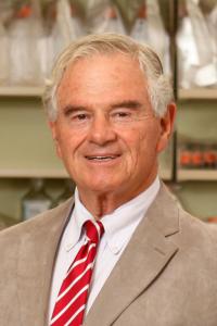 David H Sachs合作论文数Vagelos College of Physicians and Surgeons, Columbia University Irving Medical Center41
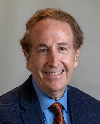 Thomas Spitzer合作论文数Mass General Cancer Center, Massachusetts General Hospital;Harvard Medical School33
 C Colby合作论文数32M Sykes合作论文数31S McAfee合作论文数21Charles J. Dimitroff合作论文数Harvard Skin Disease Research Center, Department of Dermatology, Brigham and Women's Hospital, Boston, Mass17
 Bimalangshu Ranjan Dey合作论文数Mass General Brigham;Harvard Medical School16
 Frederic I. Preffer合作论文数Harvard Medical School;Massachusetts General Hospital15Leo Buhler合作论文数14Mona M Basker合作论文数14
 Robert Fuhlbrigge合作论文数14D.K.C Cooper合作论文数13D Weymouth合作论文数12Michel Awwad合作论文数12Qing Chang合作论文数10
 Miguel Blanquer Blanquer合作论文数Hospital Morales Meseguer, Servicio de Hematología, Facultad de Medicina, Murcia, Spain10D Andrews合作论文数9Sl Mcafee合作论文数9
 Kevin B. Chandler合作论文数9
 Simon Robson合作论文数Division of Gastroenterology, Department of Medicine, Beth Israel Deaconess Medical Center;Department of Anesthesia, Beth Israel Deaconess Medical Center;Division of Medical Sciences, Harvard Griffin Graduate School of Arts and Sciences, Harvard University;Liver Center, University of Cape Town;Harvard Medical School, Harvard University;Inner Mongolian Medical College;Department of Medicine, New England Deaconess Hospital9
 Satoshi Gojo合作论文数Department of Cardiovascular Surgery, Saitama Medical Center9YH CHIN合作论文数8Jose Maria Moraleda合作论文数8
 Mariana Martins da Silva合作论文数8Jack Y. Lee合作论文数8Jd Down合作论文数8David Dombkowski合作论文数8Christiane Goepfert合作论文数8ME White-Scharf合作论文数7
 Toh Han Chong合作论文数Natl Canc Ctr7
 Thomas S. Kupper合作论文数Dana-Farber Cancer Institute, Inc.;Brigham and Women's Hospital;Harvard Medical School7
 Megan Sykes合作论文数Columbia Center for Translational Immunology7
 Mondal Nandini合作论文数The NY State Center for Excellence in Bioinformatics and Life Sciences, State University of New York7Gerald J Elfenbein合作论文数7S Treter合作论文数7
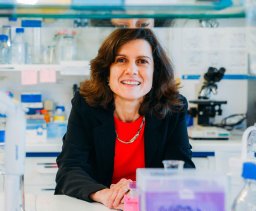 Paula Videira合作论文数Departamento De Ciencias Da Vida, Faculdade De Ciencias E Tecnologia, Universidade NOVA de Lisboa;Valvian;UCIBIO;Cellmabs;CDG & allies PPAIN7
 Ian Alwayn合作论文数Faculteit Geneeskunde, Leiden University Medical Center;Transplant Center, Leiden University Medical Center7Jungmin Lee合作论文数Cell Design Institute;University of California San Francisco;Cell Design Institute, University of California San Francisco6
 David Garcia-Bernal合作论文数Virgen de la Arrixaca Clinical University Hospital, University of Murcia6
 Karin M. Hoffmeister合作论文数Div Hematol, Brigham & Womens Hosp6
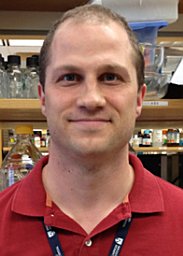 Brad Dykstra合作论文数University of Groningen, University Medical Center Groningen6Derrick J. Rossi合作论文数6
 Monica M. Burdick合作论文数Department of Chemical Engineering, Johns Hopkins University6IPJ Alwayn合作论文数5HR COLTEN合作论文数5Donnelly Conor B合作论文数5A Kraus合作论文数5Susan L Saidman合作论文数5




