This paper provides a brief description of the early use of ex vivo nuclear magnetic resonance (NMR) studies of tissue and tissue extracts performed in the laboratory of Dr. Robert G. Shulman from 1975 through 1995 at Bell Laboratories, then later at Yale University. During that period, ex vivo NMR provided critical information in support of resonance assignments and the quantitation of concentrations for magnetic resonance spectroscopy studies. The period covered saw rapid advances in magnet technology, starting with studies of microorganisms in vertical bore high-resolution NMR studies, then by 1981 studies of small mammals in a horizontal bore magnet, and then studies of humans in 1984. Ex vivo NMR played a critical role in all these studies. A general strategy developed in the lab for using ex vivo NMR to support in vivo studies is presented, as well as illustrative examples.
Since Jacob and Monod’s discovery of the lac operon ∼1960, the explanations offered for most metabolic adaptations have been genetic. The focus has been on the adaptive changes in gene expression that occur, which are often referred to as “metabolic reprogramming.” The contributions metabolism makes to adaptation have been largely ignored. Here we point out that metabolic adaptations, including the associated changes in gene expression, are highly dependent on the metabolic state of an organism prior to the environmental change to which it is adapting, and on the plasticity of that state. In support of this hypothesis, we examine the paradigmatic example of a genetically driven adaptation, the adaptation of E. coli to growth on lactose, and the paradigmatic example of a metabolic driven adaptation, the Crabtree effect in yeast. Using a framework based on metabolic control analysis, we have reevaluated what is known about both adaptations, and conclude that knowledge of the metabolic properties of these organisms prior to environmental change is critical for understanding not only how they survive long enough to adapt, but also how the ensuing changes in gene expression occur, and their phenotypes post-adaptation. It would be useful if future explanations for metabolic adaptations acknowledged the contributions made to them by metabolism, and described the complex interplay between metabolic systems and genetic systems that make these adaptations possible.
This paper provides a brief description of the early use of ex vivo nuclear magnetic resonance (NMR) studies of tissue and tissue extracts performed in the laboratory of Dr. Robert G. Shulman from 1975 through 1995 at Bell Laboratories, then later at Yale University. During that period, ex vivo NMR provided critical information in support of resonance assignments and the quantitation of concentrations for magnetic resonance spectroscopy studies. The period covered saw rapid advances in magnet technology, starting with studies of microorganisms in vertical bore high‐resolution NMR studies, then by 1981 studies of small mammals in a horizontal bore magnet, and then studies of humans in 1984. Ex vivo NMR played a critical role in all these studies. A general strategy developed in the lab for using ex vivo NMR to support in vivo studies is presented, as well as illustrative examples.
Previously we suggested that the early Warburg effect can be explained by the use by cancer cells the glycogen shunt during a rapid increase in glucose concentration. In analogy to the Crabtree effect in yeast, the shunt plays a critical role in maintaining homeostasis of glycolytic intermediate levels during these transitions. We extend this analysis here, and propose that the recently appreciated flexibility of cancer cell glucose and glycogen metabolism involves 4 metabolic states that we recently identified in metabolic control analysis studies of yeast. Under stable conditions of low glucose and normal O2 yeast, and by analogy cancer, cells are in the Respiration State in which through gene expression for oxidizing non glucose substrates. When their environment changes to high glucose with reduced O2 levels, such as occur in tumors, they transition to the Glycolysis State due to gene expression of new glycolytic enzyme isoforms such as PKM2. These isoforms optimize metabolism to sustain the Warburg effect. When the changes in glucose and O2 levels are rapid there may be insufficient time for gene expression to adapt. The metabolic flexibility conferred by 2 states of the glycogen shunt allow the cells to survive these transitions. The model explains experimental observations in cancer such as the function of the glycogen shunt and the frequent expression of PKM2 in cells undergoing the Warburg Effect. A surprising conclusion is that the function of PKM2 is to maintain glycolytic intermediate homeostasis rather than controlling the glycolytic flux. The glycogen shunt may also have an important role in cancer metabolic reprogramming by allowing cancer cells to survive large glucose and oxygen changes during the selection of mutations that lead to the Warburg phenotype.
A key issue in both molecular and evolutionary biology has been to define the roles of genes and phenotypes in the adaptation of organisms to environmental changes. The dominant view has been that an organism's metabolic adaptations are driven by gene expression and that gene mutations, independent of the starting phenotype, are responsible for the evolution of new metabolic phenotypes. We propose an alternate hypothesis, in which the phenotype and genotype together determine metabolic adaptation both in the lifetime of the organism and in the evolutionary selection of adaptive metabolic traits. We tested this hypothesis by flux-balance and metabolic-control analysis of the relative roles of the starting phenotype and gene expression in regulating the metabolic adaptations during the Crabtree effect in yeast, when they are switched from a low- to high-glucose environment. Critical for successful short-term adaptation was the ability of the glycogen/trehalose shunt to balance the glycolytic pathway. The role of later gene expression of new isoforms of glycolytic enzymes, rather than flux control, was to provide additional homeostatic mechanisms allowing an increase in the amount and efficiency of adenosine triphosphate and product formation while maintaining glycolytic balance. We further showed that homeostatic mechanisms, by allowing increased phenotypic plasticity, could have played an important role in guiding the evolution of the Crabtree effect. Although our findings are specific to Crabtree yeast, they are likely to be broadly found because of the well-recognized similarities in glucose metabolism across kingdoms and phyla from yeast to humans.
The dominant model for interpreting brain imaging experiments, which we refer to as the Standard Cognitive Model (SCM), assumes that the brain is organized in support of mental processes that control behavior. However, functional neuroimaging experiments of cognitive tasks have not shown clear anatomic segregation between mental processes originally proposed by this model. This failing has been blamed on limitations in imaging technology and non-linearity in the brain’s implementation of these processes. However, the validity of the underlying cognitive models used to describe the brain has rarely been questioned or directly tested against imaging results. We propose an alternative model of brain function, that we term the Non-cognitive Behavioral Model (NBM), which correlates observed human behavior directly with measured brain activity without making assumptions about intervening cognitive processes. Our model derives from behavioral psychology but is extended to include brain activity, in addition to behavior, as observables. A further extension is the role of neuroplasticity, as opposed to innate cognitive processes, in developing the brain’s support of cognitive behavior. We present the theoretical basis with which the SCM maps cognitive processes onto functional magnetic resonance and positron emission tomography images and compare and contrast with the NBM. We also describe how the NBM can be used experimentally to study how the brain supports behavior. Two applications are presented that support the usefulness of the NBM. In one, the NBM use of the total functional imaging signal (not just the differences between states) provides a stronger correlation of neural activity with the behavioral state of consciousness than the SCM approach in both anesthesia and coma. The second example reviews studies of facial and object recognition that provide evidence for the NBM proposal that neuroplasticity and experience play key roles in the brain’s support of recognition and other behaviors. The conclusions regarding neuroplasticity are then generalized to explain the incomplete functional segregation observed in the application of the SCM to neuroimaging.
Despite many decades of study there is a lack of a quantitative explanation for the Warburg effect in cancer. We propose that the glycogen shunt, a pathway recently shown to be critical for cancer cell survival, may explain the excess lactate generation under aerobic conditions characteristic of the Warburg effect. The proposal is based on research on yeast and mammalian muscle and brain that demonstrates that the glycogen shunt functions to maintain homeostasis of glycolytic intermediates and ATP during large shifts in glucose supply or demand. Loss of the glycogen shunt leads to cell death under substrate stress. Similarities between the glycogen shunt in yeast and cancer cells lead us here to propose a parallel explanation of the lactate produced by cancer cells in the Warburg effect. The model also explains the need for the active tetramer and inactive dimer forms of pyruvate kinase (PKM2) in cancer cells, similar to the two forms of Pyk2p in yeast, as critical for regulating the glycogen shunt flux. The novel role proposed for the glycogen shunt implicates the high activities of glycogen synthase and fructose bisphosphatase in tumors as potential targets for therapy.
Glucose is the main fuel for energy metabo- lism in the normal human brain. It is generally assumed that glucose transport into the brain is not rate-limiting for metab- olism. Since brain glucose concentrations cannot be determined directly by radiotracer techniques, we used 13C NMR spec- troscopy after infusing enriched D-(1-13C)glucose to measure brain glucose concentrations at euglycemia and at hyperglyce- mia (range, 4.5-12.1 mM) in six healthy children (13-16 years old). Brain glucose concentrations averaged 1.0 ? 0.1 Amol/ml at euglycemia (4.7 ? 0.3 mM plasma) and 1.8-2.7 ,umol/ml at hyperglycemia (7.3-12.1 mM plasma). Michaelis- Menten parameters of transport were calculated to be Kt = 6.2 ? 1.7 mM and Tmax = 1.2 + 0.1 ,umol/g-min from the relationship between plasma and brain glucose concentrations. The brain glucose concentrations and transport constants are consistent with transport not being rate-limiting for resting brain metabolism at plasma levels >3 mM.
Aerobic glycolysis in yeast and cancer cells produces pyruvate beyond oxidative needs, a paradox noted by Warburg almost a century ago. To address this question, we reanalyzed extensive measurements from (13)C magnetic resonance spectroscopy of yeast glycolysis and the coupled pathways of futile cycling and glycogen and trehalose synthesis (which we refer to as the glycogen shunt). When yeast are given a large glucose load under aerobic conditions, the fluxes of these pathways adapt to maintain homeostasis of glycolytic intermediates and ATP. The glycogen shunt uses glycolytic ATP to store glycolytic intermediates as glycogen and trehalose, generating pyruvate and ethanol as byproducts. This conclusion is supported by studies of yeast with a partial block in the glycogen shunt due to the cif mutation, which found that when challenged with glucose, the yeast cells accumulate glycolytic intermediates and ATP, which ultimately leads to cell death. The control of the relative fluxes, which is critical to maintain homeostasis, is most likely exerted by the enzymes pyruvate kinase and fructose bisphosphatase. The kinetic properties of yeast PK and mammalian PKM2, the isoform found in cancer, are similar, suggesting that the same mechanism may exist in cancer cells, which, under these conditions, could explain their excess lactate generation. The general principle that homeostasis of metabolite and ATP concentrations is a critical requirement for metabolic function suggests that enzymes and pathways that perform this critical role could be effective drug targets in cancer and other diseases.
Rodent 13C magnetic resonance spectroscopy studies show that glutamatergic signaling requires high oxidative energy in the awake resting state and allowed calibration of functional magnetic resonance imaging (fMRI) signal in terms of energy relative to the resting energy. Here, we derived energy used for glutamatergic signaling in the awake resting human. We analyzed human data of electroencephalography (EEG), positron emission tomography (PET) maps of oxygen (CMRO2) and glucose (CMRglc) utilization, and calibrated fMRI from a variety of experimental conditions. CMRglc and EEG in the visual cortex were tightly coupled over several conditions, showing that the oxidative demand for signaling was four times greater than the demand for nonsignaling events in the awake state. Variations of CMRO2 and CMRglc from gray-matter regions and networks were within ± 10% of means, suggesting that most areas required similar energy for ubiquitously high resting activity. Human calibrated fMRI results suggest that changes of fMRI signal in cognitive studies contribute at most ± 10% CMRO2 changes from rest. The PET data of sleep, vegetative state, and anesthesia show metabolic reductions from rest, uniformly >20% across, indicating no region is selectively reduced when consciousness is lost. Future clinical investigations will benefit from using quantitative metabolic measures.
Functional neuroimaging measures quantitative changes in neurophysiological parameters coupled to neuronal activity during observable behavior. These results have usually been interpreted by assuming that mental causation of behavior arises from the simultaneous actions of distinct psychological mechanisms or modules. However, reproducible localization of these modules in the brain using functional magnetic resonance imaging (MRI) and positron emission tomography (PET) imaging has been elusive other than for sensory systems. In this paper, we show that neuroenergetic studies using PET, calibrated functional magnetic resonance imaging (fMRI), (13)C magnetic resonance spectroscopy, and electrical recordings do not support the standard approach, which identifies the location of mental modules from changes in brain activity. Of importance in reaching this conclusion is that changes in neuronal activities underlying the fMRI signal are many times smaller than the high ubiquitous, baseline neuronal activity, or energy in resting, awake humans. Furthermore, the incremental signal depends on the baseline activity contradicting theoretical assumptions about linearity and insertion of mental modules. To avoid these problems, while making use of these valuable results, we propose that neuroimaging should be used to identify observable brain activities that are necessary for a person's observable behavior rather than being used to seek hypothesized mental processes.
SignificanceA near one-to-one relationship had previously been observed between increments in the fluxes of the glutamate−glutamine neurotransmitter cycle and neuronal glucose oxidation in the tricarboxylic acid (TCA) cycle. This flux relationship was consistent with a hypothesized mechanism involving glycolytic ATP in astrocytes and astrocyte-to-neuron lactate shuttling. Here, 2-fluoro-2-deoxy-d-glucose was used to evaluate the glucose flux through glycolysis and the TCA cycle in nerve terminals isolated from the brains of rats under baseline and high-activity conditions. In a direct contradiction of this hypothesis, the results show that nerve terminals metabolize significant amounts of glucose.
Contents Introduction Chapter 1.Mind and Matter Chapter 2. Biophysics: An Empirical Science Chapter 3. A Philosophical Background Chapter 4. Neuroscience: A Multidisciplinary Multi-level Field Chapter 5. The State of Cognitive Neuroscience: An Over-optimistic Theory Chapter 6. Brain Energy and Neurotransmission Chapter 7. Global Brain Energy Supports the State of Consciousness Chapter 8. Incremental Brain Energies and the Acts of Consciousness Chapter 9. A Life in Humanities and Science Index
Neuroimaging studies measure cerebral rates of glucose and oxygen consumption yet they are often interpreted to support philosophical positions about mental processes presumed to represent behavior. Cognitive Neuroscience has claimed that Neuroimaging resolves Descartes’ dualism of Mind and Matter by mapping such presumed mental processes onto the brain. Bennett & Hacker point out that this is nonsense because a person, not the brain, performs observable activities such as remembering, planning or voting. This analysis is supported by the continuing inability of neuroimaging to locate the modular brain activities postulated to perform mental activity. Pragmatist philosophy emphasizes that such postulated representations are contingent hypothesis invented to deal with the world in everyday life and are not definable as proposed by Cognitive Neuroscience. A recent philosophy of Mechanisms, reflecting what biologists actually do when explaining observable phenomena, looks for explanations not from postulated representations but from multi-level, multi-disciplinary mechanisms of metabolism and energetics which are actually measured in neuroimaging. The value of this philosophy is illustrated by the experimental relations between cerebral energetics in disorders of consciousness and the response to stimuli.
An individual, human or animal, is defined to be in a conscious state empirically by the behavioral ability to respond meaningfully to stimuli, whereas the loss of consciousness is defined by unresponsiveness. PET measurements of glucose or oxygen consumption show a widespread ≈45% reduction in cerebral energy consumption with anesthesia-induced loss of consciousness. Because baseline brain energy consumption has been shown by 13C magnetic resonance spectroscopy to be almost exclusively dedicated to neuronal signaling, we propose that the high level of brain energy is a necessary property of the conscious state. Two additional neuronal properties of the conscious state change with anesthesia. The delocalized fMRI activity patterns in rat brain during sensory stimulation at a higher energy state (close to the awake) collapse to a contralateral somatosensory response at lower energy state (deep anesthesia). Firing rates of an ensemble of neurons in the rat somatosensory cortex shift from the γ-band range (20–40 Hz) at higher energy state to <10 Hz at lower energy state. With the conscious state defined by the individual's behavior and maintained by high cerebral energy, measurable properties of that state are the widespread fMRI patterns and high frequency neuronal activity, both of which support the extensive interregional communication characteristic of consciousness. This usage of high brain energies when the person is in the “state” of consciousness differs from most studies, which attend the smaller energy increments observed during the stimulations that form the “contents” of that state.
Following the pragmatic practices of anesthesiologists, a person is defined to be in the state of consciousness by the ability to respond to stimuli. 13 C MRS studies have shown that brain energy consumption is mainly used for neuronal signaling. PET measurements have shown widespread ~45% energy reduction during anesthesia-induced loss of consciousness. We propose that high energy consumption is a necessary property of the conscious state. Additional properties are losses at deep anesthesia in fMRI activation patterns and high frequency signaling of neuronal populations. These necessary properties begin to create a physical understanding of the conscious state and differ from the causal use of brain activities that locate hypothesized mental processes assumed to be necessary and sufficient representations of consciousness..
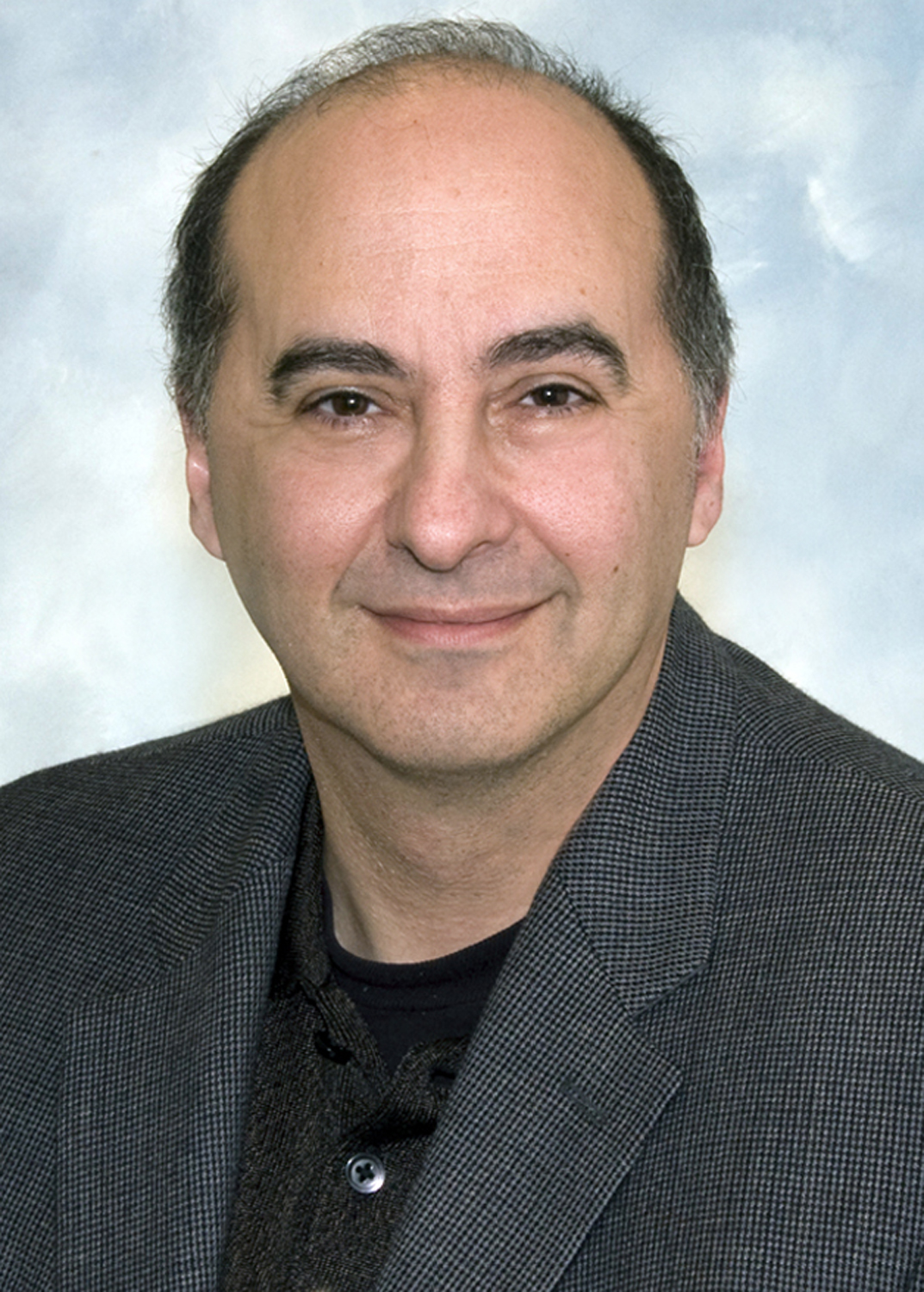 Douglas Rothman合作论文数Magnetic Resonance Research Center, Yale School of Medicine146
 Kevin Behar合作论文数School of Medicine, Yale University69
 D. S. Fahmeed Hyder合作论文数Department of Biomedical Engineering, Yale University;InnovaCyclics LLC;Magnetic Resonance Research Center, Yale University39
 James Prichard合作论文数Department of Neurology, Yale School of Medicine, Yale University34
 Jeffry Alger合作论文数Brain Research Institute, University of California, Los Angeles;Department of Radiology, University of California, Los Angeles31MJ AVISON合作论文数29
 Graeme Mason合作论文数Biomedical Engineering, School of Engineering & Applied Science, Yale University27
 Gerald I Shulman合作论文数Diabetes Research Center, School of Medicine, Yale University26OAC PETROFF合作论文数25Ja Denhollander合作论文数23
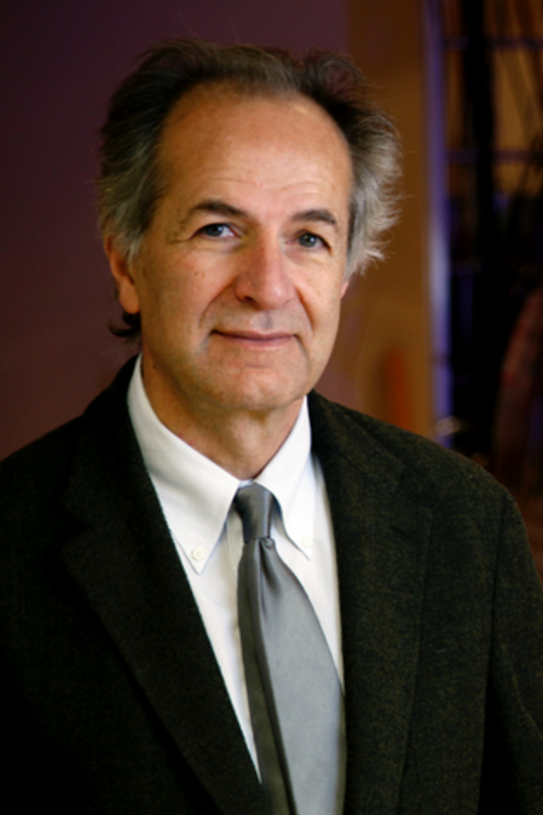 Kamil Uğurbil合作论文数Department of Radiology, Medical School, University of Minnesota;Center for Magnetic Resonance Research, University of Minnesota20
 Jan A. Den Hollander合作论文数Department of Medicine (Cardiology),, University of Alabama at Birmingham17
 Edward Novotny合作论文数Department of Neurosurgery, Yale University17NJ SIEGEL合作论文数15Andrew M. Blamire合作论文数15T OGINO合作论文数14Hoby P. Hetherington合作论文数14Brown R T合作论文数13
 Peter Michael Eisenberger合作论文数13Gunilla Thulin合作论文数11Thomas Jue合作论文数10N. R. Sibson合作论文数CR-UK/MRC Gray Institute for Radiation Oncology & Biology, University of Oxford9A Dhankhar合作论文数8
 Eugene J. Barrett合作论文数Division of Endocrinology and Metabolism, Department of Medicine, School of Medicine, University of Virginia;Diabetes Center, University of Virginia8
 Gil Navon合作论文数School of Chemistry, Tel Aviv University8
 Kurt Wüthrich合作论文数iHuman Institute, Shanghai Tech University;Department of Integrative Structural and Computational Biology, The Scripps Research Institute;Institute of Molecular Biology and Biophysics, Department of Biology, Eidgenössische Technische Hochschule Zürich8G S Brown合作论文数7P Glynn合作论文数7T Yamane合作论文数7F Arias-Mendoza合作论文数7S Ogawa合作论文数7
 Gregory McCarthy合作论文数Department of Psychology, Yale University;Human Neuroscience Lab, Yale University7B. M. Kincaid合作论文数Ernest Orlando Lawrence Berkeley National Laboratory7
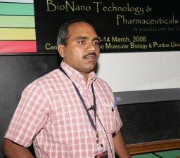 Anant B. Patel合作论文数Tata Institute of Fundamental Research7TB Price合作论文数7A Vanwaarde合作论文数6KM GAUDIO合作论文数6I Magnusson合作论文数6H Rottenberg合作论文数6
 Nicola Sibson合作论文数Department of Oncology, University of Oxford6
 Wei Chen合作论文数College of Automation, Northwestern Polytechnical University6David R. Kearns合作论文数6Susan D. Boulware合作论文数University of Connecticut Health Center6Maren R. Laughlin合作论文数NIDDK6S M Fitzpatrick合作论文数5
 Chris C Hanstock合作论文数5a m howseman合作论文数5ME STROMSKI合作论文数5W E Blumberg合作论文数5




