A finite-element-based algorithm for the in silico construction of a novel tri-tube heart valve was developed to facilitate optimization of the leaflet geometry. An anisotropic hyperelastic model fitted to high-strain rate planar equibiaxial tension and compression data was used to approximate the nonlinear and anisotropic material behavior of biologically-engineered tubes and simulate valve closure under steady back pressure and steady forward flow. Four metrics were considered to evaluate valve performance in simulated closure: coaptation area, regurgitation area, pinwheel index, and prolapse area. Response surfaces revealed competing objectives between metrics for a valve of target 24 mm diameter in terms of two design parameters, tube diameter and leaflet height. A multi-objective genetic algorithm determined an intermediate tube diameter and leaflet height (16 mm and 11 mm, respectively) of the design space as optimal. Additionally, steady flow simulations were performed using two-way fluid–structure interaction with selected designs to examine washout behind leaflets with particle tracking. One design close to the optimal point for valve closure indicated washout for particles initially distributed behind leaflets. Though comprehensive valve design optimization requires flow analysis over multiple valve cycles to capture all effects associated with flow, this methodology based on diastolic state geometry optimization followed by steady washout analysis reduces the space of design variables for further optimization.
Aims Replacement heart valves that grow with children remain an unmet need. We previously reported valves fabricated from tubes of fibroblast-derived collagenous matrix increased in size while functioning with low systolic gradients and less than moderate regurgitation over 52 weeks in most cases, when implanted as interpositional grafts in the pulmonary artery of lambs. Here, we evaluated valved conduits fabricated by including an inflow segment to the tri-tube valve allowing for myocardial anastomosis as done in a typical right ventricular outflow tract (RVOT) surgical repair, in the same growing lamb model.Methods and results In this pilot study, 19 mm valved conduits fabricated from resorbable suture were implanted into Dorset lambs (n = 3), sutured to the pulmonary annulus and distal pulmonary artery with resorbable suture after dissection of the pulmonary valve leaflets and resection of an arterial segment. Valve function and dimensions were measured with longitudinal transthoracic echocardiography. All animals exhibited an increase in valve diameter (18.2 +/- 1.8 mm at 1 week to 25.1 +/- 2.4 mm at 52 weeks) and leaflet free-edge length (21.1 +/- 2.4 mm at 1 week to 26.2 +/- 3.9 mm at 52 weeks) while functioning with at most mild regurgitation over 52 weeks. The inflow segment of the conduit grew somatically based on its unchanged thickness and increased diameter (38%) and collagen content (128%). In all three explanted conduits, the leaflets contained interstitial cells, new collagen and elastin primarily around the base, a developing endothelium on the surfaces, and they remained thin and pliable without macroscopic calcification. There was interdigitating integration of the conduit with the myocardium at the pulmonary annulus. Further, a stent was successfully placed in a valved conduit at term to evaluate feasibility of a prospective clinical intervention.Conclusion This valved conduit grows in lambs based on this pilot study and thus has clinical potential for RVOT reconstruction and long-term valve growth in children.
Anticoagulant and antiplatelet therapies are used to prevent life-threatening complications associated with thrombosis. While there are numerous clinical guidelines for antithrombotic medications, there is an incomplete understanding of whether these interventions yield similar effects in preclinical models, potentially impacting their predictive value for translational studies on the development of medical devices, therapies, and surgical techniques. Due to their close physiologic similarities to humans, we employed nonhuman primates (NHPs) using a reverse translational approach to analyze the response to clinical regimens of unfractionated heparin, low-molecular-weight heparin (LMWH) and aspirin to assess concordance with typical human responses and evaluate the predictive validity of this model. We evaluate activated clotting time (ACT) in nine rhesus and six cynomolgus macaques following the intraoperative administration of intravenous unfractionated heparin (100–300 U/kg) reflecting the clinical dose range. We observed a significant dose-dependent effect of heparin on ACT (low-dose average = 114.1 s; high-dose average = 148.3 s; p = 0.0011). LMWH and aspirin, common clinical antithrombotic prophylactics, were evaluated in three rhesus macaques. NHPs achieved therapeutic Anti-Xa levels (mean = 0.64 U/mL) and ARU (mean = 459) via VerifyNow, adhering to clinical guidance using 1.0 mg/kg enoxaparin and 81 mg aspirin. Clinical dosing strategies for unfractionated heparin, LMWH, and aspirin were safe and effective in NHPs, with no development of thrombosis or bleeding complications intraoperatively, postoperatively, or for prophylaxis. Our findings suggest that coagulation studies, performed as an integrative part of studies on biologics, bioengineered devices, or transplantation in NHPs, can be extrapolated to the clinical situation with high predictive validity.
BackgroundGrowth is the holy grail of tissue implants in pediatrics. No vascular graft currently in use for surgical repairs of congenital heart defects has somatic growth capacity.MethodsBiologically-engineered grafts (6 mm) grown from donor ovine fibroblasts in a sacrificial fibrin gel were implanted into the left pulmonary branch of 3-month old lambs for 3, 6, and 18 months. A control group of Propaten (R) PTFE grafts was implanted for 6 months.ResultsThe engineered grafts exhibit extensive site-appropriate recellularization after only 3 months and near-normal increase of diameter from the preimplant value of 6 mm to 12.9 mm and also a doubling of length from 6.0 mm to 13.0 mm at 6 months (n = 3) associated with apparent somatic graft growth (collagen content increase of 265% over 18-month, n = 2), along with excellent hemodynamics and no calcification, in contrast to the Propaten (R) grafts. The left-right flow distribution is nearly 50-50 for the engineered grafts at 6 months (n = 3) compared to about 20-80 for the Propaten (R) grafts (n = 3), which have less than one-half the diameter, a 6-fold higher pressure gradient, and stunted vascular development downstream of the graft. The engineered grafts exhibit a stable diameter over months 12-18 when the lambs become adult sheep (n = 2).ConclusionsThis study supports the use of these regenerative grafts with somatic growth capacity for clinical trial in patients born with a unilateral absent pulmonary artery branch, and it shows their potential for improving development of the downstream pulmonary vasculature. Blood vessel implants that are currently used to repair heart defects at birth do not grow with the child. This means that children need to have multiple open heart surgeries to replace implants with larger implants as they grow. We grew implants from a donor sheep's skin cells, and then completely removed the cells from the graft. We then implanted the grafts in 3-month old lambs. The lambs' cells repopulated the implants and the implants increased in size as the lambs grew. Further experiments are required first, but our preliminary findings suggest that using a similar implant in children could improve the quality of life of children with heart defects by avoiding the need for them to have multiple surgeries to replace implants as the child grows. Syedain et al. evaluate growth of biologically-engineered grafts grown from donor ovine fibroblasts in a sacrificial fibrin gel implanted into the left pulmonary branch of 3-month old lambs. The grafts exhibit extensive site-appropriate recellularization and increase in diameter and length until the lambs reach adulthood.
Introduction: A valved conduit with growth potential for pediatric patients remains an unmet need. Here we present a novel valved conduit with growth potential based on initial data in a growing lamb model. The valve is constructed from three biologically-engineered tubes of acellular collagenous matrix possessing somatic growth potential (Syedain et al., Nat Comms, 2016) using resorbable suture with each tube contributing to create a “leaflet” (Syedain et al, Sci Transl Med, 2021). This tri-tube valve previously demonstrated growth over 52 weeks when implanted interpositionally in the pulmonary artery. Methods: In this study, a fourth tube of the same matrix is placed around the tri-tube valve to create a tubular valved conduit. The inlet segment is cut diagonally to allow for direct attachment to the myocardium, emulating a clinical repair (see figure). Results: The first two lambs to date implanted in this study exhibited normal leaflet function, a pressure gradient less than 5mmHg without regurgitation or dilatation of the conduit at 12-week echocardiography (see figure). During this time the animal’s weight increased from 34±2kg to 48±1kg. The inflow pattern with this myocardial implantation was more uniform than in the previous study that used a pulmonary artery interposition implant of the just the valve (with no conduit). Discussion: Demonstrating integration of the conduit’s matrix to the myocardium is a key step toward a clinical trial. Previous pulmonary interposition implants of the valve, while achieving integration with the adjacent artery prior to dissolution of the Maxon suture (50% loss of tensile strength in ~3 weeks, complete by ~26 weeks), did not establish this clinically relevant point of myocardial integration. Conclusion: The preliminary finding is that the valved conduit exhibits excellent function at 12 weeks in the growing lamb and integration of the conduit’s matrix with the valve annulus-myocardium will likely occur. Funding: R01 HL107572
Despite its importance in physiological processes and tissue engineering, the mechanism underlying cell contact guidance in an aligned fibrillar network has defied elucidation due to multiple interdependent signals that such a network presents to cells, namely, anisotropy of adhesion, porosity and mechanical behaviour. A microstructural–mechanical model of fibril networks was used to assess the relative magnitudes of these competing signals in networks of varied alignment strength based on idealized cylindrical pseudopods projected into the aligned and orthogonal directions and computing the anisotropy of metrics chosen for adhesion, porosity and mechanical behaviour: cylinder–fibre contact area for adhesion, persistence length of pores for porosity and total force to displace fibres from the cylindrical volume as well as network stiffness experienced upon cylinder retraction for mechanical behaviour. The signals related to mechanical anisotropy are substantially higher than adhesion and porosity anisotropy, especially at stronger network alignments, although their signal to noise (S/N) values are substantially lower. The former finding is consistent with a recent report that fibroblasts can sense fibril alignment via anisotropy of network mechanical resistance, and the model reveals this can be due to either mechanical resistance to pseudopod protrusion or retraction given their signal and S/N values are similar.
Objective: There is a dire need for a heart valve that can grow with children. We fabricated tubes of biologically-engineered matrix that have been shown to regenerate and grow as a pulmonary artery replacement in lambs, and implemented a novel design for a valved conduit made from sewing three of these tubes together with degradable suture that confers valve growth potential. The study was to assess valve function and growth potential over a one-year period in the lamb model.Methods: Seven lambs were implanted with tri-tube valved conduits in sequential cohorts and compared to bioprosthetic conduits using longitudinal echocardiography and histological and mechanical characterization post-explantation.Results: Valves implanted in two lambs of the first cohort of four animals functioned with only mild regurgitation and systolic pressure drops <10 mmHg up to 52 weeks post-implantation, during which the valve diameter increased from 19 mm to a normal ~25 mm. In a second cohort of three animals, an additional tube was used to create a sleeve around the tri-tube valve to counteract faster root growth relative to the leaflets. Two valves exhibited only trivial-to-mild regurgitation at 52-weeks with similar diameter increase and systolic pressure drops of < 5 mm Hg. The same was true for the third animal until moderate regurgitation at 52 weeks correlating to hyper-increase of the valve diameter. In all explanted valves, the leaflets remained thin and pliable with only sparse, punctate microcalcification at most, and they contained interstitial cells and an endothelium progressing from the base of the leaflets.Conclusions: These results are demonstrably improved in terms of calcification and hemodynamic function compared to reported studies for clinically-used pediatric bioprosthetic valves tested in the same model. The results demonstrate the potential for long-term valve growth of this "off-the-shelf" tri-tube valved conduit in children.
Objective: Tissue-engineered valved conduits that can resist calcification and degradation while allowing somatic growth would provide an ideal solution for congenital valve replacement. The goal of this study was to assess remodeling of conduits made from acellular engineered matrix manufactured from allogeneic fibroblasts. Methods: Three 16mm diameter tubes of engineered tissue were stitched together with Maxon degradable sutures to create a novel tri-leaflet valved conduit. The valved conduit was implanted as a pulmonary replacement in 7 lambs (average age 15.5 wk) up to 12 months. Conduits explanted after 12 - 52 weeks were evaluated histologically and biochemically for matrix remodeling and calcification. Results: The valved conduits explanted at the earliest timepoint of 12 weeks showed degradation of Maxon sutures indicating matrix remodeling was providing the load-bearing properties at resorbing suture lines. The conduit increased in diameter over the implant duration. Collagen and protein concentrations in the wall of the conduit at 52 weeks were increased. Histology showed recellularization of the wall matrix with αSMA+ interstitial cells and CD31+ endothelial cells on the lumen surface. The leaflet matrix showed no change in the collagen or protein concentration compared to the implant. The cell concentration in the leaflet matrix increased from zero at implant to 34±10M cells/cm3 at the 52 week explant. Cells on the leaflet surface were also CD31+. Von Kossa stain showed no calcification other than near Maxon sutures and sparse punctate micro-calcification in two valves. Conclusions: This 1-yr study of a valved conduit implanted in a growing lamb demonstrated extensive matrix remodeling both to provide load-bearing capabilities at the degradable suture lines and new extracellular matrix to confer somatic growth. Combined with a lack of macro-calcification over the 1-yr implantation are promising outcomes to support the use of this acellular engineered matrix for potential pediatric use.
There is a need for replacement heart valves that can grow with children. We fabricated tubes of fibroblast-derived collagenous matrix that have been shown to regenerate and grow as a pulmonary artery replacement in lambs and implemented a design for a valved conduit consisting of three tubes sewn together. Seven lambs were implanted with tri-tube valved conduits in sequential cohorts and compared to bioprosthetic conduits. Valves implanted into the pulmonary artery of two lambs of the first cohort of four animals functioned with mild regurgitation and systolic pressure drops <10 mmHg up to 52 weeks after implantation, during which the valve diameter increased from 19 mm to a physiologically normal ~25 mm. In a second cohort, the valve design was modified to include an additional tube, creating a sleeve around the tri-tube valve to counteract faster root growth relative to the leaflets. Two valves exhibited trivial-to-mild regurgitation at 52 weeks with similar diameter increases to ~25 mm and systolic pressure drops of <5 mmHg, whereas the third valve showed similar findings until moderate regurgitation was observed at 52 weeks, correlating to hyperincrease in the valve diameter. In all explanted valves, the leaflets contained interstitial cells and an endothelium progressing from the base of the leaflets and remained thin and pliable with sparse, punctate microcalcifications. The tri-tube valves demonstrated reduced calcification and improved hemodynamic function compared to clinically used pediatric bioprosthetic valves tested in the same model. This tri-tube valved conduit has potential for long-term valve growth in children.
Biologically-engineered vascular grafts have the potential to provide a viable alternative to donor vessels and synthetic grafts. In congenital heart defect patients, the need is even more dire since neither has the capacity to provide somatic growth. To ensure clinically-used grafts perform to accepted standards, mechanical strength is a crucial consideration, with burst testing being considered as one key metric. While ISO 7198 standards for prosthetic vascular grafts provide multiple choices for burst testing, most studies with tissue-engineered grafts have been performed with only pressure burst testing. Here, we compare the performance of a decellularized tube of collagenous matrix grown from dermal fibroblasts, possessing circumferential fiber alignment and anisotropic tensile properties, as determined from pressure and probe burst testing. The two burst tests showed a strong correlation with each other and with tensile strength. Further, relatively weak and strong batches of grafts showed commensurate differences in pressure and probe burst values. Both probe burst and tensile strength measurements in the central and edge regions of the grafts were similar in value, consistent with homogenous collagen content and microstructure throughout the grafts as indicated by histology, in contrast to ovine femoral and carotid arteries similarly tested. Finite element analysis of the probe burst test pre-failure for a homogeneous, isotropic approximation of the matrix constitutive behavior indicated dependence of the (inferred) effective failure stress achievable on probe diameter. The results indicate a probe burst test in a sampled edge region of this biologically-engineered graft provides a representative measure of burst strength of the entire graft.
Despite the ubiquitous importance of cell contact guidance, the signal-inducing contact guidance of mammalian cells in an aligned fibril network has defied elucidation. This is due to multiple interdependent signals that an aligned fibril network presents to cells, including, at least, anisotropy of adhesion, porosity, and mechanical resistance. By forming aligned fibrin gels with the same alignment strength, but cross-linked to different extents, the anisotropic mechanical resistance hypothesis of contact guidance was tested for human dermal fibroblasts. The cross-linking was shown to increase the mechanical resistance anisotropy, without detectable change in network microstructure and without change in cell adhesion to the cross-linked fibrin gel. This methodology thus isolated anisotropic mechanical resistance as a variable for fixed anisotropy of adhesion and porosity. The mechanical resistance anisotropy |Y*| -1 - |X*| -1 increased over fourfold in terms of the Fourier magnitudes of microbead displacement |X*| and |Y*| at the drive frequency with respect to alignment direction Y obtained by optical forces in active microrheology. Cells were found to exhibit stronger contact guidance in the cross-linked gels possessing greater mechanical resistance anisotropy: the cell anisotropy index based on the tensor of cell orientation, which has a range 0 to 1, increased by 18% with the fourfold increase in mechanical resistance anisotropy. We also show that modulation of adhesion via function-blocking antibodies can modulate the guidance response, suggesting a concomitant role of cell adhesion. These results indicate that fibroblasts can exhibit contact guidance in aligned fibril networks by sensing anisotropy of network mechanical resistance.
Vascular calcification is a commonly occurring pathological process and is recognized as an independent prognostic marker for cardiovascular morbidity and mortality. Recent progress in developing novel therapies to modify vascular calcification is critically hampered due to the lack of reliable in vitro experimental models that recapitulate the structural and mechanical attributes of calcified arteries. In this study, we show the ability to model the behavior of diffuse vascular calcification in vitro using biologicallyengineered grafts approximating the composition, structure, and mechanical properties of arteries. Transmural calcification was achieved by exposing the acellular grafts of collagenous ECM to complete medium containing elevated Calcium (Ca) and Phosphate (P) concentrations. It was found that increasing the serum concentration from 2% to 10% increased the extent and degree of calcification based on histochemical, ultrastructural, chemical and thermal analyses. The presence of variably-sized spherical calcific deposits within the matrix further confirmed its morphological similarity to pathologic calcification. Mechanical testing demonstrated up to a 16-fold decrease in compliance due to the calcification, consistent with prior reports for calcified arteries. The model developed thus has potential to improve the design and development of interventional devices and therapies for the diagnosis and treatment of arterial calcification. Statement of Significance The presence of extensive vascular calcification makes angiographic/interventional procedures difficult due to reduced arterial compliance. Current attempts to develop safe and effective non-surgical adjunctive techniques to treat calcified arteries are largely limited by the lack of a physiologically relevant testing platform that mimics the structural and mechanical features of vascular calcification. Herein, we developed an off-the-shelf calcified artery model, with the goal to accelerate the pre-clinical development of novel therapies for the management of arterial calcification. To the extent of our knowledge, this is the first report of an in vitro tissue-engineered model of diffuse arterial calcification. (C) 2020 Acta Materialia Inc. Published by Elsevier Ltd. All rights reserved.
While the need for a small diameter arterial bypass graft is clear, the hemodynamics of small diameter arteries like the coronary artery have led to the failure of synthetic materials that are successful for large diameter grafts like the aorta. Many patients do not have autologous vessels suitable for use due to systemic vascular disease and previous harvest, and second site morbidity is substantial. Vascular tissue engineering has explored the use of tissue cells or stem cells combined with various types of scaffolds to make tubular constructs subjected to chemical and/or mechanical stimulation in an attempt to develop small diameter arterial bypass grafts without a proven clinical success, yet. Here, we review the use of biopolymer gels as the scaffold and preclinical successes, and we discuss their advantages and challenges.
Biologically-engineered matrix - a tissue that is grown in vitro from donor cells, decellularized, and stored prior to use as off-the-shelf allografts - offers a promising alternative to current cardiovascular biomaterials. This perspective reviews preclinical studies and clinical trials of vascular grafts and valves comprising biologically-engineered matrix, with a focus on those based on donor dermal fibroblast remodeling of fibrin gel with the capacity to heal and grow following recellularization, via animation of the matrix. It concludes with a discussion of related key clinical considerations.
Chronic venous insufficiency affects over 2 million patients in the US alone, with severe cases involving thousands of patients with chronic leg ulcers and potential amputation. Current treatment options are limited, with surgical repair of vein valves being the most effective but challenging solution. A transcatheter vein valve made from a biologically-engineered matrix possessing the ability to regenerate has the potential to provide both valve function and long-term hemocompatibility and durability because the matrix becomes endothelialized and populated with host tissue cells. We have developed a novel tissue-engineered transcatheter vein valve (TEVV) on a Nitinol stent and demonstrated function and durability in vitro. Tissue was grown from fibroblasts in fibrin gel so as to embed the stent, with a tubular extension of the engineered tissue from one end of the stent that was stitched along opposite sides and everted into the stent to form a bileaflet valve. Following decellularization, to create an "off-the-shelf" TEVV comprised of the resulting collagenous matrix, it was tested in a pulse duplicator to evaluate hydrodynamic properties for a range of flow rates. The TEVV was shown to have forward pressure drops in the range of 2-4 mmHg, low closing volume, and nil regurgitation. Further hydrodynamic tests were performed after crimping and then again after 1 million cycle durability testing, showing no degradation of valve performance or any visual damage to the matrix. The TEVV held over 600 mmHg backpressure after the durability testing, ensuring the valve would withstand pressure spikes well outside of the normal in vivo range. Catheter-based delivery into the ovine iliac vein demonstrated TEVV closing 2 weeks p.o. and en-dothelialization without thrombosis 8 weeks p.o.
Conferring antithrombogenicity to tissue-engineered vascular grafts remains a major challenge, especially for urgent bypass grafting that excludes approaches based on expanding autologous endothelial cells (ECs) that requires weeks of cell culture. Adipose-derived stem cells (ASCs) are available from most patients in sufficient number for coronary bypass graft seeding and may be effective as allogeneic cells. We thus compared the adhesion and platelet binding of human ASCs that were shear conditioned with constant and pulsatile shear stress (SS) after seeding the cells on a biologically engineered matrix suitable for arterial grafts. A monolayer of cells was maintained up to 15dyn/cm(2) constant SS and up to 15dyn/cm(2) mean pulsatile SS for 6 days of shear flow. Platelet binding was reduced from 83% to 6% of surface area and nitric oxide production was increased 23-fold with 7.5-15dyn/cm(2) constant SS, but not pulsatile SS, relative to cells cultured statically on the matrix for 6 days. The reduction in platelet binding varied from no reduction to maximum reduction over a constant shear range of approximate to 2 to 4dyn/cm(2), respectively. Collectively, the study supports the potential use of ASCs to seed the luminal surface of a vascular graft made from this biologically engineered matrix to confer an antithrombogenic surface during the development of an endothelium from the seeded cells or the surrounding blood and tissue.
Fibrin has been, and continues to be widely used in surgical applications as a tissue sealant, although this requires much higher concentrations than that of a clot which cells invade and remodel. More recently, it has been examined as a scaffold for tissue engineering. Fibrin possesses several qualities in addition to those already mentioned that make it ideal for use in tissue engineering. It is biocompatible, biodegradable, and can be produced from human serum, making it possible to use autologous sources. This entry will provide background on the structure and biochemistry of fibrin, as well as an overview of its interactions with cells. We will then finish with a discussion of the tissue engineering applications currently being pursued by researchers.
 Zeeshan H. Syedain合作论文数Departments of * Chemical Engineering and Materials Science;Biomedical Engineering;University of Minnesota;Departments of * Chemical Engineering and Materials Science, University of Minnesota42
 Victor H. Barocas合作论文数Dept Biomed Engn, Univ Minnesota35Sandra L. Johnson合作论文数18
 Edward Sander合作论文数Roy J. Carver Department of Biomedical Engineering, College of Engineering, University of Iowa12Brett C. Isenberg合作论文数Department of Chemical Engineering & Materials Science, University of Minnesota12michael neidert合作论文数11Victor K Lai合作论文数Department of Chemical Engineering, University of Minnesota-Duluth11
 Douglas A. Lauffenburger合作论文数Department of Chemical Engineering, School of Engineering, Massachusetts Institute of Technology;Department of Biology, Massachusetts Institute of Technology11T. S. Girton合作论文数11
 Richard Bianco合作论文数Department of Surgery, Medical School, University of Minnesota10Matthew Lahti合作论文数Experimental Surgical Services, Department of Surgery, University of Minnesota, Minneapolis, MN, United States9
 Richard Dickinson合作论文数Department of Chemical Engineering, University of Florida9Jay Reimer合作论文数Univ Minnesota, Dept Biomed Engn, Minneapolis, MN 55455 USA8James M. Berry合作论文数8
 Lee A. Meier合作论文数Department of Biomedical Engineering, University of Minnesota8Theodore R. Oegema合作论文数8
 Stefano Guido合作论文数Dipartimento di Ingegneria Chimica, dei Materiali e della Produzione Industriale, Università degli Studi di Napoli Federico II8Bee Haynie合作论文数7N Dubey合作论文数dsm7
 Justin S Weinbaum合作论文数7
 Ted Tower合作论文数7Kristen T. Morin合作论文数7
 Spencer Lake合作论文数Department of Orthopaedic Surgery, University of Pennsylvania6Paul C Letourneau合作论文数6Jason W. Bjork合作论文数Department of Biomedical Engineering, University of Minnesota6Jeffrey J. Ross合作论文数6Paul A.J. Enever合作论文数6Chrysanthi Williams合作论文数5Carney John合作论文数5Schmidt Jillian B合作论文数5Wendel Jacqueline S合作论文数5
 Sally Householder Zigmond合作论文数Department of Biology, University of Pennsylvania5
 Wolfgang Alt合作论文数University Bonn5Alice G. Moon合作论文数5
 David Shreiber合作论文数Department of Biomedical Engineering, School of Engineering, Rutgers, The State University of New Jersey5Frank Baaijens合作论文数4david m knapp合作论文数4Robroy MacIver合作论文数4
 Sunčica Čanić合作论文数Department of Mathematics, University of California, Berkeley4
 Ahmad Falahatpisheh合作论文数4
 Alavi S Hamed合作论文数The Edwards Lifesciences Center for Advanced Cardiovascular Technology, University of California4
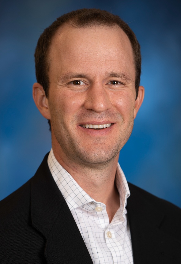 Craig Goergen合作论文数Weldon School of Biomedical Engineering, College of Engineering, Purdue University4
 Groves Elliott M合作论文数4
 Ryan C Hill合作论文数University of Colorado Denver School of Medicine, University of Colorado Denver4
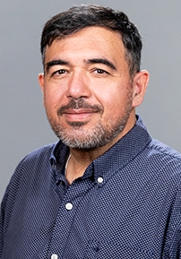 Mohammad Reza Kaazempur Mofrad合作论文数Departments of Bioengineering and Mechanical Engineering, College of Engineering, University of California, Berkeley;Molecular Cell Biomechanics Lab, University of California, Berkeley;Lawrence Berkeley National Lab4
 Catherine Verfaillie合作论文数Faculty of Medicine, KU Leuven4Grassl E D合作论文数4
 Lakshmi Dasi合作论文数Wallace H. Coulter Department of Biomedical Engineering, College of Engineering, Georgia Institute of Technology;DasiSimulations4
 Stephen H Little合作论文数Methodist DeBakey Heart Center Echocardiography Laboratory, Houston, Texas.4




