Human ecdysoneless protein (ECD) plays an essential role in regulating cell cycle progression and cell survival. ECD has previously been implicated in RNA splicing through its association with spliceosomal proteins. Here, using electrophoretic mobility shift assay, fluorescence polarization assays, and mutational analysis, we demonstrate that ECD directly binds to RNA. Enhanced CLIP-seq analysis identified a broad repertoire of mRNAs bound to ECD in cells. RNA-seq analyses revealed that ECD depletion leads to widespread splicing aberrations and altered gene expression. ECD binding to RNAs was enriched near splice sites, and a substantial fraction of ECD-bound transcripts exhibited splicing defects upon ECD depletion. ECD associates with and stabilizes the U5 small nuclear ribonucleoprotein (snRNP) complex specific proteins. While depletion of ECD reduced the levels of key U5-specific proteins, these proteins exhibited an increased association with the R2TP complex in knockout cells. Notably, we found ECD to directly bind to U5 snRNA, and an RNA binding defective mutant of ECD (Δ135-148) failed to rescue the reduced levels of U5-specific proteins or the proliferation defect induced by ECD depletion. Collectively, these findings demonstrate that ECD binds to RNAs, including the U5 snRNA, and that RNA-binding is required for ECD to stabilize the U5 snRNP and for cellular functions.
G-quadruplexes (G4s) are non-canonical DNA secondary structures enriched at promoters, yet their regulatory role in transcription remains elusive. While G4-ligand-based studies suggest transcriptional repression, their prevalence at oncogene promoters and correlation with high expression suggest a positive regulatory role. Here, we provide direct genetic evidence that promoter G4s function as positive activators of gene expression through a novel mechanism. By selectively mutating endogenous promoter G4 motifs, we demonstrate that G4 loss significantly impairs oncogene expression. Using the endogenous CXCL1 promoter G4 as an example, we revealed that loss of a single promoter G4 motif led to a marked down-regulation of CXCL1 expression as well as inhibition of cellular functions such as cell migration and invasion. Mechanistically, we identified apurinic/apyrimidinic endonuclease (APE1), a multifunctional DNA repair and redox factor, as a G4-binding protein which was recruited to promoters via its unique N-terminus. Subsequently, the redox activity of APE1 enhances transcription factor binding at G4-containing promoters, driving a pro-metastatic gene expression program. Disruption of the G4-APE1 interaction, either genetically or pharmacologically, suppresses gene expression and impairs tumor cell malignant traits. Our findings establish a direct genetic link and mechanistic basis for promoter G4s as crucial drivers of oncogene expression and tumor progression.
Signal-responsive transcriptional programs are turned on/off by cis-regulatory elements (cis-elements) acting in coordination with gene promoters. While large genomics datasets have annotated common cis-element features, interpreting and predicting specific environmental responses at the chromatin level are critical ongoing challenges. We combined signal-induced chromatin accessibility changes with transcriptomics and a comprehensive catalogue of chromatin occupancy features for predicting functional signaling responses. Using the Kit receptor tyrosine kinase pathway-a central pro-survival, pro-proliferation, and pro-differentiation pathway in hematopoiesis, erythropoiesis, and other tissues-we mapped the sequence and chromatin features at thousands of Kit signal-responsive cis-elements in the human genome. A subset of Kit-induced changes to chromatin occupancy required upregulation of early growth response-1 (EGR1), uncovering distinct EGR1-sensitive and EGR1-insensitive branches of Kit signaling. Predictions of chromatin features associated with signal responses were tested using CRISPR-mediated cis-element disruption, which impaired target gene activation even at very long ranges. These findings define subsets of Kit signaling-dependent cis-regulatory logic which can be applied to interpret how growth factor pathways and cell-type specific transcription factors communicate on chromatin to direct cell behaviors in normal and disease contexts.
How chromatin is spatially organized in the nucleus has long been studied through the lens of large-scale A/B compartments, but whether these sizes reflect true biological units or analytical artifacts has remained unclear. We find that limits imposed by the conventional eigenvector-based compartment calling have required extreme sequencing depth and coarse resolution, obscuring regulatory-scale organization. We developed CRUSH to iteratively refine compartments to 1 kb resolution without the need for extreme sequencing depth, we show that kilobase-scale A/B segregation (micro-compartments) is evident across cell types. Across these maps, we demonstrate that RNA polymerase II pausing contributes to a sub-genic compartment signature at the transcription start site and that active enhancers almost universally occupy the A compartment. We then show that fine-scale compartment maps can resolve cancer subtype-specific regulatory programs, single-cell tissue identity, and cold-adaptation regulomes in a 52,000-year-old woolly mammoth. These findings establish chromatin compartmentalization as a gene-scale regulatory feature with broad implications for development, disease, and genome evolution.
Prostate cancer (PC) is the most diagnosed cancer in males. Androgen receptor (AR) signaling is essential for PC progression. ECD (Ecdysoneless) protein participates in cell cycle progression and cell survival and is overexpressed in non-PC cancers. Current study explored the role and function of ECD in PC progression. TCGA Prostate Adenocarcinoma database was used to assess correlation between ECD and AR expression, and ECD’s expression in PC dataset. Bioinformatic analysis was performed to predict androgen response elements in the ECD promoter. Chromatin immunoprecipitation (ChIP) and Promoter-Luciferase assays were used to assess AR recruitment to the ECD promoter. ECD was overexpressed, as well as crispr knocked out in hormone-dependent LNCaP and hormone-independent C4-2B PC cell lines. RNA-seq analysis was performed to assess transcriptomics changes in ECD-overexpressing (ECD-OE) tumors. RNA immunoprecipitation (RIP) was performed to assess ECD protein association to RNAs. Glucose uptake, and glycolytic rates assays were carried out to assess glycolysis. ECD is overexpressed in PC patients’ tissues as compared to hyperplasia prostate samples, and ECD overexpression is associated with short patient survival. A significant positive correlation was observed between ECD and AR expression. Consensus androgen response elements in the ECD promoter showed androgen dependent recruitment of AR to the ECD promoter by ChIP assay. Luciferase assays showed androgen-dependent increase in ECD-luciferase activity. ECD overexpression and knockout in both LNCaP and C4-2B PC cell lines showed enhanced and decreased oncogenic traits, respectively. In vivo mice xenograft experiments showed ECD overexpression in LNCaP led to a significant increase in tumorigenesis. RNA-seq showed ECD overexpression led to increased levels of key glycolytic genes, which was confirmed by qRT-PCR. RIP assay revealed that ECD associates with the mRNA of these key glycolytic genes and enhances their stability. Functionally, ECD overexpressing PC cells showed higher glucose uptake and glycolytic rates, as compared to vector cells. Overall, our results demonstrate a novel role for androgen-regulated ECD overexpression in PC tumorigenesis through direct binding and stabilization of mRNAs of key glycolytic genes. Mohsin Raza, Asher Rajkumar Rajan, Benjamin B. Kennedy, Timothy E. Reznicek, Farshid Oruji, Sameer Mirza, M Jordan Rowley, Glen Kristiansen, Bhopal C. Mohapatra, Hamid Band, Vimla Band. Androgen regulated ECD overexpression promotes prostate cancer tumorigenesis through increased glycolysis [abstract]. In: Proceedings of the American Association for Cancer Research Annual Meeting 2025; Part 1 (Regular Abstracts); 2025 Apr 25-30; Chicago, IL. Philadelphia (PA): AACR; Cancer Res 2025;85(8_Suppl_1):Abstract nr 266.
The human ecdysoneless protein (ECD) plays an essential role in the regulation of cell cycle and cell survival. ECD has been implicated in RNA splicing through its association with the protein components of splicing complex. Here, using electrophoretic mobility shift assay and mutational analysis, we demonstrate that ECD directly binds to RNA through amino acids 135-148. Using enhanced CLIP-seq analyses, we identified a large repertoire of mRNAs bound to ECD. RNA-seq analyses revealed that ECD depletion in cells leads to widespread RNA splicing aberrations associated with alterations in gene expression. Significantly, we demonstrate that ECD mediates mRNA splicing by directly binding to RNA sequences located near splicing sites in context-dependent manner. Mechanistically, ECD associates with key proteins of the U5 snRNP complex in an RNA-independent manner and directly binds to U5 small nuclear RNA (U5 snRNA). Importantly, ECD-U5 snRNA interaction is crucial for maintaining the expression of U5-specific proteins. Furthermore, RNA binding defective mutant of ECD fails to rescue downregulated levels of U5 snRNP components or cell proliferation block induced by ECD knockout. Collectively, we provide compelling evidence that ECD regulates RNA splicing by directly associating with RNAs, and the RNA binding activity of ECD is essential for its function.
Stress erythropoiesis elevates the rate of red blood cell (RBC) production as a physiological response to stressors such as anemia or hypoxia. In acute anemia, RBC progenitors and precursors temporarily rewire their transcriptome, up- and downregulating hundreds of genes to accelerate the production of mature RBCs. Effective regeneration requires communication between critical cytokine signals (e.g., BMP4) and cis-regulatory elements on chromatin which coordinate transcriptional changes. To identify cis-regulatory changes that underlie anemia-specific gene expression and cellular responses, we analyzed chromatin accessibility in populations of cells enriched for red blood cell precursors isolated from mice at a range of time points after anemia induction. Early in the anemia response, chromatin is transiently open at AP-1-containing regions, correlated with increased Jun and Fos transcript/protein levels. Jun knockdown ex vivo decreases the percentage of KIT+ erythroid precursors after anemia induction. We observe a second rewiring event at time points consistent with anemia resolution, involving repression of GATA factor-accessible regions and activation of ETS factor-accessible regions. In both mouse in vivo models and human CD34+ cells stimulated with BMP4, accessibility changes at regions with prior associations to human blood phenotypes. Dozens of BMP4- and anemia-activated loci are sensitive to natural human variation. The representation of red blood cell trait-associated loci in ATAC-seq data remains durably elevated more than 1 month after anemia resolution. Together, these findings provide a framework to understand the early establishment and late resolution of a regeneration-dependent transcriptome in RBC precursors.
Runt-related Transcription Factor 1 (RUNX1) is essential for definitive hematopoiesis and is among the most frequently mutated genes in leukemia. Previous work from our lab demonstrated that Histone Deacetylase 1 (HDAC1), a known RUNX1 partner, is unexpectedly required for active transcription suggesting a non-histone role for HDAC1 in regulating components of the RUNX1 complex. Here, we use proteomics, genomics, and long-read transcriptomics to identify novel RUNX1 interacting partners and decipher their role in gene regulation and RNA splicing in leukemia cells. We demonstrate that Polypyrimidine Tract Binding Protein 1 (PTBP1) interacts with RUNX1 in an HDAC1-dependent manner. Chromatin profiling revealed extensive genome-wide overlap in sites occupied by RUNX1 and PTBP1, with significant enrichment at promoters of actively transcribed genes. Loss of PTBP1 in AML cells led to widespread alterations in RNA splicing and decreased expression of genes whose promoters are bound by both factors, including metabolic genes. In agreement with these findings, we found that loss of PTBP1 reduced glycolysis and glucose uptake and ultimately caused cell death. Based on our data, we propose that the interaction between RUNX1 and PTBP1 facilitates expression of metabolic proteins essential for leukemia cell growth and survival.
The ecdysoneless (ECD) mRNA and protein are overexpressed in breast cancer (BC), correlating with poor prognosis and shorter patient survival, particularly in ERBB2/HER2-positive BC. This study investigates the co-operative oncogenic mechanism of ECD and ERBB2 by deriving transgenic mice overexpressing ECD and/or ERBB2 (huHER2) in mammary epithelium under the MMTV promoter, as well as immortal human mammary epithelial cell lines (hMECs) overexpressing ECD and/or ERBB2. While the tumor latency and percentage of mice with tumors were similar between single and double-transgenic mice, we observed more and larger tumors in double transgenic mice in comparison to ECD or huHER2 single transgenic mice. Compared to huHER2Tg mice, which developed more homogenous solid nodular carcinomas, double transgenic mice (ECD;huHER2Tg) developed heterogenous and histologically aggressive mammary tumors with basal-like phenotype and epithelial mesenchymal transition (EMT) features, as seen in ECDTg mice and those reported in patients. ECD and ERBB2 overexpressing hMECs showed significant increase in oncogenic traits as compared to single gene expressing cells. Transcriptomic analysis revealed upregulation of two major oncogenic pathways, unfolded protein response (UPR) and glycolysis in ECD;huHER2Tg tumors as well as in ECD + ERBB2-overexpressing hMECs. ECD + ERBB2-overexpressing hMECs exhibited an increase in glucose uptake and enhanced glycolytic rate as compared to ECD or ERBB2-overexpressing hMECs. ECD as an RNA binding protein directly associated with mRNAs of three key glycolytic enzymes (LDHA, PKM2 and HK2) and mRNA of a major UPR regulated gene HSPA5, and increased mRNA stability. Knockdown of these genes resulted in decreased oncogenic traits of ECD + ERBB2 overexpressing hMECs. Taken together, our findings support a co-operative role of ECD and ERBB2 in oncogenesis by enhancing two major oncogenic pathways, UPR and glycolysis.
Triple-negative breast cancer (TNBC) has a relatively high metastasis rate as compared to other breast cancer subtypes. Metastasis and tumor recurrence remain major challenges for TNBC patients, resulting in poor prognosis. Therefore, it is imperative to identify and elucidate the molecular mechanisms that drive metastasis to allow the development of new therapeutic agents to inhibit TNBC metastasis and improve patients’ outcomes. Herein, we uncovered a new mechanistic connection between a multifunctional DNA repair protein APE1 and G-quadruplex (G4), a secondary DNA structure present in many metastasis-related signature genes promoters in TNBC. We found that APE1 is highly enriched at the G4 regions on the promoters of metastasis related genes and APE1 knockout or knockdown impairs the tumorigenic and pro-metastatic traits of TNBC cell lines in vitro. Intriguingly, we found that G4 is required for APE1’s enrichment at these promoters and APE1 promotes transcription factors (TFs) binding to these promoters, thereby promoting the expression of these metastasis-related genes. Deletion of APE1 impeded TFs’ binding to promoters and downregulated expression of these genes. In addition, we found that G4 deletion in some target gene promoters exerted similar inhibitory effects as APE1 knockout on cell migration, cell invasion and tumor sphere formation. We discovered that a small molecule which disrupts APE1: G4 interaction significantly disrupted APE1’s enrichment to G4 on promoters, thereby blocking TFs binding, gene expression, cell migration, invasion and tumor sphere formation in vitro. We generated orthotopic TNBC metastasis mouse model using an aggressive TNBC cell line, MDA-MB-231 and found that disrupting G4-APE1 axis either by APE1 knockout or G4 deletion or small molecules significantly inhibited the primary tumor growth and impeded TNBC distant metastasis to lungs. Overall, our study uncovered a novel functional axis of G4-APE1 in regulating TNBC growth and metastasis and identified that this regulatory axis has the potential to serve as a new therapeutic target to prevent metastasis in TNBC patients. Yingling Chen, Suravi Pramanik, Mason Tarpley, Achyuth Kalluchi, Sutapa Ray, Bhopal Mohapatra, Kyle Hewitt, Jordan Rowley, Vimla Band, Kishor K. Bhakat. A new approach to suppress triple-negative breast cancer metastasis by blocking apurinic/apyrimidinic endonuclease 1 interaction with G-quadruplex [abstract]. In: Proceedings of the American Association for Cancer Research Annual Meeting 2025; Part 1 (Regular Abstracts); 2025 Apr 25-30; Chicago, IL. Philadelphia (PA): AACR; Cancer Res 2025;85(8_Suppl_1):Abstract nr 3866.
Androgen receptor (AR)-mediated signaling is essential for PC tumorigenesis. In the TCGA database we observed a positive correlation between ECD and AR expression. Consistently, Dihydrotestosterone (DHT) treatment of PC cell lines increased ECD mRNA and protein levels, and AR knockdown (KD) reduced ECD expression. Bioinformatic analysis predicted three consensus androgen response elements in the ECD promoter, and DHT treatment increased AR occupancy at the ECD promoter, and enhanced ECD promoter activity. Enzalutamide treatment decreased ECD levels, and ECD knockout (KO) in PC cells reduced oncogenic traits, suggesting a functional role of ECD to maintain PC oncogenesis. ECD mRNA and protein are overexpressed in PC patient tissues, and its overexpression predicts shorter survival. Overexpression of ECD in PC cell lines enhanced the oncogenic traits in vitro and developed faster and larger highly proliferative xenograft tumors. RNA-seq analysis of mouse tumors revealed an increase in mRNA levels of several glycolytic genes. ECD associates with mRNA of key glycolytic genes and is required for their stability, consistent with our recent demonstration of ECD is an RNA binding protein. Higher glucose uptake and glycolysis was seen upon ECD overexpression in PC cells. Together, we demonstrate the role of a novel AR target gene ECD in PC tumorigenesis.
Analyses of ancient DNA typically involve sequencing the surviving short oligonucleotides and aligning to genome assemblies from related, modern species. Here, we report that skin from a female woolly mammoth (†Mammuthus primigenius) that died 52,000 years ago retained its ancient genome architecture. We use PaleoHi-C to map chromatin contacts and assemble its genome, yielding 28 chromosome-length scaffolds. Chromosome territories, compartments, loops, Barr bodies, and inactive X chromosome (Xi) superdomains persist. The active and inactive genome compartments in mammoth skin more closely resemble Asian elephant skin than other elephant tissues. Our analyses uncover new biology. Differences in compartmentalization reveal genes whose transcription was potentially altered in mammoths vs. elephants. Mammoth Xi has a tetradic architecture, not bipartite like human and mouse. We hypothesize that, shortly after this mammoth’s death, the sample spontaneously freeze-dried in the Siberian cold, leading to a glass transition that preserved subfossils of ancient chromosomes at nanometer scale.
Table S2 is the list of primary antibodies used for immunoblotting assays in the study.
Chromosome pairing constitutes an important level of genome organization, yet the mechanisms that regulate pairing in somatic cells and the impact on 3D chromatin organization are still poorly understood. Here, we address these questions in Drosophila, an organism with robust somatic pairing. In Drosophila, pairing preferentially occurs at loci consisting of numerous architectural protein binding sites (APBSs), suggesting a role of architectural proteins (APs) in pairing regulation. Amongst these, the anti-pairing function of the condensin II subunit CAP-H2 is well established. However, the factors that regulate CAP-H2 localization and action at APBSs remain largely unknown. Here, we identify two factors that control CAP-H2 occupancy at APBSs and, therefore, regulate pairing. We show that Z4, interacts with CAP-H2 and is required for its localization at APBSs. We also show that hyperosmotic cellular stress induces fast and reversible unpairing in a Z4/CAP-H2 dependent manner. Moreover, by combining the opposite effects of Z4 depletion and osmostress, we show that pairing correlates with the strength of intrachromosomal 3D interactions, such as active (A) compartment interactions, intragenic gene-loops, and polycomb (Pc)-mediated chromatin loops. Altogether, our results reveal new players in CAP-H2-mediated pairing regulation and the intimate interplay between inter-chromosomal and intra-chromosomal 3D interactions.
Assay for transposase-accessible chromatin with high-throughput sequencing (ATAC-seq) generates genome-wide chromatin accessibility profiles, providing valuable insights into epigenetic gene regulation at both pooled-cell and single-cell population levels. Comprehensive analysis of ATAC-seq data involves the use of various interdependent programs. Learning the correct sequence of steps needed to process the data can represent a major hurdle. Selecting appropriate parameters at each stage, including pre-analysis, core analysis, and advanced downstream analysis, is important to ensure accurate analysis and interpretation of ATAC-seq data. Additionally, obtaining and working within a limited computational environment presents a significant challenge to non-bioinformatic researchers. Therefore, we present Cloud ATAC, an open-source, cloud-based interactive framework with a scalable, flexible, and streamlined analysis framework based on the best practices approach for pooled-cell and single-cell ATAC-seq data. These frameworks use on-demand computational power and memory, scalability, and a secure and compliant environment provided by the Google Cloud. Additionally, we leverage Jupyter Notebook's interactive computing platform that combines live code, tutorials, narrative text, flashcards, quizzes, and custom visualizations to enhance learning and analysis. Further, leveraging GPU instances has significantly improved the run-time of the single-cell framework. The source codes and data are publicly available through NIH Cloud lab https://github.com/NIGMS/ATAC-Seq-and-Single-Cell-ATAC-Seq-Analysis. This manuscript describes the development of a resource module that is part of a learning platform named ``NIGMS Sandbox for Cloud-based Learning'' https://github.com/NIGMS/NIGMS-Sandbox. The overall genesis of the Sandbox is described in the editorial NIGMS Sandbox [1] at the beginning of this Supplement. This module delivers learning materials on the analysis of bulk and single-cell ATAC-seq data in an interactive format that uses appropriate cloud resources for data access and analyses.
SpiD3 synergizes with ibrutinib and elicits cytotoxic effects in ibrutinib-resistant CLL cells. A–D, Combination assays to test synergy between SpiD3 and ibrutinib (IBR; BTK inhibitor) in preclinical CLL models. HG-3 cells (A–B; n = 3 independent experiments) or CpG ODN 2006 (CpG; 3.2 µmol/L)-stimulated patient-derived CLL cells (C–D; n = 6) were treated with SpiD3, IBR, or both (1:1 ratio) for 72 hours. MTS assay was performed to detect the dose effect. A and C, Dose-effect of the single drugs and their combination (1:1 ratio). B and D, CI of the combined doses (0.5 µmol/L SpiD3 + 0.5 µmol/L IBR = 1 µmol/L on graph). CI values were calculated using the Chou-Talalay method by the software Compusyn. CI values >1 are antagonistic, CI values = 1 are additive, and CI values <1 are synergistic. E, Mitochondrial activity in parental wild-type (WT) and ibrutinib-resistant (IR) HG-3 cell lines were assessed by MTS assay following treatment with increasing concentrations of SpiD3 or IBR (72 hours; n = 6 independent experiments/cell line). IC50 values (mean ± SEM) are noted for each cell line. F and G, Representative immunoblot analyses of p-BTK (Tyr223), total BTK, p-PRAS (Thr246), total PRAS, p-ERK1/2 (Thr202/Tyr204), total ERK1/2, MYC, PARP (total and cleaved), IKKα, IKKβ, p65, and RELB in WT- and IR-HG3 cells treated with SpiD3 (1–2 µmol/L) or IBR (1 µmol/L) for 4 hours. BCR activation was induced by adding soluble α-IgM (10 µg/mL) for the last 15 minutes of treatment (n = 5 independent experiments). GAPDH served as the loading control. H, Spleen-derived malignant B cells from terminally diseased Eµ-Myc/TCL1 mice (n = 8) were stimulated ex vivo with 1X PMA/Ionomycin and treated with SpiD3 (0.25–2 µmol/L), IBR (1 µmol/L), or JQ-1 (1 µmol/L) for 48 hours. Proliferation was assessed via MTS assay and normalized to the unstimulated vehicle (dashed line). Asterisks indicate significance versus stimulated vehicle. *, P < 0.05; **, P < 0.01; ***, P < 0.001.
The three-dimensional organization of genomes plays a crucial role in essential biological processes. The segregation of chromatin into A and B compartments highlights regions of activity and inactivity, providing a window into the genomic activities specific to each cell type. Yet, the steep costs associated with acquiring Hi-C data, necessary for studying this compartmentalization across various cell types, pose a significant barrier in studying cell type specific genome organization. To address this, we present a prediction tool called compartment prediction using recurrent neural networks (CoRNN), which predicts compartmentalization of 3D genome using histone modification enrichment. CoRNN demonstrates robust cross-cell-type prediction of A/B compartments with an average AuROC of 90.9%. Cell-type-specific predictions align well with known functional elements, with H3K27ac and H3K36me3 identified as highly predictive histone marks. We further investigate our mispredictions and found that they are located in regions with ambiguous compartmental status. Furthermore, our model's generalizability is validated by predicting compartments in independent tissue samples, which underscores its broad applicability.
The human genome is not just a simple string of DNA, it is a complex and dynamic entity intricately folded within the cell's nucleus. This three-dimensional organization of chromatin, the combination of DNA and proteins in the nucleus, is crucial for many biological processes and has been prominently studied for its intricate relationship to gene expression. Indeed, the transcriptional machinery does not operate in isolation but interacts intimately with the folded chromatin structure. Techniques for chromatin conformation capture, including genome-wide sequencing approaches, have revealed key organizational features of chromatin, such as the formation of loops by CCCTC-binding factor (CTCF) and the division of loci into chromatin compartments. While much of the recent research and reviews have focused on CTCF loops, we discuss several new revelations that have emerged concerning chromatin compartments, with a particular focus on what is known about mechanistic drivers of compartmentalization. These insights challenge the traditional views of chromatin organization and reveal the complexity behind the formation and maintenance of chromatin compartments.
 Achyuth Kalluchi合作论文数33
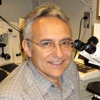 Victor Corces合作论文数Department of Human Genetics, School of Medicine, Emory University;Winship Cancer Institute, Emory University21Akshay Krishna合作论文数19Siddhartha Kumar合作论文数17
 Audrey Smith合作论文数17R. G. Bociek合作论文数17
 Alexandria Eiken合作论文数17Christopher R D'Angelo合作论文数17Sydney A Skupa合作论文数17El-Gamal Dalia合作论文数Institute of Experimental and Clinical Pharmacology, Medical University of Graz17
 Matthew Lunning合作论文数College of Medicine, University of Nebraska Medical Center17
 Amarnath Natarajan合作论文数Department of Chemistry, University of Vermont17
 Julie M. Vose合作论文数Division of Oncology and Hematology, Department of Internal Medicine, College of Medicine, University of Nebraska Medical Center17Elizabeth Schmitz合作论文数16
 Rana Sandeep合作论文数16
 Jayapal Reddy Mallareddy合作论文数Institute of Biochemistry, Hungarian Academy of Sciences16
 Aguirre de Cubas合作论文数Department of Pharmacology and Immunology, College of Medicine, Medical University of South Carolina16
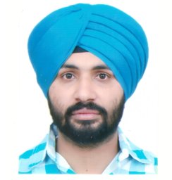 Sarbjit Singh合作论文数Department of Chemistry, Guru Nanak Dev University16
 Andrzej Wierzbicki合作论文数Department of Molecular, Cellular, and Developmental Biology, College of Literature, Science, and the Arts, University of Michigan-Ann Arbor11Hannah Harris合作论文数10
 Erez Lieberman Aiden合作论文数Baylor College of Medicine;Department of Biochemistry and Molecular Biology, The University of Texas Medical Branch;Department of Biosciences, Rice University8Vimla Band合作论文数8Timothy E. Reznicek合作论文数7
 Xiaowen Lyu (吕晓雯)合作论文数School of Medicine, Xiamen University7
 B.C. Mohapatra .合作论文数Central Institute of Freshwater Aquaculture (CIFA),, Indian Council of Agricultural Research7
 Mamta Shukla合作论文数Department of Genetics, Cell Biology and Anatomy, Nebraska Medical Center6Böhmdorfer Gudrun合作论文数6
 Hamid Band合作论文数Department of Pathology, Harvard Medical School6Christopher T. Cummings合作论文数6Michael H. Nichols合作论文数5Kyle J. Hewitt合作论文数Wisconsin Institutes for Medical Research, University of Wisconsin School of Medicine and Public Health;University of Wisconsin–Madison Blood Research Program;University of Wisconsin School of Medicine and Public Health, University of Wisconsin–Madison Blood Research Program5
 William Stafford Noble合作论文数Department of Genome Sciences, University of Washington;eScience Institute, University of Washington;Paul G. Allen School of Computer Science and Engineering, University of Washington5Omer Arina D合作论文数4Kuciński Jan合作论文数4Sameer Mirza合作论文数Department of Biochemistry, All India Institute of Medical Sciences4
 Olga Dudchenko合作论文数Center for Theoretical Biological Physics, Rice University;Mol. & Human Genetics/Aiden Lab, Baylor College of Medicine4Michael E. Talkowski合作论文数3
 Huiya Gu合作论文数3Melanie Pham合作论文数3Venkatasai Rahul Dogiparthi合作论文数3Devika Udupa合作论文数3Mozes Jacobs合作论文数3Gesine Cauer合作论文数3Masami Ando-Kuri合作论文数Dept Genet Mol, Univ Nacl Autonoma Mexico3Linda Chee合作论文数3
 Kiana Mohajeri合作论文数Harvard, Broad Institute of MIT3
 Devin Rocks合作论文数Fordham Univ, Bronx, NY 10458 USA3
 Yossi Eliaz合作论文数Dept Phys, Univ Houston3R. Eric Davis合作论文数3




