BACKGROUND:The Xoft electronic brachytherapy source is commonly used to treat superficial lesions and tumors located at shallow depths. However, uncertainties in material composition and geometry, mainly arising due to the manual assembly of the x-ray tube components, contribute to inter-source variability in the tube output spectrum. In addition, aging of the x-ray tube may lead to intra-source variability in the tube spectrum. PURPOSE:To investigate the inter- and intra-source variability of the spectrum for the Xoft S7500 model experimentally and through simulations, as well as to study the impact of this variability on dosimetry. METHODS:The Amptek X123 CdTe x-ray spectrometer was used to measure the spectrum of the Xoft S7500 source model. First, the spectrometer was calibrated using 137Cs, 152Eu, and 57Co. Second, the source output spectrum was measured on the side and at the tip of the source at a distance of 15.5 cm from the source tip. Throughout the measurement, the source was fixed to the optical table using a 3D-printed holder and was aligned using a laser beam. The intra-source variability was studied by investigating the spectrum emitted by one single source measured in five trials. Similarly, the inter-source variability was investigated by measuring five different sources of the S7500 model at the tip and the side. The symmetry of the output was investigated by comparing the side and tip spectra. The measurements were then compared with the simulated spectra using a previously developed E-Brachy software package, taking into account the range of variation in material composition reported by the manufacturer. Finally, to determine whether or not the uncertainty in the material composition and the output spectrum affect the dosimetry by a clinically significant amount, we compared the depth dose curves created by the upper and lower limits of the range of uncertainty in the material composition. RESULTS:The calibration line was obtained to correspond the detector channels to the energy in keV using linear regression. Escape peak and background corrections were performed on the spectrum, and the counts in the K-edges of cadmium (26.7 keV) and tellurium (31.8 keV) were readjusted so that the silver peaks were more clearly resolved. Intra-source variability demonstrated consistent peak resolution for tungsten, yttrium, and silver, with a coefficient of variation (CV) ranging from 0.5% to 9.8%. Inter-source variability highlighted significant differences between tip and side measurements, with up to 13.5% variation. Simulated spectra revealed the impact of material composition on the characteristic peak intensities, with the intensity of the peaks in the measured spectra lying between simulated spectra in the case of higher and lower percentage limits of yttrium and silver present in the material composition. Depth-dose simulations showed minimal differences (<1.2%) between material compositions. CONCLUSIONS:This study comprehensively compared the measured and simulated spectra of the Xoft S7500 source model. The differences in the depth-dose curves are within 2% as recommended by the AAPM TG-568 requirements, and the variations in the spectra for the source model S7500 lie within the recommended range.
Objective. Accurate determination of relative output factors (ROFs) is paramount in stereotactic radiosurgery dosimetry, particularly for small fields employed in Leksell Gamma Knife (LGK) systems. Existing guidelines for LGK film-based dosimetry lack specificity regarding the selection of a region of interest (ROI) for ROF measurements. Approach. This investigation assessed the dosimetric performance of Gafchromic TM EBT4 film, focusing on establishing a quantitative metric for the unambiguous selection of a small ROI, defined relative to the beam’s full width at half maximum (FWHM), for LGK ROF measurements. Film irradiations were performed on an LGK ICON unit using 16-, 8-, and 4 mm collimators. Calibration curves were generated by irradiating 15 films with 16 mm beams in 15 s increments, with films subsequently scanned in both portrait and landscape orientations at various resolutions. Dosimetric accuracy was assessed using the net optical density transfer function along with corresponding uncertainty propagation. The influence of detector size on ROF determination was calculated using ROIs with diameters systematically varied to include 1/10, 1/5, 1/4, 1/3, 2/5, 1/2, 2/3, and 3/4 of the beam’s FWHM. Main results. ROF measurements demonstrated high sensitivity to ROI size. A metric employing a ¼ FWHM ROI yielded ROFs values within 1% of the established Leksell GammaPlan (LGP) reference data, effectively minimizing film volume-averaging effects. Systematic dose errors, up to ±20% on average, were observed due to orientation mismatches between calibration and measurement scans. Furthermore, across all evaluated scan resolutions both FWHM and penumbra measurements consistently conformed to LGP specifications, specifically within ±0.5 mm and ±1 mm, respectively. Significance. The ¼ FWHM ROI provides a clinically practical method for LGK ROF determination, addressing the absence of a clear ROI definition in TG-178 report. This metric achieves a suitable balance for film-based measurements, ensuring a sufficient number of ROI pixels for statistical significance while minimizing volume-averaging effects.
This study analyzed the spectral response of EBT3, EBT4, and EBT-XD radiochromic films using absorption spectroscopy. The primary focus was on characterizing the evolution of spectral signatures across a range of absorbed doses, thereby elucidating the unique dose-dependent response profiles of each film type. Ten samples of each film type were subjected to open field irradiation within their designated dose ranges (1-20 Gy for EBT3 and EBT4, 1-50 Gy for EBT-XD). The corresponding absorption spectra were recorded and studied via decomposition and parameterization of dose-dependent spectral features. Lorentzian profiles were employed for spectral decomposition. Each film type displayed unique spectral signatures with distinct absorption peaks: nine for EBT3, eleven for EBT4, and twelve constituent profiles for EBT-XD. Notably, the EBT4 film demonstrated a slight difference in the blue part of the absorption spectrum and a change in the response, relative to its EBT3 predecessor. Orientation dependence of the film spectra was most pronounced for the EBT3 film type, followed by a declining trend across EBT4 and EBT-XD films. Absorption spectroscopy portrayed distinct spectral fingerprints of the studied film types, aiding the selection of the most suitable film for specific applications.
Reliable calibration is one of the major challenges in using radiochromic films (RCF) for radiation dosimetry. In this study the feasibility of using dose gradients produced by a physical wedge (PW) for RCF calibration was investigated. The aim was to establish an efficient and reproducible method for calibrating RCF using a PW. Film strips were used to capture the wedge dose profile for five different exposures and the acquired scans were processed to generate corresponding net optical density wedge profiles. The proposed method was compared to the benchmark calibration, following the guidelines for precise calibration using uniform dose fields. The results of the benchmark comparison presented in this paper showed that using a single film strip for measuring wedge dose profile is sufficient for estimating a reliable calibration curve within the recorded dose range. Furthermore, the PW calibration can be extrapolated or extended by using multiple gradients for the optimal coverage of the desired calibration dose range. The method outlined in this paper can be readily replicated using the equipment and expertise commonly found in a radiotherapy center. Once the dose profile and central axis attenuation coefficient of the PW are determined, they can serve as a reference for a variety of calibrations using different types and batches of film. This investigation demonstrated that the calibration curves obtained with the presented PW calibration method are within the bounds of the measurement uncertainty evaluated for the conventional uniform dose field calibration method.
This study aimed to establish an empirical relationship between the absorption spectrum of the EBT3 model GafChromic® film and absorbed radiation dose. Radiochromic films were irradiated with doses ranging from 1 Gy to 10 Gy using a Cobalt-60 Leksell Gamma Knife® Perfexion™ unit. Spectral analysis, conducted with the Agilent® Cary60 UV–Vis spectrometer 72 h post-irradiation, involved decomposing the net spectrum into nine Lorentzian components. Characteristic peaks remained stable within 5 nm, with the 583 nm and 635 nm absorption bands exhibiting a logistic response corresponding to the absorbed dose. Overall, the results present an empirical model showcasing dose-dependent spectrum evolution, emphasizing the potential of the ratio of two absorption band changes as a dose specifier. Furthermore, this spectroscopic approach provides key insights into radiation-induced optical changes in EBT3 films and facilitates the refinement of radiochromic film dosimetry.
BackgroundLarge reported variability in the material composition and geometrical components of the Xoft electronic high-dose-rate brachytherapy Causes inter-source discrepancy in the source output. This variability is due to the manual manufacturing and assembly of the sources.PurposeThis study aimed to develop a dosimetry software tool called E-Brachy to characterize the Xoft source and quantify the discrepancies in its photon spectrum and dosimetric properties.MethodsE-Brachy is based on the Geant4 Monte Carlo toolkit and consists of two parts. In part one, the geometry and material composition for the source received in the computer-aided design format from the vendor were converted to the geometry description markup language format using the GUIMesh Python tool and integrated into the E-Brachy software. There was a large variation in material composition and thickness for some of the tube components. The simulation started from electrons and resulted in x-ray generations in the anode region. Multithreading, a track length estimation, and the uniform bremsstrahlung splitting variance reduction techniques were used to decrease the simulation time and increase the x-ray production. The photon energy, position, and momentum were saved into a phase space file as the photon exited the source, but before interacting with the external environment. The obtained x-ray energy spectrum was compared with measurements from the National Institute of Standards and Technology (NIST). In part two, by sampling from the generated photons, the dose rates and dosimetric parameters according to the TG-43 protocol were calculated for model S7500 and compared to the ones previously calculated for model S700 source, which were deemed identical by the manufacturer.ResultsThe material composition that resulted in the most similar spectrum as the measured NIST spectrum with Pearson's correlation coefficient of 0.99 and a calculated Euclidean difference of 0.061 +/- 0.001$0.061\,\pm \,0.001$ keV was chosen for further dosimetric analysis of the model S7500 source. Characteristic peaks showed the presence of tungsten, yttrium, and silver in the source components. Differences in dose rates between the two source models surpassed 20% for polar angles theta >= 150 degrees$\theta \,\ge \,150<^>\circ$, reaching a peak at r=3$r\,=\,3$ cm and theta=175 degrees$\theta \,=\,175<^>\circ$. The differences in the radial dose function values were within 5%. The relative difference in percentage between the anisotropy function values of the two models was closer to 0 for smaller theta$\theta$ values, but at higher polar angles, they increased to 300%.ConclusionsA software package called E-Brachy was successfully developed for the characterization and dosimetry of Xoft electronic brachytherapy sources. E-Brachy can be combined with spectral measurements to investigate the inter- and intra-source variability. The software package was tested by comparing the simulated spectra from the S7500 Xoft source model with NIST measurements and its TG-43 parameters with the S700 model. The TG-43 parameters between the two sources significantly exceed the recommendations of TG-56.
BACKGROUND:In radiotherapy, it is essential to deliver prescribed doses to tumors while minimizing damage to surrounding healthy tissue. Accurate measurements of absorbed dose are required for this purpose. Gafchromic® external beam therapy (EBT) radiochromic films have been widely used in radiotherapy. While the dosimetric characteristics of the EBT3 model film have been extensively studied for photon and charged particle beams (protons, electrons, and carbon ions), little research has been done on α $\alpha$ -particle dosimetry. α $\alpha$ -emitting radionuclides have gained popularity in cancer treatment due to their high linear energy transfer, short range in tissue, and ability to spare surrounding organs at risk, thereby delivering a more localized dose distribution to the tumor. Therefore, a dose-calibration film protocol for α $\alpha$ -particles is required. PURPOSE:This study aimed to develop a dose-calibration protocol for the α $\alpha$ -particle emitting radionuclide 241Am, using Monte Carlo (MC) simulations and measurements with unlaminated EBT3 films. METHODS:In this study, a MC-based user code was developed using the Geant4 simulation toolkit to model and simulate an 241Am source and an unlaminated EBT3 film. Two simulations were performed: one with voxelized geometries of the EBT3 active volume composition and the other using water. The dose rate was calculated within a region of interest in the voxelized geometries. Unlaminated EBT3 film pieces were irradiated with the 241Am source at various exposure times inside a black box. Film irradiations were compared to a 6-MV photon beam from a Varian TrueBeam machine. The simulated dose rate was used to convert the exposure times into absorbed doses to water, describing a radiochromic-film-based reference dosimetry protocol for α $\alpha$ -particles. The irradiated films were scanned and through an in-house Python script, the normalized pixel values from the green-color channel of scanned film images were analyzed. RESULTS:The 241Am energy spectra obtained from the simulations were in good agreement with IAEA and NIST databases, having differences < $<$ 0.516% for the emitted γ $\gamma$ -rays and produced characteristic x-rays and < $<$ 0.006% for the α $\alpha$ -particles. Due to the short range of α $\alpha$ -particles, there was no energy deposition in the voxels outside the active 241Am source region projected onto the film surface. Thus, the total dose rate within the voxels covering the source was 0.847 ± $\pm$ 0.003 Gy/min within the sensitive layer of the film (LiPCDA) and 0.847 ± $\pm$ 0.004 Gy/min in water, indicating that the active volume can be considered water equivalent for the 241Am beam quality. A novel approach was employed in α $\alpha$ -film dosimetry using an exponential fit for the green channel, which showed promising results by reducing the uncertainty in dose estimation within 5%. Although the statistical analysis did not reveal significant differences between the 6-MV photon beam and the α $\alpha$ calibration curves, the dose-response curves exhibited the expected behavior. CONCLUSIONS:The developed MC user code simulated the experimental setup for α $\alpha$ -dosimetry using radiochromic film with acceptable uncertainty. Unlaminated EBT3 film is suitable for the dosimetry of α $\alpha$ -radiation at low doses and can be used in conjunction with other unlaminated GafChromic® films for quality assurance and research purposes.
This investigation aimed to optimize gradient positioning for radiochromic film calibration to facilitate a uniform distribution of calibration points. The study investigated the influence of various parameters on gradient dose profiles generated by a physical wedge, assessing their impact on the field's dose dynamic range, a scalar quantity representing the span of absorbed doses. Numerical parameterization of the physical wedge profile was used to visualize and quantify the impact of field size, depth, and energy on the dynamic range of dose gradients. This concept enabled the optimization of the gradient positioning and estimation of the necessary number of exposures for the desired calibration dose range. An optimization algorithm based on histogram bin height minimization was developed and presented. The maximum dynamic range was achieved with a 20 x$\times$ 20 cm2$\textrm {cm}<^>{2}$ field size at 5 cm depth. Optimization of wedge gradient positioning yielded the most uniform dose distribution with 7 exposures for the [1,10] Gy range and 8 exposures for the [1,20] Gy range. Film calibration using gradients centered at 1.6, 3, 3.5, and 7 Gy central axis (CAX), obtained through optimized gradient positioning, was showcased. The presented work demonstrates the potential for an improved film calibration process, with efficient material utilization and enhanced dosimetric accuracy for clinical applications. While the method was described for the use of a physical wedge, the methodology can be easily extended to the use of a more convenient dynamic wedge.
The purpose of this study was to investigate the dosimetric characteristics of the GAFCHROMIC® EBT3 film responding to alpha particle irradiation. Unlaminated GAFCHROMIC® EBT3 film pieces, were irradiated with a 30.055 kBq 241 Am alpha source, at eight different dose levels, between 0 and 509 Gy. The irradiations were performed inside an enclosed box. Epson Expression 10000 XL scanner in transmission mode was used to digitize irradiated films 24 hours post-irradiations as 16-bit RGB images in tagged image file format (TIFF). Optical density (OD) values were obtained by following the OD theorem. Raw and normalized pixel values (PV norm ) from the red, green, and blue colour channels were sampled from a 3 × 3 mm 2 region of interest. Calibration curves were created for both data sets (OD and PV norm ) and were fit accordingly. Monte Carlo simulations with the Geant4 toolkit were performed to establish a dose rate at the point within the sensitive layer of the film. An alpha dosimetry protocol for EBT3 films was obtained from the Monte Carlo calculated dose rate and dose calibration curves for alpha radiation were created. It is necessary to extend this study for different film types and compare to photon dosimetry calibration curves.
High-dose-rate endorectal brachytherapy (HDREBT) is an image guided brachytherapy treatment for patients with rectal cancer. It is based on tumor imaging with magnetic resonance in particular, which is used to choose eligible patients and improve tumor visualization. Treatment planning is performed using 3D CT simulation and treatment planning. The treatment is given on an outpatient basis and requires minimal local anesthesia. The validation of the technique was carried out through a preoperative study and is now explored as part of a radical treatment for early rectal cancer or as a boost modality. We describe technical aspects of the HDREBT and we discuss the ongoing institutional review board approved studies exploring the clinical applications of this treatment modality for patients with rectal cancer: 1) as a neoadjuvant treatment for patients with operable rectal tumor; 2) as a option to improve local control in patients with newly diagnosed rectal cancer but with previous pelvic radiation.
A novel low toxic normoxic polymer gel dosimeter containing acrylic acid monomer is prepared and characterized for dosimetry in radiotherapy. Gel samples were fabricated under normal atmospheric conditions. The gels were irradiated to different doses using a medical linear accelerator at 6 MV X-ray energy to investigate the dose response and other characteristics. The dose response was evaluated in terms of relaxation rate using NMR relaxometry, which was carried out using a 0.5 T NMR technique. The sample temperature during the NMR relaxometry was fixed at 20.0 ± 0.1 °C using a thermostatic circulating water bath connected to the NMR relaxometry. A significant improvement in the performance of the dosimeter was noticed for acrylic acid gel with the addition of MgCl2 salt. The sensitivity of gel with 0.6 M MgCl2 was about 4.6 times of the same gel without MgCl2. The total uncertainty of the R2 measurements was about 5%. Different important parameters such as dose rate, beam quality, NMR scanning temperature, and stability of the dose response were investigated in this study.
Abstract Background Rectal cancer is curable by standard surgery with Total Mesorectal Excision (TME). However, there are well known associated long-term bowel and sexual dysfunctions. Non-operative management (NOM) is an emerging treatment for patients with operable rectal cancer. There is evidence supporting dose response for tumor control in rectal adenocarcinoma. Aims In the era of modern technologies, Image-guided adaptive endorectal brachytherapy is a means to deliver local radiotherapy boost treatments. We explored its role in a randomized phase II/III trial (NCT03051464) for patients aiming to achieve cure without surgery. Total Mesorectal Excision (TME) free survival at 2 years was the primary endpoint. We now present the interim analysis upon accrual of the first 40 patients. Methods In randomized trial, patients with operable cT2-3ab N0 M0 rectal cancer received 45 Gy in 25 fractions of pelvic external beam radiotherapy (EBRT) with concurrent 5-FU/ Capecitabine. They were randomized to receive either an EBRT boost of 9 Gy in 5 fractions (Arm A), or three weekly adaptive brachytherapy boosts for a total of 30 Gy in 3 fractions (Arm B). Results Forty patients were included (20 per arm). The median age was 66 years; baseline characteristics were well balanced in terms of age, tumor location, T stage and tumor size (Table 1). The acute treatment related toxicities are similar as shown in table 2 but in arm B, there were two deaths: one patient died during his chemotherapy and external beam treatment from congestive heart failure and one patient from a heart attack after treatment prior to salvage TME surgery. The proportion of complete clinical response was 50% (n=10/20) in Arm A and 90% in Arm B (n=18/20). With a median follow-up of 2.2 years, local regrowth at 2 years occurred in 4/10 patients (40%) in Arm A and 4/18 patients (22%) in Arm B. TME-free survival rate at 2 years was 45.9% in Arm A and 85.1% in Arm B (p=0.0036) (Figure 1). Conclusions The interim analysis of this trial suggests that these two strategies of radiation dose escalation are feasible and lead to high chances of organ preservation in patients with operable rectal cancer. The Independent Monitoring Comittee (IDMC) approved the continuation of patient recruitment in the phase III study as planned. Funding Agencies Elekta
(1) Background: The introduction of total mesorectal excision (TME) for rectal cancer has led to improvement in local recurrence (LR) outcomes. Furthermore, the addition of preoperative external beam radiotherapy to TME reduces LR to less than 6%. As a trade-off to these gradual improvements in local therapies, the oncology community’s work is now focusing on mitigating treatment-related toxicities. In other words, if a small proportion of 4–6% of rectal cancer patients benefit from additional local therapy beyond TME, the burden of acute and long-term side effects must be considered with care. (2) Methods: With the introduction of better-quality imaging for tumor visualization and treatment planning, a new conformed radiation treatment was introduced with high-dose-rate endorectal brachytherapy. The treatment concept was tested in phase I and II studies: first in the pre-operative setting, and then as a boost after external beam radiation therapy, as a dose-escalation study, to achieve higher local tumor control. (3) Results: HDREBT is safe and effective in achieving a high tumor regression rate and was well tolerated in a phase II multicenter and two matched-pair studies. (4) Conclusions: HDREBT is a conformed radiation therapy that is safe and effective, and is presently explored in a phase III dose-escalation study in the NOM of patients with operable rectal cancer.
 Jan Seuntjens合作论文数Department of Medical Biophysics, Temerty Faculty of Medicine, University of Toronto;Princess Margaret Cancer Centre111
 Té Vuong合作论文数Gerald Bronfman Department of Oncology, Faculty of Medicine and Health Sciences, McGill University94
 Nada Tomic合作论文数Gerald Bronfman Department of Oncology, Faculty of Medicine and Health Sciences, McGill University66
 Francois Deblois合作论文数Jewish General Hospital, McGill University66
 Aldelaijan Saad合作论文数Department of Radiation Oncology, McGill University54
 Ervin Podgorsak合作论文数Department of Oncology Division of Radiation Oncology, McGill University Montreal36
 Natasa Tomic合作论文数33
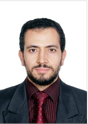 Belal Moftah合作论文数King Faisal Specialist Hospital & Research Center SA32
 Sergio Faria合作论文数Gerald Bronfman Department of Oncology, Faculty of Medicine and Health Sciences, McGill University30
 Papaconstadopoulos Pavlos合作论文数Montreal General Hospital, McGill University23
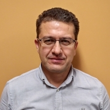 Hamed Bekerat合作论文数Gerald Bronfman Department of Oncology, Faculty of Medicine and Health Sciences, McGill University18Liheng Liang合作论文数17
 Ivan Buzurovic合作论文数Thomas Jefferson University17
 Tamim Niazi合作论文数15Michael D.C. Evans合作论文数13
 Aurelie Garant合作论文数13Lewis David F合作论文数13
 Frank Verhaegen合作论文数Dept Radiat Oncol MAASTRO, Maastricht Univ13Bekerat Hamed合作论文数12
 Ahmad Nobah合作论文数Biomedical Physics Department, King Faisal Specialist Hospital and Research Center11
 Shirin Abbasinejad Enger合作论文数Université Laval, Université Laval11
 Zoran V. Popović合作论文数Max-Planck-Institut für Festkörperforschung Heisenbergstrasse 1 D-7000 Stuttgart 80 Federal Republic of Germany10Boris Bahoric合作论文数10
 Thierry Muanza合作论文数Division of Experimental Medicine, Department of Medicine, Faculty of Medicine and Health Sciences, McGill University9
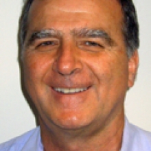 Robert Lisbona合作论文数Department of Diagnostic Radiology, Faculty of Medicine and Health Sciences, McGill University9L Souhami合作论文数8
 Patrick Charlebois合作论文数Division of General Surgery, Department of Surgery, Faculty of Medicine and Health Sciences, McGill University8
 Carolyn Freeman合作论文数Gerald Bronfman Department of Oncology, Faculty of Medicine and Health Sciences, McGill University8Han Dae Yup合作论文数8Webster Matthew J合作论文数8Soares Christopher G合作论文数8Carolyn R. Freeman合作论文数8Lorraine Portelance合作论文数McGill University Health Center8
 Luis Souhami合作论文数Gerald Bronfman Department of Oncology, Faculty of Medicine and Health Sciences, McGill University7
 Alexandre Revcolevschi合作论文数Universite Paris-Sud7Guy Dhalenne合作论文数7
 Shirley Lehnert合作论文数7
 Arman Sarfehnia合作论文数Department of Radiation Oncology, Temerty Faculty of Medicine, University of Toronto6
 Marylise Boutros合作论文数Division of General Surgery, Department of Surgery, Faculty of Medicine and Health Sciences, McGill University6Wamied Abdel-Rahman合作论文数6Daniel J. Scanderbeg合作论文数6
 Assen S. Kirov合作论文数6B. Stein合作论文数5F. Alzorkany合作论文数5Carole S. Richard合作论文数5M Shehadeh合作论文数5
 Carol-Ann Vasilevsky合作论文数5S. Liberman合作论文数5




