Objectives: Prevalence of work-related musculoskeletal disor-ders (WRMSDs) among general dental practitioners and ortho-dontists is approximated to range between 64% and 93%. Eti-ology of WRMSDs in the mentally and physically demanding occupation remains unclear, for which reason the aim of the study was to clarify the interplay of physical, psychological, and mental factors on WRMSDs. Method and materials: Of 94 orthodontists and 187 general dental practitioners (mean age = 35 years) questioned using an online survey, 84% re-ported persisting tension or pain in the back, neck, or shoul-ders. While 71% of females were employed (29% self-em-ployed), only 39% of male participants were employed. Cluster analysis was used to characterize dental practitioners accord-ing to their movement profile and the moderating effect of stress on certain WRMSDs. Results: Three movement profiles of general dental practitioners and orthodontists were signifi-cantly predictive of WRMSD. The minority could be character-ized as healthy (n = 45), whereas twice as many reported nearly twice as much pain (n = 90). Stress proved to be a strong, signif-icant moderator of WRMSDs in relation to sex, employment status, and body mass index. Conclusion: The prevalence of WRMSDs found was alarming. Given the feminization of den-tistry, and that being female, stressed, and an employee (rather than self-employed) is a significant predictor of WRMSDs, this represents a danger to the German health system
BACKGROUND:With the constantly increasing demand for metal-free solutions in dental therapy, numerous ceramic restorations have found their way into everyday clinical practice, but long-term clinical data are limited. OBJECTIVE:The aim of this prospective clinical study was to evaluate three- and four-unit fixed partial dentures in the posterior region made of zirconium dioxide frameworks veneered with feldspathic porcelain after 10 years in clinical use. METHODS:Based on the two studies published in 2009 and 2012, in which the all-ceramic FDPs were evaluated after 3 and 5 years of function, a clinical evaluation of a total of 17 restorations after 10 years with regard to their condition and long-term stability was carried out in the course of this study. The restorations were fabricated using feldspathic ceramic-veneered, yttria-stabilised, tetragonal zirconium dioxide as the framework material. The data collection was based on modified CDA criteria and included, for example, the shape, shade, surface condition and the success and survival rates of the restorations. RESULTS:The all-ceramic prostheses embodied excellent biocompatibility and colour reproduction. As a result of an increased incidence of chipping fractures, the success rate was 60%. The survival rate, however, was 88.2%, as 2 of the 17 restorations were lost. CONCLUSION:All-ceramic concepts for FDP constructions in the posterior region achieved satisfactory results in terms of durability after 10 years. The main problem was chipping, as has been generally recognised. Nevertheless, the materials were characterised by excellent aesthetics and biocompatibility, which ultimately makes them a good alternative to conventional restorative options. TRIAL REGISTRATION:This study is registered in DRKS-German Clinical Trials Register with the register number DRKS00021743.
Purpose: To date, the qualitative and quantitative recording of biomechanical processes in dental implants represents one of the greatest challenges in modern dentistry. Modern, dynamic, 3D optical measurement techniques allow highly constant and highly accurate measurement of biomechanical processes and can be superior to conventional methods. This work serves to establish a new measurement method. Materials and Methods: A comparative analysis was undertaken for two different measurement systems, two conventional strain gauges versus the 3D optical two-camera measurement system ARAMIS (GOM GmbH, Braunschweig, Germany), as they detected surface changes on an artificial bone block under masticatory force application. Two implants (Straumann Standard Implants Regular Neck, Straumann GmbH, Freiburg, Germany) were placed in the bone block, and three different three-unit bridges were fabricated. Increasing masticatory forces, from 0 to 200 N, were applied to the bone block via each of these bridges and the inserted implants. Fifteen repetitions of the test were performed using a universal testing machine. The computer unit of the ARAMIS system was used to simultaneously integrate the surface changes recorded by the strain gauges and the ARAMIS system. The areas on the bone block examined by the dynamic 3D optical measurement method corresponded exactly to the locations and extent of the strain gauges. A statistical comparative analysis was carried out separately for the strain gauges and the corresponding optical measuring surface at the defined force magnitudes. The equivalence test and the intraclass correlation served as statistical means. Results: In the case of the intraclass correlation, a clear concordance of both measurement methods could be shown for all examined cases. For the equivalence test, no significance could be shown in individual cases. Conclusion: The accuracy of the modern, dynamic, 3D optical measurement method is comparable to that of conventional strain gauges. On this basis, versatile new research approaches in the field of biomechanics of dental implants can be pursued by establishing this method.
Background Digital three-dimensional planning of dental implant treatment has proven its benefits to the implantation procedure. Hereby, 3D datasets of CBCT scans are able to be superimposed to optically obtained data of the oral situation in order to reproduce and consider the soft tissue situation during the planning process. The quality of this procedure depends on the used scan datasets and their immanent failure. Aim/Hypothesis This study aimed to compare the accuracy of the superimposition of CBCT images with digital intraoral scanning datasets of a polymethacrylate model on the one hand and with lab-side scan datasets of the same model on the other hand. Subsequently, a recommendation for the workflow should be given. Material and Methods A highly precise polymethacrylate model of an upper jaw with full dentition was scanned with a CBCT unit (3D eXam, Kavo dental GmbH, Germany) for the digital superimposition with digital intraoral scanning data and lab-side scanning data. In this context, the same model was scanned with the TRIOS digital intraoral scanner (3Shape A S, Denmark) first and with the D640 model scanner (lab-side scanning unit, 3Shape A S) second. Files obtained with TRIOS were assigned to group 1 and files obtained with D640 model scanner to group 2. Each scan of both groups was superimposed to the CBCT dataset throughout two different ways of local fitting (local-best-fit) after conversion into STL-files. In a first step, every scan was superimposed to the CBCT image by using the virtual structures of the teeth. Afterwards, the hard palate was used to define the relation. 3D deviations of every image pair were calculated for every locus over the teeth using ATOS professional software (GOM GmbH, Germany). Results This study clearly showed that the scanning procedure which is used to reproduce the oral hard and soft tissue situation leads to different deviations in the superimposition with a CBCT dataset. Using a digital intraoral scanning unit, mean deviation was limited to 0.075 mm. The lab-side scanning revealed a mean deviation of 0.129 mm. By comparing the two ways of local-best-fit strategies, the relation to the teeth showed a mean deviation of 0.039 mm and the relation to the hard palate revealed a mean deviation of 0.165 mm. The combination of these two examined parameters lead to the lowest mean deviation for the TRIOS scan datasets superimposed to the CBCT scan by using the local-best-fit over the teeth with 0.022 mm and the highest for the lab-side scan superimposed in relation to the hard palate with 0.203 mm. All differences between groups and local-best-fit relations and combinations were statistically significant (statistical evaluation performed with SPSS Statistics 23). Conclusions and Clinical Implications The present data implicate a huge impact of the scanning method of the oral hard and soft tissue on the accuracy of digital superimposition to corresponding CBCT images. As immanent failure has to be reduced in clinical processes such as digital implant planning, the superimposition of a CBCT dataset with digital intraoral scanning datasets can be recommended regarding this workflow by using a local-best-fit relation to the virtual structure of the teeth.
We investigated the oral stereognostic ability (OSA) of dentate individuals, conventional complete denture patients, and maxillary implant-supported denture patients with bar attachments. Stereognosis tests were carried out, and the relationship between OSA and patient satisfaction was assessed with a satisfaction survey: the Turkish version of the oral health impact profile-14 (OHIP-TR-14). To compare differences in oral sensory function among individuals with natural dentition, complete denture wearers, and maxillary implant-supported denture wearers, tactile awareness (thickness perception threshold) and pressure awareness (threshold of lateral loading) were assessed. According to the results, the oral perception level of natural dentate patients was higher than that of the complete denture group and maxillary implant-supported complete denture group at the end of the study. There was no statistically significant difference in OHIP-TR-14 scores between the maxillary implant-supported complete denture group and the complete denture group (P < .05). The control group perceived the applied lateral and vertical forces statistically earlier than the other groups (P < .001). There was no statistically significant difference between the complete denture and maxillary implant-supported denture groups in terms of the lateral pressure threshold or thickness tactile threshold (P > .05). At the end of the study it was concluded that there was no correlation between oral perception levels and satisfaction in patients with complete dentures and patients with maxillary implant-supported complete dentures. The control group, compared with the complete denture and maxillary implant-supported complete denture groups, perceived the lateral and vertical forces statistically earlier than the other groups.
AIMS:Complete maxillary edentulism and prosthetic rehabilitation with removable full dentures are known to affect speech intelligibility. The aim of this study was to prospectively investigate the long-term effect of time on speech intelligibility in patients being rehabilitated with newly fabricated full maxillary dentures. MATERIALS AND METHODS:Speech was recorded in a group of 14 patients (male = 9, female = 5; mean age ± standard deviation [SD] = 66.14 ± 7.03 years) five times within a mean period of 4 years (mean ± SD: 47.50 ± 18.16 months; minimum/maximum: 24/68 months) and in a control group of 40 persons with healthy dentition (male = 30, female = 10; mean age ± SD = 59 ± 12 years). All 14 participants had their inadequate removable full maxillary dentures replaced with newly fabricated dentures. Speech intelligibility was measured by means of a polyphone-based speech recognition system that automatically computed the percentage of accurately spoken words (word accuracy [WA]) at five different points in time: 1 week prior to prosthetic maxillary rehabilitation (both with and without inadequate dentures in situ) and at 1 week, 6 months, and a mean of 48 months after the insertion of newly fabricated full maxillary dentures. RESULTS:Speech intelligibility of the patients significantly improved after 6 months of adaptation to the new removable full maxillary dentures (WA = 66.93% ± 9.21%) compared to inadequate dentures in situ (WA = 60.12% ± 10.48%). After this period, no further significant change in speech intelligibility was observed. After 1 week of adaptation, speech intelligibility of the rehabilitated patients aligned with that of the control group (WA = 69.79% ± 10.60%) and remained at this level during the examination period of 48 months. CONCLUSION:The provision of new removable full maxillary dentures can improve speech intelligibility to the level of a healthy control group on a long-term basis.
Acceptance of new technology is influenced by a number of situational and social factors. So far, only limited data are available on the influence of the teaching staff's gender on the acceptance of virtual dental implant planning by students. This study aimed at assessing the influence of the teaching staff's gender on the acceptance of a virtual implant planning course by male and female undergraduate dental students and their general attitude toward implantology.Two groups of third-year dental students (group 1, 9 males, 22 females; group 2, 12 males, 20 females) attended a virtual dental implant planning course. For the first group the teaching staff was all-male, while the teaching staff was all-female for the second group. After completion of the course the students filled in a technology acceptance questionnaire.An all-female teaching staff led to a degree of technology acceptance that did not differ significantly for male and female students. When the teaching staff was all-male, significant differences for technology acceptance occurred between male and female students.However, male as well as female students attributed the practice of implantology to both genders of dentists, equally, without statistically significant difference independent of the gender of the teaching staff.The more evenly distributed degree of technology acceptance of students of both genders being taught by a female staff is a favorable effect which may be explained by the more egalitarian style of women. Therefore, while feminization in dentistry proceeds, adequate measures should be taken to increase the number of female teachers.
The present clinical case history report describes an interdisciplinary treatment protocol that combines maxillary tumor resection with immediate reconstruction to achieve functional rehabilitation. A fibula flap that received four dental implants and a split-thickness graft epithelial layer was prefabricated for a 31-year-old man. The flap was designed so that it could be adapted to fit in different extents of tumor resection. Resection and immediate reconstruction were successfully performed 6 weeks after flap prefabrication, with the final bar-retained dental prosthesis delivered 4 weeks later.
The objectives were to assess the sinus anatomy and alveolar ridge dimensions and to identify clinical factors and morphological relationships that may predict the characteristics of the sinus anatomy. A total 336 edentulous molar and 168 edentulous premolar regions were analyzed. The mediolateral maxillary sinus (5 mm and 10 mm above alveolar bone), lateral maxillary sinus wall, ostium height, and alveolar ridges were measured. Measurement regions were defined by radiopaque simulation of the restoration goal. Mean molar mediolateral dimensions were >10 mm: first molar, 11.8 mm (range 3.8-21.1 mm); second molar, 12.2 mm (range 7.9-20.1 mm). The extent of the sinus differed significantly between premolars and molars (P < 0.001). The first molar region had the highest mean value for the lateral sinus wall (2.4 mm, range 0.2-7.9 mm) and the greatest distance between the alveolar crest and simulated restorative goal (8.1 mm, range 1.1-16.3 mm). Septum prevalence was 46% in the molar region and 27% in the premolar region. A lower alveolar ridge height was associated with a wider transverse extent of maxillary sinus and consequently longer distances between the crest and restorative goal. Systematic three-dimensional analysis of the maxillary sinus yields precise preoperative information about sinus configurations. There were significant variations and relationships among characteristics of the maxillary sinus, ridge, and the restorative goal.
For full-arch reconstruction of an atrophied cleft maxilla with missing premaxilla, a prefabricated microvascular free bony flap is a relevant option. A fibula flap was prefabricated in a cleft patient who received six dental implants and an epithelial layer. Six weeks later, maxillary reconstruction was performed. The inpatient period could be confined to 2 weeks. A fixed provisional prosthesis was delivered after an additional 2 weeks. A prefabricated flap allows for the reduction of the interval without a dental prosthesis to only a few weeks, even when a complex full-arch reconstruction of the maxilla is required.
OBJECTIVES:Three-dimensional radiological imaging data play an increasingly role in planning, simulation, and navigation in oral and maxillofacial surgery. The aim of this study was to establish a new, highly precise, in vitro measurement technology for the evaluation of the geometric accuracy down to the micrometric range of digital imaging data.MATERIAL AND METHODS:A macerated human mandible was scanned optically with an industrial, non-contact, white light scanner, and a three-dimensional (3D) model was obtained, which served as a master model. The mandible was then scanned 10 times by cone beam computed tomography (CBCT), and the generated 3D surface bone model was virtually compared with the master model. To evaluate the accuracy of the CBCT scans, the standard deviation and the intraclass coefficient were determined.RESULTS:A total of 19 measurement points in 10 CBCT scans were investigated, and showed an average value of 0.2676 mm with a standard deviation of 0.0593 mm. The standard error of the mean was 0.0043 mm. The intraclass correlation coefficient (ICC) within the 10 CBCT scans was 0.9416.CONCLUSIONS:This highly precise measuring technology was demonstrated to be appropriate for the evaluation of the accuracy of digital imaging data, down to the micrometric scale. This method is able to exclude human measurement errors, as the software calculates the superimposition and deviation. Thus inaccuracies caused by measurement errors can be avoided. This method provides a highly precise determination of deviations of different CBCT parameters and 3D models for surgical, navigational, and diagnostic purposes. Thus, surgical procedures and the post-operative outcomes can be precisely simulated to benefit the patient.
In the case of horizontal bone defects of the jaw, bone splitting is considered one of the safest and most efficient augmentation techniques. Major advantages are the one-stage approach and the absence of donor site morbidity. The indication for bone splitting is not limited to horizontal bone defects. In daily practice, it is extended to a majority of combined defects with small and medium vertical bone defects, for which a compensation using implant prosthetics is intended. Because clinical examination and conventional radiographic diagnostics provide little information on the transverse dimensions of the jaw, it is difficult to select the right patients based on this information. A minimally invasive approach is also not feasible because the alveolar ridge must be exposed during surgery. Three-dimensional bone splitting allows a reliable setting of the indication for bone splitting as well as a minimally invasive approach based on the transfer of the virtual planning results using a guided template and a splitting vector.
OBJECTIVE:The purpose of this prospective and randomized clinical study was to assess differences in patient morbidity between minimally invasive lateral sinus elevation (study group, n = 14) and conventional one-stage lateral sinus elevation (control group, n = 12). It was hypothesized that trauma to soft tissue was reduced in the study group. MATERIALS AND METHODS:Optical 3-D imaging was assessed blinded on days 1 and 7 after surgery to determine the visible soft tissue swelling of the upper lip and cheeks. Postoperative pain and discomfort were evaluated by a visual analogue scale (VAS; scale 0-10). RESULTS:Immediately and on day 1 after surgery, the study group patients rated pain and discomfort as 2.4 [SD 1.7] and 3.1 [SD 2.1], respectively, on the VAS; while, the controls rated 4 [SD 1.6] and 5.6 [SD 1.7], respectively. The mean facial soft tissue volume change, assessed on day 1 after surgery, was 5.0 cm(3) [range: 0.2-9.2] for the study group and 15.5 cm(3) (9.3-21.55) for the controls (p = 0.00). CONCLUSIONS:Despite the small number of patients in this prospective study, 3-D based minimally invasive one-stage lateral sinus elevation resulted in favourable patient morbidity with less postoperative visible facial soft tissue volume changes.
BackgroundLingual undercuts are common in the edentulous mandible and pose the risk of perforating the lingual cortical bone during insertion of dental implants, which may lead to hemorrhage or infections of the parapharyngeal space. The aim of our study was to determine the occurrence and extent of lingual undercuts in the molar and premolar/canine region of the mandible.MethodsWe analyzed 716 cross-sections of the edentulous molar region and 215 cross-sections of the edentulous first premolar/canine region. Mandibular morphology was classified into a U-configuration (undercut), P-configuration (parallel), and C-configuration (convex), depending on the shape of the alveolar ridge. Depth of the lingual concavity, concavity angle, and further parameters were measured to describe the mandibular morphology.ResultsLingual undercuts had a prevalence of 68% in the molar region. The prevalence was significantly higher in the second molar region (90%) than in the first molar region (56%). A deep position of the inferior alveolar nerve close to the basal cortical bone was significantly associated with the presence of lingual undercuts.ConclusionLingual undercuts are a frequent in the edentulous mandible. Cross-sectional analysis of three-dimensional radiographs provides the opportunity to determine a lingual undercut and to prevent complications of perforating the lingual cortical bone.
Für den langfristigen Erfolg von Implantatversorgungen spielt die Passgenauigkeit der Suprakonstruktion eine wesentliche Rolle. Die Weiterentwicklung der CAD / CAM-Technologie ermöglicht die Fertigung von präzise sitzenden Stegen. Dabei kommt es besonders auf das möglichst exakte Übertragen der Implantatposition in ein virtuelles Modell an. Neben der Abformmethode liefert auch die Gipsmodellherstellung eine mögliche Fehlerquelle. Deshalb wurde in dieser Studie die konventionelle Polyetherabformung mit dem direkten Scannen des Abdruckmaterials verglichen.
This report describes a complex, surgical, and prosthodontic hemimaxillary rehabilitation with a prefabricated vascularized free fibula graft. The treatment was based on 3-dimensional skull and fibula models but avoided software-based virtual planning of the reconstructive procedure or the implant positions. Cast-based surgical guides were used for implant placement during prefabrication of the fibula graft and for the positioning of the prefabricated graft in the maxilla during the reconstructive procedure. This procedure allowed the definitive prosthesis to be attached to the implants of the reconstructed maxilla during the inpatient period.
PURPOSE Tooth loss and its prosthetic rehabilitation significantly affect speech intelligibility. However, little is known about the influence of speech deficiencies on oral health-related quality of life (OHRQoL). The aim of this study was to investigate whether speech intelligibility enhancement through prosthetic rehabilitation significantly influences OHRQoL in patients wearing complete maxillary dentures. Speech intelligibility by means of an automatic speech recognition system (ASR) was prospectively evaluated and compared with subjectively assessed Oral Health Impact Profile (OHIP) scores. MATERIALS AND METHODS Speech was recorded in 28 edentulous patients 1 week prior to the fabrication of new complete maxillary dentures and 6 months thereafter. Speech intelligibility was computed based on the word accuracy (WA) by means of an ASR and compared with a matched control group. One week before and 6 months after rehabilitation, patients assessed themselves for OHRQoL. RESULTS Speech intelligibility improved significantly after 6 months. Subjects reported a significantly higher OHRQoL after maxillary rehabilitation with complete dentures. No significant correlation was found between the OHIP sum score or its subscales to the WA. CONCLUSION Speech intelligibility enhancement achieved through the fabrication of new complete maxillary dentures might not be in the forefront of the patients' perception of their quality of life. For the improvement of OHRQoL in patients wearing complete maxillary dentures, food intake and mastication as well as freedom from pain play a more prominent role.
OBJECTIVE:In 2008, a consensus meeting of the Association for Dental Education in Europe stated that dental implant treatment represents a popular treatment alternative and, therefore, it should be an integral part of dental student pre-graduate education. In 2009, the Friedrich-Alexander University, Erlangen-Nuremberg implemented a voluntary, structured, education program for pre-graduate dental students, called 'i.lect'. The present study evaluated the effectiveness of the i.lect program.METHODS:Concurrent to the dental curriculum, the i.lect program provides 200 hours in 3 years of theoretical and practical education on all aspects of modern dental implantology. We recruited dental students in the i.lect program and additionally students from 15 other universities that voluntarily participated in a single, 3-day implantology camp (R = 58). To demonstrate their knowledge on dental implant issues, both student groups completed a written examination that covered 'Basic information and materials of implantology', 'Implant planning' and 'Soft tissue management'.RESULTS:The students in the i.lect program achieved higher scores than students from the implantology camp on questions concerning 'basic implantology' and 'implant planning'.CONCLUSION:The results indicated that the structured i.lect undergraduate curriculum enhanced individual knowledge in the specific field of implantology. This program could have pilot character for use in other universities.
The study assessed the use of a new xenograft material. The aim of the present in vivo experiment was to obtain information on the clinical results and the rate of bone formation using two new experimental bone substitutes compared with an established bovine bone substitute and the autogenous graft, which is considered the gold standard for grafting. In the study miniature pigs were used as an animal model as they represent the highest similarities to human bone regarding anatomy, morphology and remodelling and as a species are considered to be a close representation of human bone considering the process of bone formation. In a surgical procedure the second premolars of 20 animals were extracted in all 4 quadrants and defects created which were filled with the different materials. After 30 days the first group was sacrificed and the second group after 60 days. To evaluate the rate of new bone formation sections were ground down for toluidine blue staining. Furthermore, the technique of fluorochrome sequential labelling was used to obtain information about the dynamics of new bone formation and remodelling processes in the loaded area at different times. A greater part of the defects loaded with the experimental blocks could not be integrated in the statistical analysis because the animals either lost the grafts a few weeks after transplantation or the healing was unsuccesssful. Histological examination revealed about 50% lower evidence of bone induction of the experimental blocks compared to the bovine reference material. Fluorescence microscopy did not offer any significant differences between the bone biopsies. The results of this study demonstrate that the use of the experimental blocks was less successful considering healing and amount of new bone formation compared to the established bovine bone substitutes grafts. Nevertheless, the study showed that the combination of fluorescence polarization and histological analysis is an efficient method to obtain information about bone remodelling and fibrous reactions on top of the basal bone by bone grafting experiments.
 Manfred Wichmann合作论文数47
 Stefan Holst合作论文数24
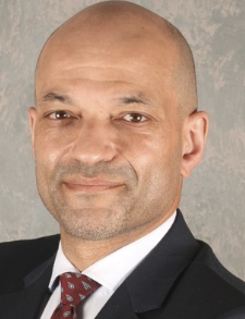 Emeka Nkenke合作论文数Department of Oral and Maxillofacial Surgery, Medical University of Vienna17
 Hans-Joachim Nickenig合作论文数16
 Karl Andreas Schlegel合作论文数15Andreas Schlegel合作论文数10M Wichmann合作论文数8
 Markus B. Blatz合作论文数Department of Preventive and Restorative Sciences, School of Dental Medicine, University of Pennsylvania8
 Florian Stelzle合作论文数7Jörg Hamel合作论文数6Bauer Julia合作论文数6Vairaktaris Elefterios合作论文数6
 Ragai Matta合作论文数6Christian Knipfer合作论文数5Sven Reich合作论文数5Zöller Joachim E合作论文数4Schmitt Johannes合作论文数4Sokol Biljana合作论文数4Anne Bauersachs合作论文数3Michael Bergler合作论文数3
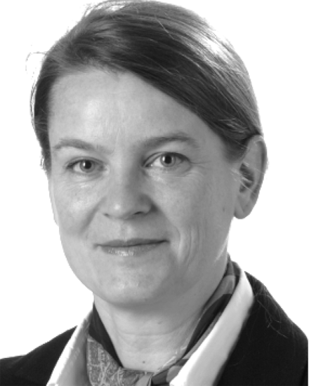 Maria Schuster合作论文数3
 Elmar Nöth合作论文数Department of Computer Science, Friedrich-Alexander-Universität;Pattern Recognition Lab, Friedrich-Alexander-Universität3
 Werner Adler合作论文数Department of Medical Informatics, Biometry and Epidemiology, Friedrich-Alexander University Erlangen-Nürnberg3
 Friedrich Wilhelm Neukam合作论文数Department of Oral and Cranio-Maxillofacial Surgery, Dental School, Erlangen-Nuremberg University;Department of Maxillofacial Surgery, Friedrich-Alexander-University of Erlangen-Nürnberg3Matthias Karl合作论文数3Hubertus Spiekermann合作论文数3Matthias Kreppel合作论文数Department for Oral and Craniomaxillofacial Plastic Surgery, University of Cologne, Cologne, Germany.3
 Tobias Bocklet合作论文数SRI International3Constantin Motel合作论文数2Hendrik Doering合作论文数2N Beck合作论文数2Michael M Lell合作论文数2Heckmann Josef G合作论文数2
 Cornelius Von Wilmowsky合作论文数2
 Christian Martin Schmitt合作论文数2
 Max Riemann合作论文数Implaneo Dental Clinic, Munich2Nickenig H-J合作论文数2Frieder Graef合作论文数2
 Stefan Schultze-Mosgau合作论文数2
 Rainer Lutz合作论文数Fachbereich Psychologie, Phillips-Universität Marburg2C Knipfer合作论文数1Hans-Günter Schaller合作论文数1
 Ravza Eraslan合作论文数1Winfried Neuhuber合作论文数1Simon Paul Stelzer合作论文数1
 Lara Berger合作论文数1Tilo Schlittenbauer合作论文数1
 Manuel Olmos合作论文数1




