Background and aims: Molecular heterogeneity in hepatocellular carcinoma (HCC) is ill-defined since trunk drivers (early events; common to all cells), branch drivers (later events; present in a subset of cells) and passenger mutations (not relevant), have not been thoroughly described. Most FDA/EMA approved molecular drugs target trunk drivers. We explored heterogeneity by analyzing trunk vs branch mutations in different HCC regions within single and multinodular tumours. Methods: Intra-tumoral heterogeneity was assessed in 21 patients with single HCCs (size > 4cm; 2 regions/tumour: 42 samples) and inter-tumoral heterogeneity was studied in 17 patients with multinodular HCCs (2-3 nodules/patient; total: 39 samples). Gene expression profiling, SNP array and deep-sequencing (coverage ∼850x) assessing 6 oncodrivers (TERT promoter, TP53, CTNNB1, ARID1A, AXIN1-2 by TruSeqAmplicon, validated by sanger) were explored. Clonality differentiating metastatic (clonal) vs synchronic (non-clonal) tumours was defined by SNP profiles. Trunk mutations were defined as present in a) all regions of a given tumour, or b) in all nodules of metastatic-clonal tumours; all other were considered as branch. Results: Intra-tumoral heterogeneity assessed by sequencing identified at least 1 oncodriver in 19/21 patients with single tumours. Among those, trunk mutations accounted for 17/19 (90%), and branch for 2/19 cases. Overall 63 mutations were identified, 56 (90%) were identical in different tumoral regions (i.e. truncal; TERT promoter most prevalent). Inter-tumoral heterogeneity explored by SNP profiles defined metastases in 35% (6/17 multinodular cases) and synchronous tumors in 65% (11/17 cases). Genetic proximity confirmed clonality in all metastatic nodules. Regarding molecular subclasses, half of clonal tumours retained identical molecular fingerprint, but the other half switched to more aggressive subclass. All non-clonal tumours belonged to distinct molecular subclasses. Driver oncogenes were explored in 9 patients (5 metastasis and 4 synchronic). Metastatic tumours showed 13 mutations, among which 11 (85%) were truncal. Mutations in non-clonal synchronic tumours were distinct. Conclusions: Single large HCCs shared common trunk drivers at distinct regions (90%). Similarly, 40% of multinodular tumours were clonal (metastasis) and shared common trunk oncodrivers, while 60% were synchronic, with distinct genomic profile/oncodrivers. Further studies at single-cell sequencing level are recommended. Citation Format: Daniela Sia, Andrew Neelis Harrington, Sara Torrecilla, Zhongyang Zhang, Genis Camprecios, Agrin Moeini, Sara Toffanin, Maria Isabel Fiel, Ke Hao, Monica Higuera, Laia Cabellos, Helena Cornella, Milind Mahajan, Yujin Hoshida, Augusto Villanueva, Sander Florman, Myron Schwartz, Josep Maria Llovet. Molecular heterogeneity and trunk driver mutations in hepatocellular carcinoma. [abstract]. In: Proceedings of the 107th Annual Meeting of the American Association for Cancer Research; 2016 Apr 16-20; New Orleans, LA. Philadelphia (PA): AACR; Cancer Res 2016;76(14 Suppl):Abstract nr 2388.
Methods: CD133 + cells were isolated by magnetic bead sorting after Huh-7 cells were genetically labeled with green fluorescent protein (GFP) or red fluorescent protein (RFP).In this scheme, CD133 + cells were labeled with GFP and CD133 -cells were labeled with RFP.The same number of GFP CSCs and the RFP non-CSCs were mixed and injected subcutaneously or in the spleen of nude mice.Results: CSCs had higher proliferative potential compared to non-CSCs in vitro.CSCs performed a higher in vitro proliferative potential and lower mRNA expressions of mature hepatocyte markers, glutamine synthetase and cytochrome P450 3A4, than non-CSCs.When either CD133 + or CD133 -cells were subcutaneously injected into SCID mice, CD133 + cells formed tumors, whereas CD133 -cells induced either a very small number of tumors or none at all.GFP-CSCs were highly tumorigenic and metastatic as well as highly resistant to chemotherapy in vivo compared to RFP-non-CSCs.Conclusions: The identification of CD133 + cells could thus be a potentially powerful tool to investigate the tumorigenic process in the hepatoma system and to also develop effective therapies targeted against hepatocellular carcinoma.The ability to specifically distinguish CSCs in vivo in real time provides a visual target for prevention of metastasis and drug resistance.
Guichard C, Amaddeo G, Imbeaud S, et al. Integrated analysis of somatic mutations and focal copy-number changes identifies key genes and pathways in hepatocellular carcinoma. Nat Genet 2012;44:694–698. Hepatocellular carcinoma (HCC) is a major health problem worldwide. With an incidence of 700,000 new cases per year, it is the 6th most common neoplasm (CA Cancer J Clin 2011;61:69–90). Its mortality rates are high and it is now considered the 3rd most deadly tumor worldwide, trailing only lung and stomach cancers. Hepatocarcinogenesis is the foremost cause of death in cirrhotic patients (Lancet 2012;379:1245–1255), and its incidence is steeply growing. Several studies have depicted the molecular heterogeneity of HCC. A broad variety of pathways activated in HCC have been reported including the Wnt/β-catenin, Ras/MAPK, IGF, PI3K/Akt signaling among many others (Semin Liver Dis 2007;27:55–76). Depending on the specific alterations, distinct molecular subclasses of HCC have been defined (Hepatology 2004;40:667–676; Hepatology 2007;45:42–52; Cancer Res 2008;68:6779–6788). In addition, several structural alterations have been characterized so far. Mutations in TP53 (30%), CTNNB1 (25%), and AXIN1 (10%) and high-level amplifications in 11q13 (5%–7%) and 6p21 (5%) are the most relevant ones (Cancer Res 2008;68:6779–6788; Semin Liver Dis 2007;27:55–76). Interestingly, FGF19 has recently been pointed as an oncogene and potential target in patients with high-level amplification in 11q13 (Cancer Cell 2011;19:347–358). Unfortunately, in contrast with other cancers (eg, BCR-ABL fusion protein in chronic myeloid leukemia, Her2/nu amplification in breast cancer, B-RAF mutation in melanoma), there is not a clear oncogenic addiction loop characterizing this neoplasm. To provide a comprehensive landscape of molecular alterations in HCC, Guichard et al performed whole-exome sequencing of tumor and surrounding nontumoral liver in 24 patients with HCC. These results were validated with Sanger sequencing and integrated with copy number changes and clinical data in an independent cohort of 125 samples. They identified 994 functional somatic mutations affecting 906 genes and 135 homozygous deletions (HD) and predicted their functional consequences. This set of alterations consisted of 74% missense mutations, 12% nonsense modifications, and 14% short coding indels. As previously reported in HCC patients, copy-number alterations analyzed in the validation set showed chromosomal gains at 1q, 5, 6p, 7, 8q, 17q, and 20, and losses at 1p, 4q, 6q, 8p, 13q, 16, 17p, and 21. Focal chromosome amplification in 32% of the tumors and HD in 40% were also described. The presence of HD was associated with poor prognosis. An additional screen was performed to determine which gene mutations were well-represented throughout the entire cohort. Selected genes included those present in ≥3 of 24 tumors that underwent exome sequencing or those known to be highly altered in HCC or hepatocellular adenoma. The screen found that 4 of these genes (CTNNB1, TP53, ARID1A, and AXIN1) were altered in >10% of the cohort. Integrative analysis of sequencing and SNP results with transcriptomic data enabled the authors to point out 5 key pathways of HCC: Wnt/β-catenin, p53, chromatin remodeling complex, PI3K/Ras, and oxidative stress signaling. Not surprisingly, the most altered pathway was the Wnt/β-catenin pathway with activating mutations in CTNNB1 gene (32.8%) or, less frequently, inactivating mutations in the tumor suppressors AXIN1 (15.2%) or APC (1.6%). As expected, tumors with CTNNB1 mutations were characterized by the presence of the G5-G6 signatures, previously reported to reflect activation of the canonical Wnt/β-catenin pathway. Aberration of the p53 pathways was the second most common alteration observed in HCC, with inactivating mutation in the TP53 gene in about 21% of cases. In addition, the authors reported undescribed mutations in IRF2, an interacting partner of the p53 inhibitor MDM2, in approximately 5% of cases. Modulation of IRF2 expression in animal models suggested IRF2 acts as a tumor suppressor, and its function as an important regulator of the p53 pathway was validated through in vitro assays. Interestingly, mutations in IRF2 and TP53 were mutually exclusive and associated with G1–3 signatures, which correspond with activation of proliferative pathways. Mutations in genes involved in chromatin remodeling were reported in >24% of HCC, particularly in ARID1A (16.8%) and ARID2 (5.6%) genes. Finally, less frequent mutations were described in genes related to the PI3K/Ras pathway (eg, KRAS, 1.6%; PIK3CA, 1.6%; RPS6KA3, 9.6%) and in genes involved in the oxidative stress signaling, such as NFE2L2 (6.4%). Mutations in RPS6KA3 were first described and occurred in tumors emerging on noncirrhotic livers, which might point toward a specific mechanism of activation of this pathway. Over the past few years, there has been a revolutionary advance in genome analysis predominated by the development of next-generation sequencing (NGS). This technology enables, among other purposes, the sequencing of the coding region ("exome sequencing") or the entire genome ("whole-genome sequencing") at high depth and physical coverage (Nat Rev Genet 2010;11:685–696). The advent of NGS technology has changed the way we study the cancer genome, mainly through providing the capability to generate a large amount of data in a fast, automatic, and relatively inexpensive fashion. The possibility of handling such an amount of data (in some cases exceeding a billion short reads for each instrument run) represents the primary step to understand the basics of cancer. Thanks to this remarkable technological advancement, we have the capacity to discover the molecular events marking the difference between a tumor and its corresponding normal counterpart as well as describe the most frequent alterations in a specific type of tumor compared with other neoplastic conditions. In this setting, the study by Guichard et al represents the result of a successful application of this cutting-edge technology in HCC. The strength of this study mostly lies in its ability to catalogue HCC-related mutations and subsequently highlight the alterations with both functional impact and high frequency using a training and validation set. In agreement with previous findings, the Wnt/β-catenin pathway was the most altered pathway in HCC, strengthening the link between this signaling and the development of this disease. Thanks to NGS, the authors were able to discover novel mutations in HCC, such as inactivating mutations in chromatin regulators ARID1A and ARID2. In parallel, Fujimoto et al performed whole-genome sequencing of 27 HCCs from patients with hepatitis C (HCV)- or B (HBV)-related tumors and identified mutations in multiple chromatin regulators, including ARID1A, ARID1B, ARID2, MLL, and MLL3 in approximately 50% of the tumors (Nat Genet 2012;44:760–764). Mutations were confirmed in independent cohort of 120 samples. Notably, the reported frequencies of predicted inactivating mutations in ARID1A and ARID2 genes were very similar between both the studies by Guichard et al and Fujimoto et al (16.8% and 10% for (5.6% for ARID1A; 5.8% for ARID2) despite the differences in the underlying liver disease of the cohorts (predominantly alcohol-related vs virus-associated HCCs). Additionally, another recent study reported ARID2 inactivation mutations in 18.2% of individuals with HCV-associated HCC in the United States and Europe (Nat Genet 2011;43:828–829), providing additional evidence that the family of ARID genes might act as tumor suppressor in HCC. ARID1A and ARID2 are part of the SWItch/Sucrose Non-Fermentable (SWI/SNF)-related chromatin remodeling complex that regulates the interaction between specific promoter regions with transcription factors. The SWI/SNF complex is responsible for regulation of many genes involved in cell-cycle control and proliferation (Nat Rev Cancer 2011;11:481–492). The presence of inactivating mutations within ARID1 and ARID2 genes points to the conclusion that restoration of their function may be beneficial in a subset of HCC patients. Nevertheless, restoring the function of tumor suppressor genes as antineoplastic strategy remains challenging. Improved comprehension of the network of molecules involved in the formation of the SWI/SNF complex will help in the identification of possible druggable targets in tumors with mutations in ARID1 or ARID2. Furthermore, the manuscript by Guichard et al highlights other potential therapeutic targets in HCC including RPS6KA3 and IRF2. RPS6KA3 (RSK2) is a kinase that acts downstream of MAPK/ERK signaling pathway and exerts feedback inhibition on the ERK pathway by phosphorylation and consequent inactivation of SOS. On the other side, RPS6KA3 has been shown to activate the mTORC1 signaling via inactivation of TSC2, a regulator of mTORC1 (J Biol Chem 2011;286:27111–27122). An inhibitor blocking RPS6KA3 activity has been developed and shown to decrease invasiveness of breast cancer cells (Nat Chem Biol 2012;8:471–476). In contrast, in the paper commented on here, mutations in RPS6KA3 were predicted to inactivate its function. Taking into account the dual function on the regulation of the MAPK/ERK and mTOR signaling, a deep investigation of the implications of RPS6KA3 mutations in HCC is required. Finally, the authors provided experimental evidence that IRF2 is involved in the regulation of p53 pathway and acts as a tumor suppressor in HBV-related HCCs with high chromosomal instability. As speculated for ARID1 and ARID2, the reactivation of the function of a tumor suppressor gene is considered a tricky approach in the current panorama of therapeutic strategies. Gene therapy approaches might be attractive but the high frequency of random integration events owing to the use of viral vectors raises important concerns about their safety. Therefore, new alternatives need to be provided. In this setting, a very recent study described a compound that selectively targeted p53 mutant cancer cells by inducing a conformational switch into p53 molecule from an inactive isoform to a more functional one (Cancer Cell 2012;21:614–625). Undoubtedly, the findings reported by Guichard et al represent a major step in the molecular understanding of HCC heterogeneity. Few studies have applied NGS technology in HCC, including a clonality study of the evolution of tumors in a patient with multi-focal HCC (Proc Natl Acad Sci U S A 2011;108:12042–12047) and the first whole-genome sequencing analysis in 1 HCV-positive HCC case (Nat Genet 2011;43:464–469). In the near future, we need to be prepared to handle the large amount of data that will be obtained using NGS technology in a relentlessly increasing number of samples. The most challenging issue will be how researchers will translate these data from bench to bedside. There are several concepts that need to be considered. The identification of specific mutations in HCC with aggressive behavior will help in the discovery of biomarkers of poor prognosis, which can be monitored in the clinical setting. Furthermore, the access to the "personal" profile of cancer-related mutations will lead to a more stratified treatment in case the targets are actionable. In fact, the detection of mutations with functional consequences in novel genes (eg, ARID1, IRF2) will point out new potential therapeutic targets. As highlighted in the manuscript, the role of the chromatin remodeling signaling might be important in a subset of HCCs, supporting the rationale for designing new drugs interfering with this process. Further studies are needed to overcome technical limitations associated with this NGS technology (eg, the use of formalin-fixed, paraffin-embedded tissues is still limited). The massive flow of information arising from NGS studies will increase in the next years: we are just at the dawn of an exciting era in the field of HCC research.
PRESENTATIONSliver apoptosis or steatosis and were completely protected from hepatocarcinogenesis, whereas 100% of NEMO Dhepa mice developed hepatocellular carcinoma.Conclusions: Ablation of Casp8 protects from receptor-mediated apoptosis but may trigger RIP-dependent programmed necrosis in an inflammatory environment.However, inhibition of Casp8 has beneficial effects in chronic liver injury with dominant liver apoptosis as it protects from hepatocarcinogenesis.
POSTERSas well as in rabbit with VX2 carcinoma in the liver, comparing with free doxorubicin (FD) and a commercial liposome drug DOXIL ® .Each formula was administered intravenously at a dose of 2 mg/kg as doxorubicin three times with 5 day intervals, and anticancer efficacy was confirmed by MRI (T2) and hematoxylineosin staining.Results: On the end of the study (Day 20th), the relative tumor volume of YCC group was decreased three and four fold compared with the FD and DOXIL ® group, respectively.Nevertheless, the loss of body weight as a side effect in YCC group was lower than other groups.The results from TEM confirmed that YCC was distributed in the liver, especially in tumor region rather than non-tumor region.In rabbit model with VX2 carcinoma in the liver, the relative tumor volume in YCC group was decreased two and four fold compared with the FD and DOXIL ® group, respectively, as a result of MRI and tissue staining.In both rat and rabbit model, YCC showed the MRI sensitivity comparable to a conventional MRI contrast agent (Resovist ® ) even in its lower iron content.Conclusion: Taken together, a novel nanoparticle YCC had anticancer efficacy better than conventional doxorubicin formulas (free doxorubicin and DOXIL ® ) with the function of MR imaging in rat and rabbit model with carcinoma in the liver.It was considered to a good candidate for the treatment of liver cancer monitoring the progress of the cancer using MRI.
 Josep M. Llovet合作论文数Faculty of Medicine, University of Barcelona7
 Laia Cabellos合作论文数4
 Helena Cornella合作论文数4A Villanueva合作论文数4Daniela Sia合作论文数3Yujin Hoshida合作论文数3A. Lachenmayer合作论文数2Sara Torrecilla合作论文数2M. Higuera合作论文数2
 Alsinet Clara合作论文数HCC Translat Res Lab, Univ Barcelona2
 Zhang Zhongyang合作论文数Icahn Institute for Genomics and Multiscale Biology, Icahn School of Medicine at Mount Sinai2Moeini Agrin合作论文数2
 Genis Camprecios合作论文数2Harrington Andrew N合作论文数2
 Myron E Schwartz合作论文数Recanati/Miller Transplantation Institute, Mount Sinai;The Mount Sinai Medical Center;Icahn School of Medicine at Mount Sinai2
 Milind C Mahajan合作论文数2M. Isabel Fiel合作论文数2
 Sander S Florman合作论文数Recanati/Miller Transplantation Institute, The Mount Sinai School of Medicine2Samar Shah合作论文数1ML Berres合作论文数1
 John Powers合作论文数Division of Pharmacology and Toxicology, College of Pharmacy, University of Texas at Austin1M Solé合作论文数1
 Analisa DiFeo合作论文数Department of Pathology, Medical School, University of Michigan1
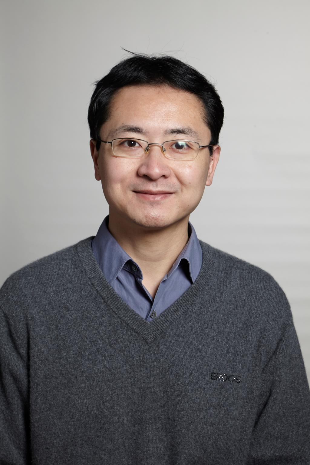 Ke Hao (郝柯)合作论文数College of Environmental Science and Engineering, Tongji University1
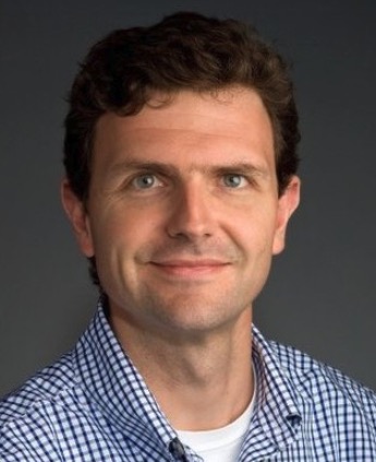 Charles G. Mullighan合作论文数Department of Pathology, St. Jude Children's Research Hospital;Comprehensive Cancer Center, St. Jude Children's Research Hospital1
 Miriam Merad合作论文数Precision Immunology Institute, Icahn School of Medicine at Mount Sinai1A. Harrington合作论文数1
 Jun Lu (陆军)合作论文数School of Life Sciences, Northeast Normal University1
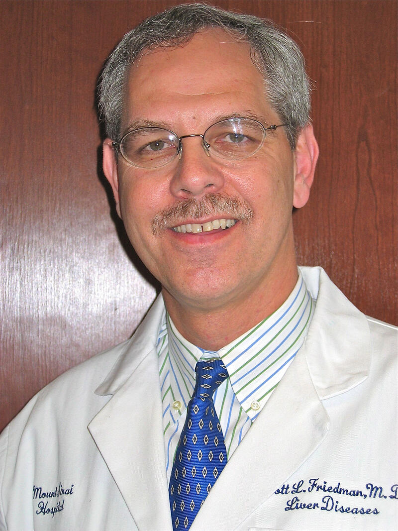 Scott L. Friedman合作论文数Division of Liver Diseases, Mount Sinai School of Medicine1Judit Peix合作论文数1Hui Dong合作论文数1
 Todd R. Golub合作论文数Broad Institute;Dana-Farber Cancer Institute;Harvard Medical School1
 Jordi Bruix合作论文数Barcelona Clinic Liver Cancer Group, Hospital Clinic of Barcelona;University of Barcelona1Victoria Tovar合作论文数1
 Pedro Melgar-Lesmes合作论文数Institut d’Investigacions Biomèdiques August Pi i Sunyer (IDIBAPS),, University of Barcelona1Beatriz Minguez合作论文数Tisch Cancer Institute, Transplant Institute1Swan N Thung合作论文数1
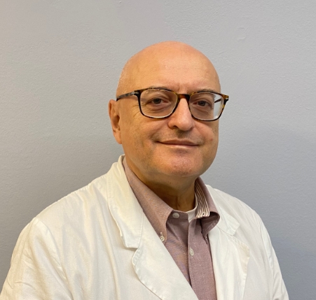 Mazzaferro Vincenzo Maria合作论文数Department of Oncology and Hematology-Oncology, Università degli Studi di Milano1
 Rameen Beroukhim合作论文数Division of Medical Sciences, Harvard Medical School;Dana-Farber Cancer Institute1P. Manikya Reddy合作论文数1
 Tony Ng合作论文数Department of Pathology and Laboratory Medicine, Faculty of Medicine, The University of British Columbia1
 Costica Aloman合作论文数Westchester Medical Center Health Network;Division of Gastroenterology and Hepatobiliary Diseases, School of Medicine, New York Medical College1




