Accurate sister chromatid segregation requires remodeling chromosome architecture, decatenation, and attachment to the mitotic spindle. Some of these events are initiated during S-phase, but they accelerate and conclude during mitosis. Here we describe SRBD1 as a histone and nucleic acid binding protein that prevents DNA damage in interphase cells, localizes to nascent DNA during replication and the chromosome scaffold in mitosis, and is required for chromosome segregation. SRBD1 inactivation causes micronuclei, chromatin bridges, and cell death. Inactivating SRBD1 immediately prior to mitotic entry causes anaphase failure, with a reduction in topoisomerase IIα localization to mitotic chromosomes and defects in properly condensing and decatenating chromosomes. In contrast, SRBD1 is not required to complete cell division after chromosomes are condensed. Strikingly, depleting condensin II reduces the severity of the anaphase defects in SRBD1-deficient cells by restoring topoisomerase IIα localization. Thus, SRBD1 is an essential genome maintenance protein required for mitotic chromosome organization and segregation.
Pds5 is required for sister chromatid cohesion, and somewhat paradoxically, to remove cohesin from chromosomes. We found that Pds5 plays a critical role during DNA replication that is distinct from its previously known functions. Loss of Pds5 hinders replication fork progression in unperturbed human and mouse cells. Inhibition of MRE11 nuclease activity restores fork progression, suggesting that Pds5 protects forks from MRE11-activity. Loss of Pds5 also leads to double-strand breaks, which are again reduced by MRE11 inhibition. The replication function of Pds5 is independent of its previously reported interaction with BRCA2. Unlike Pds5, BRCA2 protects forks from nucleolytic degradation only in the presence of genotoxic stress. Moreover, our iPOND analysis shows that the loading of Pds5 and other cohesion factors on replication forks is not affected by the BRCA2 status. Pds5 role in DNA replication is shared by the other cohesin-removal factor Wapl, but not by the cohesin complex component Rad21. Interestingly, depletion of Rad21 in a Pds5-deficient background rescues the phenotype observed upon Pds5 depletion alone. These findings support a model where loss of either component of the cohesin releasin complex perturbs cohesin dynamics on replication forks, hindering fork progression and promoting MRE11-dependent fork slowing.
Identifying proteins that function at replication forks is essential to understanding DNA replication, chromatin assembly, and replication-coupled DNA repair mechanisms. Combining quantitative mass spectrometry in multiple cell types with stringent statistical cutoffs, we generated a high-confidence catalog of 593 proteins that are enriched at replication forks and nascent chromatin. Loss-of-function genetic analyses indicate that 85% yield phenotypes that are consistent with activities in DNA and chromatin replication or already have described functions in these processes. We illustrate the value of this resource by identifying activities of the BET family proteins BRD2, BRD3, and BRD4 in controlling DNA replication. These proteins use their extra-terminal domains to bind and inhibit the ATAD5 complex and thereby control the amount of PCNA on chromatin.
Abasic sites are one of the most common DNA lesions. All known abasic site repair mechanisms operate only when the damage is in double-stranded DNA. Here, we report the discovery of 5-hydroxymethylcytosine (5hmC) binding, ESC-specific (HMCES) as a sensor of abasic sites in single-stranded DNA. HMCES acts at replication forks, binds PCNA and single-stranded DNA, and generates a DNA-protein crosslink to shield abasic sites from error-prone processing. This unusual HMCES DNA-protein crosslink intermediate is resolved by proteasome-mediated degradation. Acting as a suicide enzyme, HMCES prevents translesion DNA synthesis and the action of endonucleases that would otherwise generate mutations and double-strand breaks. HMCES is evolutionarily conserved in all domains of life, and its biochemical properties are shared with its E. coli ortholog. Thus, HMCES is an ancient DNA lesion recognition protein that preserves genome integrity by promoting error-free repair of abasic sites in single-stranded DNA.
In E. coli, replisomes duplicate only a fraction of the chromosome before being ejected from the DNA due to collisions with damaged DNA or nucleoprotein complexes. Premature replisome ejection leaves an abandoned replication fork that must be reinitiated by DNA replication restart proteins (PriA, PriB, PriC, and DnaT in E. coli). PriC can mediate helicase loading without the help of other replication restart proteins in vitro and it directly interacts with the DnaB/DnaC (helicase/loader) complex. Interestingly, ΔpriC strains appear wild‐type but have synthetic lethal relationships with genes not implicated in DNA replication restart. Our poster describes recent structural efforts to probe the PriC/DnaB/DnaC complex and a transposon‐sequencing approach to identify genes linked to priC and other DNA replication restart genes in E. coli.Support or Funding InformationNIH R01GM098885.This abstract is from the Experimental Biology 2018 Meeting. There is no full text article associated with this abstract published in The FASEB Journal.
Multi-protein DNA replication complexes called replisomes perform the essential process of copying cellular genetic information prior to cell division. Under ideal conditions, replisomes dissociate only after the entire genome has been duplicated. However, DNA replication rarely occurs without interruptions that can dislodge replisomes from DNA. Such events produce incompletely replicated chromosomes that, if left unrepaired, prevent the segregation of full genomes to daughter cells. To mitigate this threat, cells have evolved 'DNA replication restart' pathways that have been best defined in bacteria. Replication restart requires recognition and remodeling of abandoned replication forks by DNA replication restart proteins followed by reloading of the replicative DNA helicase, which subsequently directs assembly of the remaining replisome subunits. This review summarizes our current understanding of the mechanisms underlying replication restart and the proteins that drive the process in Escherichia coli (PriA, PriB, PriC and DnaT).
s for the 41st Human Genetics Society of Australasia Annual Scientific Meeting Brisbane, Queensland
RAD51 promotes homology-directed repair (HDR), replication fork reversal, and stalled fork protection. Defects in these functions cause genomic instability and tumorigenesis but also generate hypersensitivity to cancer therapeutics. Here we describe the identification of RADX as an RPA-like, single-strand DNA binding protein. RADX is recruited to replication forks, where it prevents fork collapse by regulating RAD51. When RADX is inactivated, excessive RAD51 activity slows replication elongation and causes double-strand breaks. In cancer cells lacking BRCA2, RADX deletion restores fork protection without restoring HDR. Furthermore, RADX inactivation confers chemotherapy and PARP inhibitor resistance to cancer cells with reduced BRCA2/RAD51 pathway function. By antagonizing RAD51 at forks, RADX allows cells to maintain a high capacity for HDR while ensuring that replication functions of RAD51 are properly regulated. Thus, RADX is essential to achieve the proper balance of RAD51 activity to maintain genome stability.
To ensure efficient genome duplication, cells have evolved numerous factors that promote unperturbed DNA replication and protect, repair and restart damaged forks. Here we identify downstream neighbor of SON (DONSON) as a novel fork protection factor and report biallelic DONSON mutations in 29 individuals with microcephalic dwarfism. We demonstrate that DONSON is a replisome component that stabilizes forks during genome replication. Loss of DONSON leads to severe replication-associated DNA damage arising from nucleolytic cleavage of stalled replication forks. Furthermore, ATM- and Rad3-related (ATR)-dependent signaling in response to replication stress is impaired in DONSON-deficient cells, resulting in decreased checkpoint activity and the potentiation of chromosomal instability. Hypomorphic mutations in DONSON substantially reduce DONSON protein levels and impair fork stability in cells from patients, consistent with defective DNA replication underlying the disease phenotype. In summary, we have identified mutations in DONSON as a common cause of microcephalic dwarfism and established DONSON as a critical replication fork protein required for mammalian DNA replication and genome stability.
Collisions between DNA replication complexes (replisomes) and barriers such as damaged DNA or tightly bound protein complexes can dissociate replisomes from chromosomes prematurely. Replisomes must be reloaded under these circumstances to avoid incomplete replication and cell death. Bacteria have evolved multiple pathways that initiate DNA replication restart by recognizing and remodeling abandoned replication forks and reloading the replicative helicase. In vitro, the simplest of these pathways is mediated by the single-domain PriC protein, which, along with the DnaC helicase loader, can load the DnaB replicative helicase onto DNA bound by the single-stranded DNA (ssDNA)-binding protein (SSB). Previous biochemical studies have identified PriC residues that mediate interactions with ssDNA and SSB. However, the mechanisms by which PriC drives DNA replication restart have remained poorly defined due to the limited structural information available for PriC. Here, we report the NMR structure of full-length PriC from Cronobacter sakazakii. PriC forms a compact bundle of α-helices that brings together residues involved in ssDNA and SSB binding at adjacent sites on the protein surface. Disruption of these interaction sites and of other conserved residues leads to decreased DnaB helicase loading onto SSB-bound DNA. We also demonstrate that PriC can directly interact with DnaB and the DnaB·DnaC complex. These data lead to a model in which PriC acts as a scaffold for recruiting DnaB·DnaC to SSB/ssDNA sites present at stalled replication forks.
Collisions between cellular DNA replication machinery (replisomes) and damaged DNA or immovable protein complexes can dissociate replisomes before the completion of replication. This potentially lethal problem is resolved by cellular "replication restart" reactions that recognize the structures of prematurely abandoned replication forks and mediate replisomal reloading. In bacteria, this essential activity is orchestrated by the PriA DNA helicase, which identifies replication forks via structure-specific DNA binding and interactions with fork-associated ssDNA-binding proteins (SSBs). However, the mechanisms by which PriA binds replication fork DNA and coordinates subsequent replication restart reactions have remained unclear due to the dearth of high-resolution structural information available for the protein. Here, we describe the crystal structures of full-length PriA and PriA bound to SSB. The structures reveal a modular arrangement for PriA in which several DNA-binding domains surround its helicase core in a manner that appears to be poised for binding to branched replication fork DNA structures while simultaneously allowing complex formation with SSB. PriA interaction with SSB is shown to modulate SSB/DNA complexes in a manner that exposes a potential replication initiation site. From these observations, a model emerges to explain how PriA links recognition of diverse replication forks to replication restart.
ABSTRACT DNA helicases have important roles in genome maintenance. The RecD helicase has been well studied as a component of the heterotrimeric RecBCD helicase-nuclease enzyme important for double-strand break repair in Escherichia coli . Interestingly, many bacteria lack RecBC and instead contain a RecD2 helicase, which is not known to function as part of a larger complex. Depending on the organism studied, RecD2 has been shown to provide resistance to a broad range of DNA-damaging agents while also contributing to mismatch repair (MMR). Here we investigated the importance of Bacillus subtilis RecD2 helicase to genome integrity. We show that deletion of recD2 confers a modest increase in the spontaneous mutation rate and that the mutational signature in Δ recD2 cells is not consistent with an MMR defect, indicating a new function for RecD2 in B. subtilis . To further characterize the role of RecD2, we tested the deletion strain for sensitivity to DNA-damaging agents. We found that loss of RecD2 in B. subtilis sensitized cells to several DNA-damaging agents that can block or impair replication fork movement. Measurement of replication fork progression in vivo showed that forks collapse more frequently in Δ recD2 cells, supporting the hypothesis that RecD2 is important for normal replication fork progression. Biochemical characterization of B. subtilis RecD2 showed that it is a 5′-3′ helicase and that it directly binds single-stranded DNA binding protein. Together, our results highlight novel roles for RecD2 in DNA replication which help to maintain replication fork integrity during normal growth and when forks encounter DNA damage.
Frequent collisions between cellular DNA replication complexes (replisomes) and obstacles such as damaged DNA or frozen protein complexes make DNA replication fork progression surprisingly sporadic. These collisions can lead to the ejection of replisomes prior to completion of replication, which, if left unrepaired, results in bacterial cell death. As such, bacteria have evolved DNA replication restart mechanisms that function to reload replisomes onto abandoned DNA replication forks. Here, we define a direct interaction between PriC, a key Escherichia coli DNA replication restart protein, and the single-stranded DNA-binding protein (SSB), a protein that is ubiquitously associated with DNA replication forks. PriC/SSB complex formation requires evolutionarily conserved residues from both proteins, including a pair of Arg residues from PriC and the C terminus of SSB. In vitro, disruption of the PriC/SSB interface by sequence changes in either protein blocks the first step of DNA replication restart, reloading of the replicative DnaB helicase onto an abandoned replication fork. Consistent with the critical role of PriC/SSB complex formation in DNA replication restart, PriC variants that cannot bind SSB are non-functional in vivo. Single-molecule experiments demonstrate that PriC binding to SSB alters SSB/DNA complexes, exposing single-stranded DNA and creating a platform for other proteins to bind. These data lead to a model in which PriC interaction with SSB remodels SSB/DNA structures at abandoned DNA replication forks to create a DNA structure that is competent for DnaB loading.
DNA replication restart is an essential process in bacteria that reloads DNA replication complexes (replisomes) that have ejected prematurely from replication forks due to encounters with impassable DNA damage or protein complexes. In E. coli and related bacterial species, the “replication restart proteins” (PriA, PriB, PriC, DnaT, and Rep) catalyze this activity. The 20‐kDa PriC protein is unique among these proteins in that it can mediate in vitro replisome reloading in the absence of other replication restart proteins. Precisely how PriC recognizes abandoned DNA replication fork structures and directs replisome reloading is currently unknown. We have found that the single‐strand DNA‐binding protein (SSB) forms a direct complex with PriC that we hypothesize is important for directing PriC reloading activities to abandoned DNA replication forks. PriC variants that fail that to bind SSB (but that retain DNA binding) were identified to test this hypothesis. Consistent with an essential role for the PriC/SSB interaction, these variants cannot catalyze the first step of replication restart (loading the DnaB helicase onto an SSB‐bound synthetic replication fork) in vitro. On‐going experiments aim to determine the solution structure of PriC and to assess the importance of complex formation with SSB in PriC‐mediated DNA replication restart in vivo. Research supported by GM068061 and T32‐GM07215.
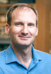 David K. Cortez合作论文数Department of Biochemistry, School of Medicine, Vanderbilt University;Vanderbilt-Ingram Cancer Center, Vanderbilt University8
 James L. Keck合作论文数Department of Biomolecular Chemistry, School of Medicine and Public Health, University of Wisconsin-Madison7
 Audrey Vernet合作论文数3Michael A. Simpson合作论文数3Rita Fischetto合作论文数3
 Carol A Wise合作论文数3Angela L Duker合作论文数3Alexander Brean合作论文数3
 Zygimante Tarnauskaite合作论文数MRC Human Genet Unit, Univ Edinburgh3
 Sateesh Maddirevula合作论文数King Faisal Specialist Hosp & Res Ctr, Ctr Genom Med, Dept Translat Genom, Riyadh, Saudi Arabia3Hannah Bye合作论文数Royal Marsden NHS Fdn Trust, Sutton, Surrey, England3Gökhan Yigit合作论文数Univ Med Ctr Gottingen, Inst Human Genet, Gottingen, Germany3Hessa S Alsaif合作论文数King Faisal Specialist Hosp & Res Ctr, Dept Genet, Riyadh, Saudi Arabia3
 Ferda Percin合作论文数Gazi University3
 Helen S Cox合作论文数Department of Pathology, University of Cape Town;Wellcome Centre for Infectious Disease Research in Africa, University of Cape Town;Institute of Infectious Disease and Molecular Medicine, University of Cape Town;University of Cape Town, University of Cape Town3
 Grace Yoon合作论文数Departments of Paediatrics and Molecular Genetics, Faculty of Medicine, University of Toronto;Division of Clinical & Metabolic Genetics, The Hospital for Sick Children;Division of Neurology, The Hospital for Sick Children;SickKids Research Institute;Neurogenetics Clinic3
 Natalie Prescott合作论文数School of Basic & Medical Biosciences, Faculty of Life Sciences & Medicine, King’s College London3
 Janine Altmüller合作论文数Center for Molecular Medicine Cologne;Berlin Institute of Health at Charité, Universitätsmedizin Berlin;Max Delbrück Center for Molecular Medicine3
 von Kriegsheim Alex合作论文数Syst Biol Ireland, Univ Coll Dublin3
 mohammed zain Seidahmed合作论文数3
 Angela Peron合作论文数3
 Seema Thakur合作论文数Department of Genetic Medicine, Sir Ganga Ram Hospital3Agaadir Almoisheer合作论文数3
 Christopher Mathew合作论文数King’s Global Health Institute, School of Basic & Medical Biosciences, Faculty of Life Sciences and Medicine, Kings College London;Department of Medical and Molecular Genetics, School of Basic & Medical Biosciences, Faculty of Life Sciences and Medicine, Kings College London;Sydney Brenner Institute for Molecular Bioscience, Faculty of Health Sciences, Witwatersrand University3Leitch Andrea合作论文数3
 Al-Aama Jumana Yousuf合作论文数Princess Al-Jawhara Albrahim Center of Excellence in Research of Hereditary Disorders, King Abdulaziz University;Faculty of Medicine, King Abdulaziz University;King Abdulaziz University, King Abdulaziz University3Brady Angela合作论文数3
 Paula Carroll合作论文数3Amar Ariella合作论文数3Heller Raoul合作论文数3
 Murina Olga合作论文数Institute of Molecular Cancer Research, University of Zurich3Challis Rachel C合作论文数3Murray Jennie合作论文数3Skouloudaki Kassiani合作论文数3
 Peter Nürnberg合作论文数Cologne Center for Genomics, University of Cologne3
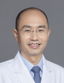 Yun Li (李运)合作论文数Peking University People's Hospital3Runxiang Zhao合作论文数3
 John Joseph Reynolds合作论文数3
 Mohammed Al Balwi合作论文数Department of Pathology and Laboratory Medicine, King Abdulaziz Medical City3
 Ranad Shaheen合作论文数Department of Applied Chemistry and Microbiology, University of Helsinki3
 Abdulrahman Alswaid合作论文数National Guard Health Affairs;King Abdulaziz Medical City3
 Malcolm Taylor合作论文数Department of Cancer and Genomic Sciences, University of Birmingham3
 Clare V Logan合作论文数3
 Fowzan S. Alkuraya (Fowzan Alkuraya)合作论文数College of Medicine, Alfaisal University;Department of Translational Genomics, King Faisal Specialist Hospital and Research Center3
 Pavel Tomancak合作论文数Max Planck Institute of Molecular Cell Biology and Genetics;Central European Institute of Technology3
 Steven J. Sandler合作论文数Morrill Science Center, University of Massachusetts at Amherst3
 Dimitrios K Papadopoulos合作论文数3
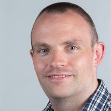 Martin R. Higgs合作论文数Institute of Cancer and Genomic Sciences, College of Medical and Dental Sciences, University of Birmingham3Bertram D Henderson合作论文数2




