Prospective, multi-institutional surgical data collection in pediatric neuro-oncology remains limited despite substantial variation in operative and perioperative management across institutions. To address this, we are developing the NeuroPoint Alliance (NPA) Quality Outcomes Database (QOD) Pediatric Tumor Surgery Registry. Here, we used a modified Delphi process to define a core outcome set for the registry. A modified Delphi study was conducted among pediatric neurosurgeons serving as site principal investigators for the proposed registry. Candidate data elements were rated on a 9-point Likert scale. Consensus for inclusion was predefined as ≥70
BACKGROUND:In children with Chiari type I malformation and syringomyelia, neurosurgical posterior fossa decompression (PFD) provides clinical improvement, but whether duraplasty (incising the dura and placing a dural graft) improves outcomes is unclear. METHODS:We conducted a multicenter, cluster-randomized, controlled trial of PFD with duraplasty (PFD-D) as compared with PFD alone. Persons 21 years of age or younger with cerebellar tonsillar ectopia of at least 5 mm and a maximum syrinx diameter of 3.0 to 9.9 mm were enrolled at 38 centers. Centers were cluster-randomized: all the participants within each center underwent the same intervention. The primary outcome was surgical complications within 6 months. Secondary outcomes were clinical improvement, syrinx reduction, and repeat decompression at 10 to 24 months and the change in overall health-related quality of life at 6 to 24 months. RESULTS:A total of 162 participants were included in the trial, of whom 78 were assigned to undergo PFD-D and 84 to undergo PFD alone. The percentage of participants with complications within 6 months was 14% with PFD-D and 6% with PFD (adjusted odds ratio, 2.59; 95% confidence interval [CI], 0.86 to 7.84; P = 0.11). At 24 months, the percentage of participants with clinical improvement was 58% with PFD-D and 46% with PFD; the mean (±SD) syrinx reduction was 3.08±2.33 mm and 1.22±1.79 mm, respectively; and the percentage of participants with repeat decompression was 3% and 14%. Changes in health-related quality of life were similar in the two groups. CONCLUSIONS:The percentage of participants with surgical complications did not differ significantly between those who underwent PFD-D and and those who underwent PFD alone. Larger trials are needed to determine the relative benefits and risks of these two procedures. (Funded by the Patient-Centered Outcomes Research Institute and others; ClinicalTrials.gov number, NCT02669836.).
OBJECTIVE:The choice of surgical technique for decompression of the cervicomedullary junction remains controversial. The authors report their experience with using subpial tonsillectomy for Chiari malformation with and without associated syringohydromyelia. METHODS:Children from two institutions who underwent the subpial tonsillar resection technique for decompression of the craniocervical junction from 2014 to 2024 were included. Demographic information, relevant presurgical symptoms, surgical history, perioperative radiographic characteristics, postoperative outcomes, complications, reoperations, and 90-day readmissions were collected. Two outcomes were assessed at last follow-up: 1) status of presenting symptom(s); and 2) radiographic status of syringohydromyelia or isolated presyrinx edema, if present preoperatively. RESULTS:A total of 109 patients were identified with a mean age of 10.8 years. Seventy-eight (71.6%) of these patients presented with headaches, and 23 (21.1%) had medullary or lower cranial nerve dysfunction. Radiographically, 86 (78.9%) patients had Chiari type 1 and 23 (21.1%) had Chiari type 1.5. The mean (range) preoperative cerebellar tonsillar herniation was 14.0 (4.5-40) mm, and 49 (45%) patients had syringohydromyelia or presyrinx edema. The mean operative time was 152 minutes, and the mean length of stay was 3.49 days. Fourteen patients were readmitted within 90 days of surgery, 9 within the first 30 days, and 5 after 30 days. Of these 14 readmissions, 8 (57%) were for reasons attributable to the index operation. Over a mean follow-up of 3.70 years, headaches resolved or improved in 68 of 78 (87.2%) patients, and 18 of 23 (78.3%) patients with brainstem or lower cranial nerve dysfunction demonstrated improvement. Of the patients with syringohydromyelia who had follow-up (n = 45), 39 (86.7%) had complete resolution of their syringohydromyelia (n = 16) or > 50% reduction in diameter of the syrinx (n = 23). One patient was deemed a treatment failure, eventually needing a syringopleural shunt. CONCLUSIONS:Subpial tonsillar resection for Chiari malformations is a safe and effective method to maximally decompress the craniocervical junction and immediately reestablish pulsatile cerebrospinal fluid flow out of the fourth ventricle. Most patients will experience improvement or resolution of their associated symptoms and significant reduction (> 50% diameter reduction) or resolution of their syringohydromyelia.
Background Skull base approaches are utilized to improve microsurgical treatment of cerebral aneurysms. Advantages include early proximal and distal control, increased visualization, and minimal brain retraction. Orbitozygomatic (OZ) craniotomies via pterional incision are commonly used for the treatment of anterior communicating artery (ACoA) aneurysms. A smaller, less invasive OZ craniotomy performed through an eyebrow incision may provide several advantages over a standard OZ approach.Objective We compare surgical outcomes of the standard and eyebrow OZ for the treatment of ACoA aneurysms.Design All patients who underwent microsurgical treatment for ACoA aneurysms by a single surgeon over an 8-year period were included in this retrospective analysis. Patient demographics and clinical data were collected.Participants Thirty-seven consecutive patients were identified, with 15 receiving eyebrow OZ and 22 receiving standard OZ.Main Outcome Measures Data were collected on patient demographics, pathology, intraoperative and perioperative data, and 30-day morbidity.Results A total of 100% of the eyebrow OZ group and 95.5% of the standard OZ group had complete aneurysmal occlusion. Four eyebrow OZ and six standard OZ patients had an intraoperative rupture. All were managed without complication. Two eyebrow OZ and one standard OZ patient died within 30 days of surgery. No patients in either group had aneurysm recurrence, required retreatment, or were limited intraoperatively by exposure.Conclusions The OZ approach via an eyebrow incision has similar outcomes to a standard OZ approach and is a safe option for the treatment of ACoA aneurysms.
Objective: Ventriculoperitoneal (VP) shunt placement requires a concurrent abdominal procedure. For peritoneal access laparoscopic or open approach may be utilized. Our aim was to compare patient/procedure characteristics and outcomes by peritoneal approach for VP shunts in children.Methods: NSQIP-Pediatric procedure targeted cerebral spinal fluid shunt Participant Use Data Files from 2016 to 2020 were queried. Patients were grouped into laparoscopic vs open abdominal approach. Patient demographics, procedure characteristics and 30-day outcomes were compared.Results: 7742 NSQIP-Pediatric patients underwent VP shunt placement. Patients undergoing laparoscopic approach were older and required less preoperative support. Mean operative time was longer with laparoscopy (mean(SD): 74.2(48.1) vs. 64.6(39) minutes, p < 0.0001) but had shorter hospital LOS. There was no difference in SSI, readmissions, or reoperation rates.Conclusion: Patients undergoing laparoscopy for distal VP shunts are older with less support needs preoperatively. While laparoscopic approach had a shorter hospital LOS, there was no demonstratable difference in SSI, readmissions or reoperations between approaches. Further studies are needed to assess long-term outcomes.
OBJECTIVE:Congenital anomalies of the atlanto-occipital articulation may be present in patients with Chiari malformation type I (CM-I). However, it is unclear how these anomalies affect the biomechanical stability of the craniovertebral junction (CVJ) and whether they are associated with an increased incidence of occipitocervical fusion (OCF) following posterior fossa decompression (PFD). The objective of this study was to determine the prevalence of condylar hypoplasia and atlas anomalies in children with CM-I and syringomyelia. The authors also investigated the predictive contribution of these anomalies to the occurrence of OCF following PFD (PFD+OCF). METHODS:The authors analyzed the prevalence of condylar hypoplasia and atlas arch anomalies for patients in the Park-Reeves Syringomyelia Research Consortium database who underwent PFD+OCF. Condylar hypoplasia was defined by an atlanto-occipital joint axis angle (AOJAA) ≥ 130°. Atlas assimilation and arch anomalies were identified on presurgical radiographic imaging. This PFD+OCF cohort was compared with a control cohort of patients who underwent PFD alone. The control group was matched to the PFD+OCF cohort according to age, sex, and duration of symptoms at a 2:1 ratio. RESULTS:Clinical features and radiographic atlanto-occipital joint parameters were compared between 19 patients in the PFD+OCF cohort and 38 patients in the PFD-only cohort. Demographic data were not significantly different between cohorts (p > 0.05). The mean AOJAA was significantly higher in the PFD+OCF group than in the PFD group (144° ± 12° vs 127° ± 6°, p < 0.0001). In the PFD+OCF group, atlas assimilation and atlas arch anomalies were identified in 10 (53%) and 5 (26%) patients, respectively. These anomalies were absent (n = 0) in the PFD group (p < 0.001). Multivariate regression analysis identified the following 3 CVJ radiographic variables that were predictive of OCF occurrence after PFD: AOJAA ≥ 130° (p = 0.01), clivoaxial angle < 125° (p = 0.02), and occipital condyle-C2 sagittal vertical alignment (C-C2SVA) ≥ 5 mm (p = 0.01). A predictive model based on these 3 factors accurately predicted OCF following PFD (C-statistic 0.95). CONCLUSIONS:The authors' results indicate that the occipital condyle-atlas joint complex might affect the biomechanical integrity of the CVJ in children with CM-I and syringomyelia. They describe the role of the AOJAA metric as an independent predictive factor for occurrence of OCF following PFD. Preoperative identification of these skeletal abnormalities may be used to guide surgical planning and treatment of patients with complex CM-I and coexistent osseous pathology.
BACKGROUND:Nonmissile penetrating spinal cord injury (NMPSCI) with a retained foreign body (RFB) is rare and usually results in permanent neurological deficits. In extremely rare cases, patients can present without significant neurological deficits despite an RFB that traverses the spinal canal. Given the rarity of these cases, a consensus has not yet been reached on optimal management. In a patient with an RFB and a neurologically normal clinical examination, the risk of open surgical exploration may outweigh the benefit and direct withdrawal may be a better option.OBSERVATIONS:A 10-year-old female suffered an NMPSCI to the thoracic spine with an RFB that bisected the spinal canal but remained neurologically intact. Direct withdrawal of the RFB was chosen instead of open surgical exploration, leading to an excellent clinical outcome. The literature was reviewed to find other examples of thoracic NMPSCI with RFB and neurologically normal examinations. Management strategies were compared.LESSONS:For NMPSCI with RFB and without significant neurological deficits, direct withdrawal is a viable and possibly the best treatment option. The use of fast-acting anesthesia without intubation minimizes patient manipulation, speeds up recovery, and allows early assessment of neurological status after removal.
Pediatric basilar artery aneurysms are rare and challenging to treat. Microsurgical options and standard endovascular coiling are often undesirable choices for treatment of this pathology. Additional endovascular strategies are needed. Presentation, diagnosis, and management of pediatric basilar aneurysms were reviewed, with an emphasis on endovascular treatment strategies. Our case series of 2 patients was presented in detail, one treated with flow diversion and vessel sacrifice and one treated with stent-assisted coiling. An extensive review of the literation was performed to find other examples of pediatric basilar artery aneurysms treated with endovascular techniques. Twenty-nine studies met inclusion criteria. Fifty-nine aneurysms in 58 patients were treated using endovascular techniques. Mortality rate was 10.3
Background The occipital interhemispheric transtentorial (OITT) approach is frequently used for accessing the pineal region. There are scarce reports of using the OITT to access superior cerebellar lesions. This approach affords the patient several advantages over traditional posterior fossa approaches. Purpose This study is to describe and evaluate clinical outcomes in a single surgeon case series of the OITT approach for pediatric patients with lesions of the superior cerebellum. Methods All pediatric patients who underwent an OITT craniotomy for a superior cerebellar lesion by a single surgeon over a 5-year period were included in this retrospective analysis. Patient demographics and clinical data were collected. Results Thirteen pediatric patients were identified. Cases included twelve tumors and one arteriovenous malformation. Gross total resection was achieved in 92% of cases. No patients developed posterior fossa syndrome. Two patients had transient homonymous hemianopsia that resolved by 1 month post-operatively. There were no permanent neurological deficits. Conclusion For superomedial cerebellar lesions presenting to the tentorial surface of the superior cerebellum in patients with normal to steep tentorial angles, the OITT approach is effective and safe. This approach has a low risk of posterior fossa syndrome and permanent visual deficits when applied appropriately. Patient selection is critical for maximizing the advantages of the OITT for superior cerebellar lesions.
Background Cerebral vasospasm (CV) is a common complication of aneurysmal subarachnoid hemorrhage (aSAH) and is associated with significant morbidity and mortality. Objective To report our institutional experience with intrathecal (IT) nicardipine for treatment of moderate to severe CV. Methods A retrospective study was performed for all patients treated with intrathecal nicardipine from October 2014-May 2017. The primary goal was to evaluate the safety of IT nicardipine to treat subarachnoid induced CV. Results. 48 patients with aSAH were treated with IT nicardipine for moderate or severe CV. The average age of patients was 49.6 years (range 14–77 years) with mean Hunt Hess of 3.2 and mean Fisher scores of 3.1. Patients were treated with different dosing regiments ranging from 2 mg q12 to 4 mg q8hr. The average total dose was 35.1 mg (range 6 to 112 mg). The average initiation of treatment was post bleed day 6 (range day 2–15) and length of treatment was 5.9 days (range 1–15 days.) 10/48 (20.8%) patients underwent TBA with only 4/48 (8.3%) treated after initiation of IT nicardipine. Three patients (6.3%) developed meningitis/ventriculitis. Three (6.3%) patients required ventriculoperitoneal shunt. Favorable outcome (Glasgow Outcome Scale 4 or 5) was achieved in 56.3% of patients. Conclusions The use of intrathecal nicardipine should be considered a safe option in the treatment of moderate to severe cerebral vasospasm. The use of intrathecal nicardipine has resulted in a near discontinuation of balloon angioplasty in our institution. Randomized trials to identify the optimal dosing, timing, and length of treatment is warranted.
Background The eyebrow orbitozygomatic craniotomy is a minimally invasive approach that can access a wide variety of lesions. Unintentional breach of the frontal sinus frequently occurs and has been cited as a reason to avoid this approach. Lack of access to a large pericranial graft and the inability to completely cranialize the sinus requires alternate techniques of sinus repair. We describe a technique for repairing an opened frontal sinus and retrospectively reviewed complications related to this approach. Methods All patients, who underwent an orbitozygomatic craniotomy via an eyebrow incision by a single surgeon from August 1, 2012 to August 31, 2018, were included in this retrospective analysis. Data were collected on patient demographics, pathology treated, operative details, and perioperative morbidity. Follow-up ranged from 6 weeks to 6 years. Results Total 50 patients with a wide variety of pathologies underwent analysis. Frontal sinus breach occurred in 21 patients. All were repaired by the described technique. One patient (ruptured aneurysm) had a suspected cerebrospinal fluid (CSF) leak postoperatively that resolved without any additional intervention. One patient developed a pneumomeningocele 4 years postoperatively that required reoperation. No patient suffered any infection or delayed CSF leak. Conclusion Breach of the frontal sinus is common during eyebrow craniotomies. Despite reduced options for local repair, these patients have experienced no CSF leaks requiring intervention and no infections in our series. Long-term mucocele risk is not reliably determined with our length of follow-up. Breach of the frontal sinus is not a contraindication to the eyebrow approach.
BACKGROUND:Occipital-cervical fusion (OCF) and ventral decompression (VD) may be used in the treatment of pediatric Chiari-1 malformation (CM-1) with syringomyelia (SM) as adjuncts to posterior fossa decompression (PFD) for complex craniovertebral junction pathology. OBJECTIVE:To examine factors influencing the use of OCF and OCF/VD in a multicenter cohort of pediatric CM-1 and SM subjects treated with PFD. METHODS:The Park-Reeves Syringomyelia Research Consortium registry was used to examine 637 subjects with cerebellar tonsillar ectopia ≥ 5 mm, syrinx diameter ≥ 3 mm, and at least 1 yr of follow-up after their index PFD. Comparisons were made between subjects who received PFD alone and those with PFD + OCF or PFD + OCF/VD. RESULTS:All 637 patients underwent PFD, 505 (79.2%) with and 132 (20.8%) without duraplasty. A total of 12 subjects went on to have OCF at some point in their management (PFD + OCF), whereas 4 had OCF and VD (PFD + OCF/VD). Of those with complete data, a history of platybasia (3/10, P = .011), Klippel-Feil (2/10, P = .015), and basilar invagination (3/12, P < .001) were increased within the OCF group, whereas only basilar invagination (1/4, P < .001) was increased in the OCF/VD group. Clivo-axial angle (CXA) was significantly lower for both OCF (128.8 ± 15.3°, P = .008) and OCF/VD (115.0 ± 11.6°, P = .025) groups when compared to PFD-only group (145.3 ± 12.7°). pB-C2 did not differ among groups. CONCLUSION:Although PFD alone is adequate for treating the vast majority of CM-1/SM patients, OCF or OCF/VD may be occasionally utilized. Cranial base and spine pathologies and CXA may provide insight into the need for OCF and/or OCF/VD.
OBJECTIVE The aim of this study was to determine differences in complications and outcomes between posterior fossa decompression with duraplasty (PFDD) and without duraplasty (PFD) for the treatment of pediatric Chiari malformation type I (CM1) and syringomyelia (SM). METHODS The authors used retrospective and prospective components of the Park-Reeves Syringomyelia Research Consortium database to identify pediatric patients with CM1-SM who received PFD or PFDD and had at least 1 year of follow-up data. Preoperative, treatment, and postoperative characteristics were recorded and compared between groups. RESULTS A total of 692 patients met the inclusion criteria for this database study. PFD was performed in 117 (16.9%) and PFDD in 575 (83.1%) patients. The mean age at surgery was 9.86 years, and the mean follow-up time was 2.73 years. There were no significant differences in presenting signs or symptoms between groups, although the preoperative syrinx size was smaller in the PFD group. The PFD group had a shorter mean operating room time (p < 0.0001), fewer patients with > 50 mL of blood loss (p = 0.04), and shorter hospital stays (p = 0.0001). There were 4 intraoperative complications, all within the PFDD group (0.7%, p > 0.99). Patients undergoing PFDD had a 6-month complication rate of 24.3%, compared with 13.7% in the PFD group (p = 0.01). There were no differences between groups for postoperative complications beyond 6 months (p = 0.33). PFD patients were more likely to require revision surgery (17.9% vs 8.3%, p = 0.002). PFDD was associated with greater improvements in headaches (89.6% vs 80.8%, p = 0.04) and back pain (86.5% vs 59.1%, p = 0.01). There were no differences between groups for improvement in neurological examination findings. PFDD was associated with greater reduction in anteroposterior syrinx size (43.7% vs 26.9%, p = 0.0001) and syrinx length (18.9% vs 5.6%, p = 0.04) compared with PFD. CONCLUSIONS PFD was associated with reduced operative time and blood loss, shorter hospital stays, and fewer postoperative complications within 6 months. However, PFDD was associated with better symptom improvement and reduction in syrinx size and lower rates of revision decompression. The two surgeries have low intraoperative complication rates and comparable complication rates beyond 6 months.
OBJECTIVE:Scoliosis is common in patients with Chiari malformation type I (CM-I)-associated syringomyelia. While it is known that treatment with posterior fossa decompression (PFD) may reduce the progression of scoliosis, it is unknown if decompression with duraplasty is superior to extradural decompression. METHODS:A large multicenter retrospective and prospective registry of 1257 pediatric patients with CM-I (tonsils ≥ 5 mm below the foramen magnum) and syrinx (≥ 3 mm in axial width) was reviewed for patients with scoliosis who underwent PFD with or without duraplasty. RESULTS:In total, 422 patients who underwent PFD had a clinical diagnosis of scoliosis. Of these patients, 346 underwent duraplasty, 51 received extradural decompression alone, and 25 were excluded because no data were available on the type of PFD. The mean clinical follow-up was 2.6 years. Overall, there was no difference in subsequent occurrence of fusion or proportion of patients with curve progression between those with and those without a duraplasty. However, after controlling for age, sex, preoperative curve magnitude, syrinx length, syrinx width, and holocord syrinx, extradural decompression was associated with curve progression > 10°, but not increased occurrence of fusion. Older age at PFD and larger preoperative curve magnitude were independently associated with subsequent occurrence of fusion. Greater syrinx reduction after PFD of either type was associated with decreased occurrence of fusion. CONCLUSIONS:In patients with CM-I, syrinx, and scoliosis undergoing PFD, there was no difference in subsequent occurrence of surgical correction of scoliosis between those receiving a duraplasty and those with an extradural decompression. However, after controlling for preoperative factors including age, syrinx characteristics, and curve magnitude, patients treated with duraplasty were less likely to have curve progression than patients treated with extradural decompression. Further study is needed to evaluate the role of duraplasty in curve stabilization after PFD.
OBJECTIVE Posterior fossa decompression with duraplasty (PFDD) is commonly performed for Chiari I malformation (CM-I) with syringomyelia (SM). However, complication rates associated with various dural graft types are not well established. The objective of this study was to elucidate complication rates within 6 months of surgery among autograft and commonly used nonautologous grafts for pediatric patients who underwent PFDD for CM-I/SM. METHODS The Park-Reeves Syringomyelia Research Consortium database was queried for pediatric patients who had undergone PFDD for CM-I with SM. All patients had tonsillar ectopia ≥ 5 mm, syrinx diameter ≥ 3 mm, and ≥ 6 months of postoperative follow-up after PFDD. Complications (e.g., pseudomeningocele, CSF leak, meningitis, and hydrocephalus) and postoperative changes in syrinx size, headaches, and neck pain were compared for autograft versus nonautologous graft. RESULTS A total of 781 PFDD cases were analyzed (359 autograft, 422 nonautologous graft). Nonautologous grafts included bovine pericardium (n = 63), bovine collagen (n = 225), synthetic (n = 99), and human cadaveric allograft (n = 35). Autograft (103/359, 28.7%) had a similar overall complication rate compared to nonautologous graft (143/422, 33.9%) (p = 0.12). However, nonautologous graft was associated with significantly higher rates of pseudomeningocele (p = 0.04) and meningitis (p < 0.001). The higher rate of meningitis was influenced particularly by the higher rate of chemical meningitis (p = 0.002) versus infectious meningitis (p = 0.132). Among 4 types of nonautologous grafts, there were differences in complication rates (p = 0.02), including chemical meningitis (p = 0.01) and postoperative nausea/vomiting (p = 0.03). Allograft demonstrated the lowest complication rates overall (14.3%) and yielded significantly fewer complications compared to bovine collagen (p = 0.02) and synthetic (p = 0.003) grafts. Synthetic graft yielded higher complication rates than autograft (p = 0.01). Autograft and nonautologous graft resulted in equal improvements in syrinx size (p < 0.0001). No differences were found for postoperative changes in headaches or neck pain. CONCLUSIONS In the largest multicenter cohort to date, complication rates for dural autograft and nonautologous graft are similar after PFDD for CM-I/SM, although nonautologous graft results in higher rates of pseudomeningocele and meningitis. Rates of meningitis differ among nonautologous graft types. Autograft and nonautologous graft are equivalent for reducing syrinx size, headaches, and neck pain.
BACKGROUND:A transition is underway in neurosurgery to perform relatively safe surgeries outpatient, often at ambulatory surgery centers (ASC). We sought to evaluate whether simple intracranial endoscopic procedures such as third ventriculostomy and cyst fenestration can be safely and effectively performed at an ASC, while comparing costs with the hospital.METHODS:A retrospective chart review was performed for patients who underwent elective intracranial neuroendoscopic (NE) intervention at either a quaternary hospital or an affiliated ASC between August 2014 and September 2017. Groups were compared on length of stay, perioperative and 30-day morbidity, as well as clinical outcome at last follow-up. The total cost for these procedures were compared in relative units between all ASC cases and a small subset of hospital cases.RESULTS:In total, 16 NE operations performed at the ASC (mean patient age 29.8 years) and 37 at the hospital (mean age 15.4 years) with average length of stay of 3.5 hours and 23.1 hours respectively (P < 0.05). There were no acute complications in either cohort or morbid events requiring hospitalization within 30 days. Surgical success was noted for 75% of the ASC patients and 73% of the hospital cohort. The mean cost of 5 randomly selected hospital operations with same-day discharge and 5 with overnight stay was 3.4 and 4.1 times that of the ASC cohort, respectively (P < 0.05).CONCLUSIONS:Elective endoscopic third ventriculostomy and other simple NE procedures can be safely and effectively performed at an ASC for appropriate patients with significantly reduced cost compared with the hospital.
Background and Importance We present a case of a patient with a residual intraosseous sphenoid wing meningioma presenting with proptosis, orbital pain, and monocular vision loss for 8 months who underwent decompression of the optic canal, orbital contents, and orbital reconstruction resulting in significant improvement in her vision loss with full resolution of proptosis and orbital pain. Clinical Presentation A 43-year-old female presented with a 1 year history of headache, peri-orbital pain, proptosis, and severe vision loss. She had previously undergone subtotal resection of a large Simpson Grade 1 spheno-orbital meningioma 3 years prior at an outside institution. Workup at our institution revealed hyperostosis of the left greater wing of the sphenoid bone and narrowing of the optic canal along with bony enhancement concerning for residual tumor. The patient was given the recommendation from outside institutions for radiation, presumably due to the chronicity of her visual loss. Our institution recommended resection of the residual osseous tumor with orbital reconstruction. Less than 2 weeks after surgery, the patient noted significant improvement in orbital pain and vision. At 3 months, she had regained full and symmetric orbital appearance with no orbital pain. Her visual acuity improved to 20/30 with full visual fields. Conclusion Surgical decompression of the optic canal and orbital contents for tumor related sphenoid wing hyperostosis should be strongly considered, despite an extended duration of visual change and loss. This case report shows that vision can be significantly restored even after symptoms have been present for greater than 6 months.
Background: Traumatic tension pneumocephalus is a rare complication after craniofacial fractures that can cause devastating neurologic deficits if not managed promptly and effectively. Case Report: A 38-year-old man with no past medical history presented to the Emergency Department (ED) after a motor vehicle crash. He was noted to have an open frontal scalp laceration. Computed tomography (CT) revealed a right frontal subdural hematoma and right medial frontal contusion. There was also a frontal bone fracture extending through the frontal sinus with mild underlying pneumocephalus. He was monitored for cerebrospinal fluid (CSF) leak and was subsequently discharged on postinjury day 9. He re-presented to the ED 14 days post injury with lethargy, confusion, headache, and swelling around his scalp laceration. ACT scan was obtained that revealed a large-volume intraparenchymal pneumocephalus (pneumocerebri) with mass effect and midline shift. The patient was started on 100% oxygen and admitted to the intensive care unit. He was taken to the operating room for evacuation of the pneumocerebri, repair of dural defect, placement of a vascularized pericranial graft, and placement of a lumbar drain. His lumbar drain was removed on postoperative day 3 and he was discharged home neurologically intact on postoperative day 6. At 1 month follow-up he had no evidence of CSF leak and was neurologically intact. Why Should an Emergency Physician Be Aware of This?: This case is presented to increase awareness among emergency physicians that traumatic tension pneumocephalus, and in this case, pneumocerebri, is a rare life-threatening neurosurgical emergency in patients with severe craniofacial fractures after blunt or penetrating head trauma. Early temporizing measures in the ED, such as 100% oxygen via nonrebreather face mask, and urgent neurosurgical consultation are indicated to prevent neurologic deterioration. Published by Elsevier Inc.
 Brendan D. Killory合作论文数15
 Peter Nakaji合作论文数Department of Neurosurgery, Banner – University Medical Center Phoenix10Daniel E. Couture合作论文数9Mark Iantosca合作论文数8Greg Olavarria合作论文数8Tae Sung Park合作论文数8
 Cormac O. Maher合作论文数Department of Adult Neurosurgery, Stanford University8
 Jeffrey P. Greenfield合作论文数Neurological Surgery, Weill Cornell Medicine, Cornell University8
 Nathan R Selden合作论文数Dept Neurol Surg, Oregon Hlth & Sci Univ8
 Francesco T. Mangano合作论文数Division of Pediatric Neurosurgery, Cincinnati Children's Hospital Medical Center;Department of Neurosurgery, University of Cincinnati;College of Medicine, University of Cincinnati8
 Johnston James M合作论文数8Daniels David J合作论文数8
 Stephanie Greene合作论文数8Durham Susan R合作论文数8
 Strahle Jennifer M合作论文数Department of Neurosurgery, University of Michigan8
 P. David Adelson合作论文数Department of Emergency Medicine, College of Medicine - Tucson, University of Arizona;Children's Neuroscience Institute8
 Chern Joshua J合作论文数8Matthew D. Smyth合作论文数Epilepsy Research Laboratory, University of California at San Francisco8John C. Wellons合作论文数Pediatric Neurosurgery Children’s Hospital8
 Gerald Arthur Grant合作论文数Department of Neurosurgery, School of Medicine, Stanford University;Department of Neurosurgery, Duke University8
 David D Limbrick合作论文数8
 Ramin Eskandari合作论文数Department of Neurosurgery, University of Utah8
 Francisco Antonio Ponce合作论文数8
 Robert F Keating合作论文数Department of Neurosurgery, Children’s National Medical Center8
 Douglas L. Brockmeyer合作论文数Division of Pediatric Neurosurgery, Primary Children's Hospital, University of Utah8
 Tord D Alden合作论文数St. Louis Children's Hospital, Washington University School of Medicine8
 Richard C. E. Anderson合作论文数8John Ragheb合作论文数8
 Brent R O'Neill合作论文数8
 Manish N Shah合作论文数7
 Bermans Iskandar合作论文数Department of Neurological Surgery, School of Medicine and Public Health, University of Wisconsin-Madison7
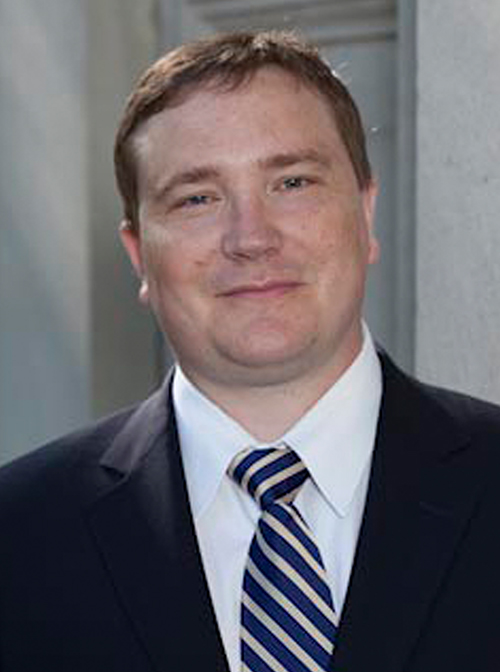 Gregory G. Heuer合作论文数Department of Neurosurgery, Perelman School of Medicine, University of Pennsylvania7
 Hankinson Todd C合作论文数School of Medicine, University of Colorado Anschutz Medical Campus7
 Chevis Shannon合作论文数American Society for Reproductive Medicine7
 Richard G. Ellenbogen合作论文数Department of Radiology, University of Washington;Department of Neurological Surgery, School of Medicine, University of Washington;Department of Global Health, University of Washington7Gross Naina L合作论文数7
 J Gordon Mc Comb合作论文数Keck School of Medicine, University of Southern California7Arnold H. Menezes合作论文数7Mark D. Krieger合作论文数7
 Scellig Stone合作论文数University of Toronto, University Health Network7Daniel J. Guillaume合作论文数7
 Philipp R Aldana合作论文数Department of Neurosurgery, University of Florida College of Medicine, Jacksonville, FL7
 Gregory W. Albert合作论文数College of Medicine, University of Arkansas for Medical Sciences7
 Harold L. Rekate合作论文数St. Joseph’s Hospital and Medical Center, Barrow Neurological Institute7Jeffrey R. Leonard合作论文数7David F. Bauer合作论文数7David R. Peters合作论文数6Patrick C Graupman合作论文数6Curtis A. Dickman合作论文数6




