The Mount Galili Formation in the Afar region, Ethiopia, samples a critical time in hominin evolution, 4.4 to 3.8 Ma, documenting the last appearance of Ardipithecus and the origin of Australopithecus. This period is also important in the evolution of cercopithecids, especially the origin of Theropithecus in general and Theropithecus oswaldi lineage in particular. Galili has provided a total of 655 cercopithecid specimens that include crania, mandibles, isolated teeth and postcrania. All the fossils were recovered from the Lasdanan (5.3-4.43 Ma), Dhidinley (4.43-3.9 Ma) and Shabeley Laag (similar to 3.92-3.8 Ma) Members. Here, we described and analyzed 362 fossils employing both qualitative and quantitative methods. Descriptions of the material were supplemented with dental metrics and cranial shape analysis using three-dimensional geometric morphometrics. Results indicate the presence of at least six cercopithecid taxa: Theropithecus oswaldi serengetensis (n = 28), Theropithecus sp. (n = 2), three non- Theropithecus papionin groups (n = 134) and one colobine-size group (n = 58). The T. o . serengetensis represents the earliest form of the lineage, documented from similar to 3.9 Ma Galili sediments. The three Galili papionins include a smaller taxon, a medium-sized taxon comparable to Pliopapio alemui and a large papionin overlapping in size with Soromandrillus , Gorgopithecus and Dinopithecus . The majority of Galili colobines have closest affinities to Kuseracolobus aramisi and some overlap with other taxa. Papionins dominate the Galili cercopithecid collection, although colobines are still fairly common (approximately 25% of the sample). Thus, Galili sample is like Kanapoi (4.2-4.1 Ma) and Gona (5.2-3.9 Ma) localities but distinct from Aramis, suggesting paleoecological similarity to the former sites. On the other hand, Theropithecus is less abundant at Galili than geologically younger Hadar (3.4-3.2 Ma) and Woranso-Mille (3.8-3.6 Ma) sites. Whether this difference is due to sampling, time or landscape variation requires further investigation.
Paleontological field research conducted during recent decades has led to the discovery of many new hominin fossils that document the remarkable taxonomic diversity and distinctive paleobiology of our earliest ancestors. The anatomical and behavior transition to bipedality by hominins required significant reorganization of the musculoskeletal system that is unique among mammals. Australopithecus africanus was the first truly ancient hominin known when it was discovered in 1924. Australopithecus bahrelghazali is notable for expanding the range of australopiths beyond eastern and South Africa into the paleo-Lake Chad basin in North–Central Africa. The biogeography of the earliest hominins remains poorly known and requires paleontological prospecting in new areas throughout Africa – work that is best identified through the use of remote imaging and geospatial data analysis.
SignificanceHumans have the proportionately smallest male canines among all anthropoids and little canine sexual dimorphism. However, the evolutionary emergence of this defining condition remains unclear because until now we have lacked a reliable method of determining dimorphism in weakly dimorphic fossil species. Using a probability-based method we recently developed, we estimated canine size dimorphism in the ∼4.5 million-year-oldArdipithecus ramidusand found it to be weak and comparable to that of modern humans. Our analysis of >300 fossils spanning 6 million years shows that male canine size reduction occurred early in human evolution, broadly coincident with the adoption of bipedality. This suggests a profound and evolutionarily deep sociobehavioral shift that minimized male–male aggression, most likely mediated by female choice.
Body and canine size dimorphism in fossils inform sociobehavioral hypotheses on human evolution and have been of interest since Darwin's famous reflections on the subject. Here, we assemble a large dataset of fossil canines of the human clade, including all available Ardipithecus ramidus fossils recovered from the Middle Awash and Gona research areas in Ethiopia, and systematically examine canine dimorphism through evolutionary time. In particular, we apply a Bayesian probabilistic method that reduces bias when estimating weak and moderate levels of dimorphism. Our results show that Ar. ramidus canine dimorphism was significantly weaker than in the bonobo, the least dimorphic and behaviorally least aggressive among extant great apes. Average male-to-female size ratios of the canine in Ar. ramidus are estimated as 1.06 and 1.13 in the upper and lower canines, respectively, within modern human population ranges of variation. The slightly greater magnitude of canine size dimorphism in the lower than in the upper canines of Ar. ramidus appears to be shared with early Australopithecus, suggesting that male canine reduction was initially more advanced in the behaviorally important upper canine. The available fossil evidence suggests a drastic size reduction of the male canine prior to Ar. ramidus and the earliest known members of the human clade, with little change in canine dimorphism levels thereafter. This evolutionary pattern indicates a profound behavioral shift associated with comparatively weak levels of male aggression early in human evolution, a pattern that was subsequently shared by Australopithecus and Homo.
Accurate characterization of sexual dimorphism is crucial in evolutionary biology because of its significance in understanding present and past adaptations involving reproductive and resource use strategies of species. However, inferring dimorphism in fossil assemblages is difficult, particularly with relatively low dimorphism. Commonly used methods of estimating dimorphism levels in fossils include the mean method, the binomial dimorphism index, and the coefficient of variation method. These methods have been reported to overestimate low levels of dimorphism, which is problematic when investigating issues such as canine size dimorphism in primates and its relation to reproductive strategies. Here, we introduce the posterior density peak (pdPeak) method that utilizes the Bayesian inference to provide posterior probability densities of dimorphism levels and within-sex variance. The highest posterior density point is termed the pdPeak. We investigated performance of the pdPeak method and made comparisons with the above-mentioned conventional methods via 1) computer-generated samples simulating a range of conditions and 2) application to canine crown-diameter datasets of extant known-sex anthropoids. Results showed that the pdPeak method is capable of unbiased estimates in a broader range of dimorphism levels than the other methods and uniquely provides reliable interval estimates. Although attention is required to its underestimation tendency when some of the distributional assumptions are violated, we demonstrate that the pdPeak method enables a more accurate dimorphism estimate at lower dimorphism levels than previously possible, which is important to illuminating human evolution.
OBJECTIVES To examine the correlation between spheno-occipital synchondrosis fusion stages and the hand-wrist skeletal maturity index. MATERIALS AND METHODS Digital records of 164 individuals (77 males, 87 females) aged 10 to 18 years old were examined. Three-dimensional CBCT scans and hand-wrist two-dimensional radiographs were scored for the spheno-occipital synchondrosis fusion stages and hand-wrist skeletal maturity index, respectively. Statistical analyses were performed for associations using R software with a significance threshold of P< .01. RESULTS A significant positive relationship was demonstrated between spheno-occipital synchondrosis fusion stages and hand-wrist skeletal maturity in both sexes. The Kendall's rank correlation τ between hand-wrist skeletal maturity index and spheno-occipital synchondrosis fusion percentage were high and positive in males and females (r = .74 and r = .71, respectively). CONCLUSIONS The significant, positive relationship between the hand-wrist skeletal maturity index and spheno-occipital synchondrosis fusion stages support the idea of using spheno-occipital synchondrosis fusion as a biological indicator for craniofacial and mandibular growth spurt prediction.
Runners frequently experience pain in the hip, pelvis, and thigh. The differential diagnosis is broad, and pain in these regions often presents a diagnostic and therapeutic challenge. Common conditions include muscle strain, proximal hamstring tendinopathy, greater trochanteric pain syndrome, snapping hip syndrome, iliopsoas bursitis/tendonitis, and femoroacetabular impingement. A detailed understanding of potential etiologies as well as performing a detailed history and physical examination is critical to establish a diagnosis and to initiate appropriate treatment. Physical therapy and running gait analysis with retraining are essential in treating hip, pelvic, and thigh pain in the runner. More high-quality and runner-specific research is needed to determine optimal treatments.
Although stone tools generally co-occur with early members of the genus Homo, they are rarely found in direct association with hominins. We report that both Acheulian and Oldowan artifacts and Homo erectus crania were found in close association at 1.26 million years (Ma) ago at Busidima North (BSN12), and ca. 1.6 to 1.5 Ma ago at Dana Aoule North (DAN5) archaeological sites at Gona, Afar, Ethiopia. The BSN12 partial cranium is robust and large, while the DAN5 cranium is smaller and more gracile, suggesting that H. erectus was probably a sexually dimorphic species. The evidence from Gona shows behavioral diversity and flexibility with a lengthy and concurrent use of both stone technologies by H. erectus, confounding a simple "single species/single technology" view of early Homo.
The Early Pliocene Sagantole Fm. in the Gona Project area, Afar State, Ethiopia, is noted for discoveries of the early hominin Ardipithecus ramidus. A large series of fossil cercopithecid primates dated to between 4.8 and 4.3 Ma has also been collected from these sediments. In this paper, we use qualitative analysis and standard dental and postcranial measures to systematically describe the craniodental remains and tentatively allocate postcrania to taxa where we are able to. We then use these data to compare these specimens to fossil assemblages from contemporary sites, interpret their paleobiology, and discuss implications for the paleoecology of the Gona Sagantole Fm. We recognize three cercopithecid species in the Gona Sagantole Fm. Pliopapio alemui makes up approximately two-thirds of the identifiable specimens; nearly all of the rest are allocated to Kuseracolobus aramisi, and a single molar indicates the presence of a second, somewhat larger but morphologically distinct papionin. Among the Early Pliocene cercopithecids from Gona are also a number of postcranial elements. None of the postcranial remains are directly associated with any of the cranial material. Nonetheless, some of the distal humeri and proximal femora can be tentatively allocated to either Pl. alemui or K. aramisi based on a combination of size, as the latter is approximately 50% larger than the former, and morphology. If these assignments are correct, they suggest K. aramisi was primarily arboreal and similar to most extant colobines, whereas Pl. alemui was more mixed in its substrate use, being more terrestrially adapted than K. aramisi, but less so than extant Papio or Theropithecus. Thus, we interpret the predominance of Pl. alemui over K. aramisi is consistent with a somewhat more open environment at Gona than at Aramis.
Background Several sacroiliac joint (SIJ) provocative tests used to assess posterior pelvic pain involve moving and stressing the hip. It is unknown if there is a subgroup of patients with posterior pelvic pain who have underlying hip deformity that could potentially influence performance and interpretation of these tests. Objective To describe the prevalence of radiographic hip deformity and hip osteoarthritis in a group of adults 40 years old and under who met the clinical diagnostic criteria for treatment of posterior pelvic pain with an image guided intra‐articular SIJ injection. Design Retrospective cohort study. Setting Tertiary university orthopedic department Patients (or Participants) One hundred and forty‐eight patients were evaluated (83% (123/148) female; mean age 31.3 ± 6.2 years). All had completed a trial of comprehensive noninvasive treatment for posterior pelvic pain and had a minimum of three positive SIJ provocative tests on physical examination. Methods Retrospective review identified patients undergoing SIJ injection for pain recommended and performed by seven physiatrists between 2011 and 2017. Hip radiographs were read by a physician with expertise in hip measurements with previously demonstrated excellent intrarater reliability. Main Outcome Measurements Percentage of patients with hip deformity findings. Results No patients meeting the inclusion criteria had significant radiographic hip osteoarthritis (Tonnis ≥ 2 indicating moderate or greater radiographic hip osteoarthritis) and 4/148 (3%) were found to have mild radiographic hip osteoarthritis. Prearthritic hip disorders were identified in 123 (83%, 95% CI: 76, 89%) patients. For those patients with prearthritic hip disorders, measurements consistent with femoroacetabular impingement (FAI) were seen in 61 (41%) patients, acetabular dysplasia in 49 (33%) patients, and acetabular retroversion in 85 (57%) patients. Acetabular retroversion was identified in 43% (crossover sign) and 39% (prominent ischial spine) of patients. Conclusions Approximately 57% of adult patients under the age of 40 years with the clinical symptom complex of SIJ pain were found to have radiographic acetabular retroversion. This is a higher percentage than the 5%‐15% found in asymptomatic people in the current literature. Further study is needed to assess links between hip structure, hip motion, and links to pelvic pain including peri and intra‐articular SIJ pain. Level of Evidence III
Functional analyses of the 4.4 Ma hominin Ardipithecus ramidus postcrania revealed a previously unknown and unpredicted locomotor pattern combining arboreal clambering and a form of terrestrial bipedality. To date, all of the fossil evidence of Ar. ramidus locomotion has been collected from the Aramis area of the Middle Awash Research Project in Ethiopia. Here, we present the results of an analysis of additional early Pliocene Ar. ramidus fossils from the Gona Project study area, Ethiopia, that includes a fragmentary but informative partial skeleton (GWM67/P2) and additional isolated manual remains. While we reinforce the original functional interpretations of Ar. ramidus of having a mixed locomotor adaptation of terrestrial bipedality and arboreal clambering, we broaden our understanding of the nature of its locomotor pattern by documenting better the function of the hip, ankle, and foot. The newly recovered fossils document a greater adaptation to bipedality in the Ar. ramidus ankle and hallux than previously recognized. In addition, a newly discovered scaphoid bone with a fusing os centrale provides further evidence about the nature of hominin hand evolution.
Poster presentado en: 88th Annual Meeting of the American Association of Physical Anthropologists, 27-30 de marzo 2019, Cleveland
Among living mammals, only the African apes and some anteaters adopt knuckle-walking as their primary locomotor behavior. That Pan and Gorilla both knuckle-walk has been cited as evidence of their common ancestry and a primitive condition for a combined Homo, Pan, and Gorilla clade. Recent research on forelimb ontogeny and anatomy, in addition to recently described hominin fossils, indicate that knuckle-walking was independently acquired after divergence of the Pan and Gorilla lineages. Although the large-bodied, largely suspensory orangutan shares some aspects of the African ape bauplan, it does not regularly knuckle-walk when terrestrial. While many anatomical correlates of knuckle-walking have been identified, a functional explanation of this unusual locomotor pattern has yet to be proposed. Here, we argue that it was adopted by African apes as a means of ameliorating the consequences of repetitive impact loadings on the soft and hard tissues of the forelimb by employing isometric and/or eccentric contraction of antebrachial musculature during terrestrial locomotion. Evidence of this adaptation can be found in the differential size and fiber geometry of the forearm musculature, and differences in torso shape between the knuckle-walking and non-knuckle-walking apes (including humans). We also argue that some osteological features of the carpus and metacarpus that have been identified as adaptations to knuckle-walking are consequences of cartilage remodeling during ontogeny rather than traits limiting motion in the hand and wrist. An understanding of the functional basis of knuckle-walking provides an explanation of the locomotor parallelisms in modern Pan and Gorilla. Anat Rec, 301:496-514, 2018. © 2018 Wiley Periodicals, Inc.
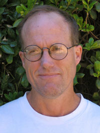 Jay Quade合作论文数Department of Geosciences, College of Sciences, The University of Arizona31
 Sileshi Semaw合作论文数Stone Age Institute and CRAFT Research Center, Indiana University31
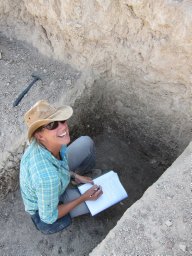 Naomi E. Levin合作论文数Department of Geology and Geophysics, University of Utah22
 C. Owen Lovejoy合作论文数Department of Orthopaedic Surgery, School of Medicine, University of Pittsburgh;Department of Anthropology, College of Arts & Sciences, Kent State University;Cleveland Museum of Natural History19
 Michael J. Rogers合作论文数Department of Anthropology, Southern Connecticut State University12Katherine F. Russell合作论文数10
 Tim D. White合作论文数Department of Integrative Biology, College of Letters & Science, University of California, Berkeley;Human Evolution Research Center, The University of California at Berkeley9
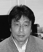 Gen Suwa (諏訪元)合作论文数The University Museum, The University of Tokyo9
 Bruce Latimer合作论文数Department of Anthropology, College of Arts and Sciences, Case Western Reserve University;Department of Anatomy, School of Medicine, Case Western Reserve University;Department of Cognitive Science, College of Arts and Sciences, Case Western Reserve University9William C. Mcintosh合作论文数8
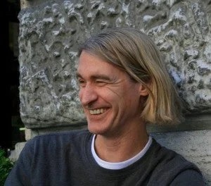 Stephen Frost合作论文数College of Arts and Sciences, University of Oregon8
 Richard S Meindl合作论文数8
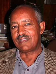 Berhane Asfaw合作论文数Rift Valley Research Service;Centre for Research and Conservation of Cultural Heritage, Ethiopian Ministry of Culture;National Museum of Ethiopia7
 Margaret J. Schoeninger合作论文数Department of Anthropology, University of California, San Diego;Center for Academic Research & Training in Anthropogeny5
 Dale L. Hutchinson合作论文数Institute of Arts and Humanities, The University of North Carolina at Chapel Hill;Department of Anthropology, College of Arts and Sciences, The University of North Carolina at Chapel Hill5
 Darin A. Croft合作论文数5
 Clark Spencer Larsen合作论文数Bioarchaeology Research Laboratory, Department of Anthropology, College of Arts and Sciences, The Ohio State University5
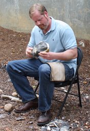 Dietrich Stout合作论文数Institute of Archaeology, University College London5
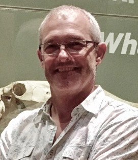 Michael Rogers合作论文数School of Clinical Medicine, University of New South Wales Sydney5
 Robert Butler合作论文数University of Portland5Hailay G. Reda合作论文数4Meghan M Cotter合作论文数4
 Christopher J. Hernandez合作论文数Sibley School of Mechanical and Aerospace Engineering, College of Engineering, Cornell University4Kleinsasser Lynnette合作论文数4
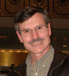 Christopher B. Ruff合作论文数Center for Functional Anatomy & Evolution, School of Medicine, Johns Hopkins University4Melanie Everett合作论文数3Tomohiko Sasaki合作论文数Univ Tokyo, Univ Museum, Bunkyo Ku, Hongo 7-3-1, Tokyo 1130033, Japan3Michael S Selby合作论文数3
 Yonas Beyene合作论文数Department of Archeology and Anthropology, C.R.C.C.H.3Reiko T. Kono合作论文数3Nelia W. Dunbar合作论文数Earth and Environmental Science Department, New Mexico Institute of Mining and Technology3
 Nicholas Toth合作论文数College of Arts and Sciences, Indiana University;Center for Research into the Anthropological Foundations of Technology, Indiana University;Stone Age Institute3
 Lars Werdelin合作论文数Department of Palaeobiology, Naturhistoriska Riksmuseet3
 Kathy D. Schick合作论文数Department of Anthropology, University of California3d stokes合作论文数2df su合作论文数2Charles A Kunos合作论文数2David A. Loomis合作论文数2Brendan J. Goodwine合作论文数2ni chonchuir合作论文数2ME BEDFORD合作论文数2r j fitzgerald合作论文数2
 Anwar Alhazmi合作论文数Dept Prevent Dent Sci, Jazan Univ2Jeanne Gallagher合作论文数School of Biology and Environmental Science/Earth Institute, University College Dublin2Hasen Said合作论文数2
 Evan A. Simons合作论文数Dept Anthropol Courtesy Researcher, Univ Oregon2
 Daniel Simpson合作论文数The Walter and Eliza Hall Institute of Medical Research Parkville Victoria Australia2
 Gary E. Stinchcomb合作论文数Dept Earth & Environm Sci, Murray State Univ2
 Naomi Levin合作论文数Department of Earth and Environmental Sciences, College of Literature, Science, and the Arts, University of Michigan-Ann Arbor2




