Background: Induction of immunogenic cell death (ICD) in tumors can enhance antitumor immunity and modulate immunosuppression in the tumor microenvironment (TME). Objective: In the current study, we investigated the effect of silibinin, a natural compound with anticancer activity, and its polymer-based nanoformulations on the induction of apoptosis and ICD in cancer cells. Methods: Free and nanoparticulate silibinin were evaluated for their growth-inhibitory effects using an MTT assay. Annexin V/PI staining was used to analyze apoptosis. Calreticulin (CRT) expression was measured by flow cytometry. Western blotting was conducted to examine the levels of elf2α, which plays a role in the ICD pathway. The HSP90 and ATP levels were determined using specific detection kits. Results: Compared to the free drug, silibinin-loaded nanocarriers significantly increased the induction of apoptosis and ICD in B16F10 cells. ICD induction was characterized by significantly increased levels of ICD biomarkers, including CRT, HSP90, and ATP. We also observed an increased expression of p-elf-2α/ elf-2α in B16F10 cells treated with silibinin-loaded micelles compared to cells that received free silibinin. Conclusion: Our findings showed that the encapsulation of silibinin in polymeric nanocarriers can potentiate the effects of this drug on the induction of apoptosis and ICD in B16F10 melanoma cells.
Aims The aim of this study was to synthesize two positively charged surfactants Stearoylcholine and Oleoylcholine from choline or vitamin B4, saturated and mono-unsaturated fatty acids to modify solid lipid nanoparticles (SLNs) in order to enhance cancer cell uptake.Methods These surfactants were synthesized by using the esterification method and then SLN formulations of unmodified and modified SLNs containing docetaxel were prepared by emulsification technique. Cytotoxicity of the SLNs was investigated in A549 and MCF7 cancer cells and their cell uptake was assessed by using fluorescent microscope and flow cytometry.Results The results of our study revealed that SLNs pose a mean particle size range of 69-133 nm with spherical morphology. In vitro release study demonstrated a slow-release pattern for all three kinds of DTX-loaded SLNs. Stearoylcholine-containing SLNs showed the highest cytotoxic effect on both cells while cytotoxicity of Oleoylcholine SLNs exhibited a dose-dependent manner which may be due to the effect of saturated and mono-unsaturated parts of surfactants. According to flow cytometric analysis, OC and SC containing SLNs showed the highest uptake into A549 and MCF7 cells, respectively.Conclusion In conclusion, choline-based surfactants could effectively increase the A549 and MCF7 uptake of modified SLNs, which may be due to cationic surface, choline transporters, and special receptors and mediators.
Triple-negative breast cancer (TNBC) is an aggressive subtype of breast cancer. Radiation therapy (RT) is a modality for TNBC management. Radiosensitizers can mitigate the adverse effects of RT. Applying green-synthesized silver nanoparticles (Ag-NPs) from biological sources such as plants is a potential strategy to sensitize cancer cells to radiotherapy due to the low toxicity. Therefore, identifying novel natural sources for synthesizing stable and broadly applicable green-Ag-NPs has gained more attention in cancer therapy. In the present study, we synthesized green- Ag-NPs from pumpkin peel extract and elucidated the impact of green-synthesized Ag-NPs as a radiosensitizer in MDA-MB 231 cells (a model of TNBC). The prepared Ag-NPs had a spherical shape with an average size of 81 nm and a zeta potential of − 9.96 mV. Combination of green-synthesized Ag-NPs with RT exhibited synergistic anticancer effects with an optimum combination index (CI) of 0.49 in MDA-MB-231 cells. Green-synthesized Ag-NPs synergistically potentiated RT-induced apoptosis in MDA-MB-231 cells compared to the corresponding monotherapies. Morphological features of apoptosis were further confirmed by the DAPI–TUNEL staining assay. HIF-1α expression was decreased in cells subjected to combination therapy. Bax and p53 expression increased, whereas Bcl-2 genes decreased. Combination therapy significantly increased the protein level of PERK and CHOP while decreasing cyclin D1 and p-ERK/total ERK levels compared to monotherapies. These findings indicate the potential effect of green-synthesized Ag-NPs as a radiosensitizer for TNBC treatment.
Chrysin is a natural flavonoid with anti-cancer effects. Despite its beneficial effects, little information is available regarding its immunogenic cell death (ICD) properties. In this work, we hypothesized that chrysin can potentiate radiotherapy(RT)-induced immunogenicity in melanoma cell line (B16-F10). We examined the effects of chrysin alone and in combination with radiation on ICD induction in B16-F10 cells. Cell viability was assessed using an MTT assay. Cell apoptosis and calreticulin (CRT) exposure were determined using flow cytometry. Western blotting and ELISA assay were employed to examine changes in protein expression. Combination therapy exhibited a synergistic effect, with an optimum combination index of 0.66. The synergistic anti-cancer effect correlated with increased cell apoptosis in cancer cells. Compared to the untreated control, chrysin alone and in combination with RT induced higher levels of DAMPs, such as CRT, HSP70, HMGB1, and ATP. The protein expression of p-STAT3/STAT3 and PD-L1 was reduced in B16-F10 cells exposed to chrysin alone and in combination with RT. Conditioned media from B16-F10 cells exposed to mono-and combination treatments elicited IL-12 secretion in dendritic cells (DCs), inducing a Th1 response. Our findings revealed that chrysin could induce ICD and intensify the RT-induced immunogenicity.
Hematopoietic stem cells (HSCs) are tightly regulated by specific microenvironments called niches to produce an appropriate number of mature blood cell types. Self-renewal and differentiation are two hallmarks of hematopoietic stem and progenitor cells, and their balance is critical for proper functioning of blood and immune cells throughout life. In addition to cell-intrinsic regulation, extrinsic cues within the bone marrow niche and systemic factors also affect the fate of HSCs. Despite this, many paracrine and endocrine factors that influence the function of hematopoietic cells remain unknown. In hematological malignancies, malignant cells remodel their niche into a permissive environment to enhance the survival of leukemic cells. These events are accompanied by loss of normal hematopoiesis. It is well known that extracellular vehicles (EVs) mediate intracellular interactions under physiological and pathological conditions. In other words, EVs transfer biological information to surrounding cells and contribute not only to physiological functions but also to the pathogenesis of some diseases, such as cancers. Therefore, a better understanding of cell-to-cell interactions may lead to identification of potential therapeutic targets. Recent reports have suggested that EVs are evolutionarily conserved constitutive mediators that regulate hematopoiesis. Here, we focus on the emerging roles of EVs in normal and pathological conditions, particularly in hematological malignancies. Owing to the high abundance of EVs in biological fluids, their potential use as biomarkers and therapeutic tools is discussed.
BackgroundThe discovery of novel cancer therapeutic strategies leads to the development of nanotechnology-based methods for cancer treatment. Silver nanoparticles (Ag-NPs) have garnered considerable interest owing to their size, shape, and capacity to modify chemical, optical, and photonic properties. This study aimed to investigate the impact of Ag-NPs on inducing of apoptosis in MDA-MB 231 cells by examining specific signaling pathways.Materials and methodsThe cytotoxicity of Ag-NPs was determined using an MTT assay in MDA-MB 231 cells. The apoptotic effects were assessed using the Annexin-V/PI assay. Real-time PCR and western blotting were conducted to analyze the expression of apoptosis-related genes and proteins, respectively. Levels of ERK1/2 and cyclin D1 were measured using ELISA. Cell cycle assay was determined by flow cytometry. Cell migration was evaluated by scratch assay.ResultsThe results revealed that Ag-NPs triggered apoptosis and cell cycle arrest in MDA-MB 231 cells. The expression level of Bax (pro-apoptotic gene) was increased, while Bcl-2 (anti-apoptotic gene) expression was decreased. Increased apoptosis was correlated with increased levels of p53 and PTEN. Additionally, notable alterations were observed in protein expression related to the Janus kinase/Signal transducers (JAK/STAT) pathway, including p-AKT. Additionally, reduced expression of h-TERT was observed following exposure to Ag-NPs. ELISA results demonstrated a significant reduction in p-ERK/Total ERK and cyclin D1 levels in Ag-NPs-exposed MDA-MB 231 cells. Western blotting analysis also confirmed the reduction of p-ERK/Total ERK and cyclin D1. Decreased level of cyclin D is associated with suppression of cell cycle progression. The migratory ability of MDA-MB-231 cells was reduced upon treatment with Ag-NPs.ConclusionsOur findings revealed that Ag-NPs influenced the proliferation, apoptosis, cell cycle, and migration in MDA-MB 231 cells, possibly by modulating protein expression of the AKT/ERK/Cyclin D1 axis.
Introduction: Multiple sclerosis (MS) is a chronic neuroinflammatory diseases characterized by demyelination of the nerve fibers. Immunogenic cell death (ICD) is a process, during which damaged and stressed cells release danger-associated molecular patterns (DAMPs) activating immune responses. This study aimed to elucidate the induction of ICD in MS diseases. Methods: To achieve this goal, the level of DAMPs including Annexin A1 (ANXA1), calreticulin and HMGB1 was measured in the cerebrospinal fluid (CSF) of a secondary progressive multiple sclerosis (SPMS) patient in comparison to control group. Results: Results showed significant upregulation (more than two-fold) of ANXA1, calreticulin (CRT) and HMGB1 in the CSF of the patient. Conclusion: Although further studies are suggested in this regard, this data could imply induction of ICD in MS. The proposed ICD might trigger immune response against neural cells resulting in neuroinflammation and demyelination in CNS in MS. Our observation could suggest inclusion of ICD interfering treatments in routine MS therapy.
Chrysin, a natural phytochemical compound found in various plant sources, possesses diverse pharmacological benefits, including anticancer, antioxidant, antidiabetic, neuroprotective, cardioprotective, hepatoprotective, immunoregulatory, and anti-inflammatory properties. Despite its well-documented biological activities, chrysin's low water solubility and bioavailability hinder its clinical development. This review explores the application of nanocarriers as a strategic approach to overcome these challenges and enhance the delivery of chrysin. Nanocarriers, including polymer-based nanoparticles (NPs), lipid-based NPs, and inorganic nanocarriers, have shown promise in improving the solubility, bioavailability, and tumor-targeted delivery of chrysin. The paper discusses chrysin's anticancer effects on different types of human cancers, elucidating its impact on crucial signaling pathways involved in tumorigenesis. The review categorizes and analyzes various nanocarriers, providing insights into their structural properties and drug release profiles. Among the nanocarriers, polymer-based NPs, especially those utilizing PLGA, emerge as promising strategies for chrysin encapsulation, demonstrating improvements in drug release, stability, and bioavailability. Lipid-based NPs and inorganic nanocarriers also exhibit potential in enhancing chrysin delivery. The comprehensive insights provided contribute to a deeper understanding of chrysin's pharmacological properties and its potential clinical applications, offering valuable perspectives for future research and translation into clinical settings. The review underscores the importance of selecting suitable structures for chrysin encapsulation to enhance its physicochemical properties and anticancer effects, paving the way for innovative nanomedicine approaches in cancer therapy.
A growing body of evidence indicates that the anticancer effect of the immune system can be activated by the immunogenic modulation of dying cancer cells. Cancer cell death, as a result of the activation of an immunomodulatory response, is called immunogenic cell death (ICD). This regulated cell death occurs because of increased immunogenicity of cancer cells undergoing ICD. ICD plays a crucial role in stimulating immune system activity in cancer therapy. ICD can therefore be an innovative route to improve anticancer immune responses associated with releasing damage-associated molecular patterns (DAMPs). Several conventional and chemotherapeutics, as well as preclinically investigated compounds from natural sources, possess immunostimulatory properties by ICD induction. Natural compounds have gained much interest in cancer therapy owing to their low toxicity, low cost, and inhibiting cancer cells by interfering with different mechanisms, which are critical in cancer progression. Therefore, identifying natural compounds with ICD-inducing potency presents agents with promising potential in cancer immunotherapy. Naturally derived compounds are believed to act as immunoadjuvants because they elicit cancer stress responses and DAMPs. Acute exposure to DAMP molecules can activate antigen-presenting cells (APCs), such as dendritic cells (DCs), which leads to downstream events by cytotoxic T lymphocytes (CTLs) and natural killer cells (NKs). Natural compounds as inducers of ICD may be an interesting approach to ICD induction; however, parameters that determine whether a compound can be used as an ICD inducer should be elucidated. Here, we aimed to discuss the impact of multiple ICD inducers, mainly focusing on natural agents, including plant-derived, marine molecules, and bacterial-based compounds, on the release of DAMP molecules and the activation of the corresponding signaling cascades triggering immune responses. In addition, the potential of synthetic agents for triggering ICD is also discussed.
Introduction: Silibinin is a natural flavonoid compound known to induce apoptosis in cancer cells. Despite silibinin's safety and efficacy as an anticancer drug, its effects on inducing immunogenic cell death (ICD) are largely unknown. Herein, we have evaluated the stimulating effects of silibinin on ICD in cancer cells treated with silibinin alone or in combination with chemotherapy. Methods: The anticancer effect of silibinin, alone or in combination with doxorubicin or oxaliplatin (OXP), was assessed using the MTT assay. Compusyn software was used to analyze the combination therapy data. Western blotting was conducted to examine the level of STAT3 activity. Flow cytometry was used to analyze calreticulin (CRT) and apoptosis. The heat shock protein (HSP70), high mobility group box protein1 (HMGB1), and IL-12 levels were assessed by ELISA. Results: Compared to the negative control groups, silibinin induced ICD in CT26 and B16F10 cells and significantly enhanced the induction of this type of cell death by doxorubicin, and these changes were allied with substantial increases in the level of damage-associated molecular patterns (DAMPs) including CRT, HSP70, and HMGB1. Furthermore, conditioned media from cancer cells exposed to silibinin and doxorubicin was found to stimulate IL-12 secretion in dendritic cells (DCs), suggesting the link of this treatment with the induction of Th1 response. Silibinin did not augment the ICD response induced by OXP. Conclusion: Our findings showed that silibinin can induce ICD and it potentiates the induction of this type of cell death induced by chemotherapy in cancer cells.
BACKGROUND:Prostate cancer is among the most common cancers in men with an increasing incidence rate. Radiation therapy (RT) is a therapeutic strategy for the management of prostate cancer after surgery; nonetheless, it has different side effects on neighboring healthy cells/tissues. Moreover, radioresistance has been an increasing phenomenon in the recent years. Therefore, there is an urgent need for the introduction of a safe and effective radiosensitizing agent. Accordingly, the recent trend in the development of novel drugs is accompanied by a push toward natural compounds. Our study evaluated the effects of betanin combined with RT as a potential radiosensitizing agent in the PC-3 cell line.METHODS AND RESULTS:MTT assay was utilized to determine the growth inhibitory impact of betanin. The possible synergistic effect was evaluated with CompuSyn software upon Trypan blue exclusion test. Apoptosis-related gene expression was evaluated via Real-time PCR and the protein expression of P21 was determined using western blotting. A synergistic anticancer effect with an optimal combination index of 0.61 was achieved by treating PC-3 cells with betanin and RT. The results pointed out that betanin synergistically triggered RT-mediated apoptosis and cell cycle arrest through modulating gene and protein expression in comparison with each of the monotherapies.CONCLUSION:These findings shed light on the synergistic antitumor effect of betanin and RT in prostate cancer, indicating the potential use of betanin as a radiosensitizer agent.
Metabolic reprogramming is a common hallmark of cancer cells. Cancer cells exhibit metabolic flexibility to maintain high proliferation and survival rates. In other words, adaptation of cellular demand is essential for tumorigenesis, since a diverse supply of nutrients is required to accommodate tumor growth and progression. Diversity of carbon substrates fueling cancer cells indicate metabolic heterogeneity, even in tumors sharing the same clinical diagnosis. In addition to the alteration of glucose and amino acid metabolism in cancer cells, there is evidence that cancer cells can alter lipid metabolism. Some tumors rely on fatty acid oxidation (FAO) as the primary energy source; hence, cancer cells overexpress the enzymes involved in FAO. Carnitine is an essential cofactor in the lipid metabolic pathways. It is crucial in facilitating the transport of long-chain fatty acids into the mitochondria for β-oxidation. This role and others played by carnitine, especially its antioxidant function in cellular processes, emphasize the fine regulation of carnitine traffic within tissues and subcellular compartments. The biological activity of carnitine is orchestrated by specific membrane transporters that mediate the transfer of carnitine and its derivatives across the cell membrane. The concerted function of carnitine transporters creates a collaborative network that is relevant to metabolic reprogramming in cancer cells. Here, the molecular mechanisms relevant to the role and expression of carnitine transporters are discussed, providing insights into cancer treatment.
Triple-negative breast cancer (TNBC) is the most aggressive form of breast cancer, accounting for 20
Induction of immunogenic cell death (ICD) is a promising strategy for cancer immunotherapy. Chrysin, which has potential anticancer effects, faces limitations in clinical applications due to its poor water solubility. This study aimed to formulate chrysin with PEG-poly(α-benzylcarboxylate-ε-caprolactone) (PBCL) nanoparticles (NPs) and assess their anticancer and ICD-inducing potency in melanoma cells, comparing with free chrysin. The co-solvent evaporation method was employed to develop chrysin-loaded NPs. UV spectroscopy, dynamic light scattering, and the dialysis bag method were used to evaluate the encapsulation efficiency (EE), particle size, polydispersity index (PDI), and drug release profile, respectively. The anticancer effects of the drugs were assessed using the MTT and trypan blue exclusion assays. Flow cytometry was employed to evaluate apoptosis and calreticulin (CRT) expression. ELISA and western blotting were used to detect heat shock protein 90 (HSP90), Annexin A1, GRP78 (Glucose-related protein78), and activated protein kinase R-like endoplasmic reticulum kinase (p-PERK). Chrysin-loaded PEG-PBCL NPs (chrysin-PEG-PBCL) showed an EE of 97 ± 1
Cisplatin is the most often used chemotherapy in the treatment of ovarian cancer (OC), however long-term usage leads to drug resistance and treatment failure. Silibinin is a sparingly water-soluble natural compound with well-known anticancer effects. The use of lipid-based delivery systems is a potential approach for enhancing silibinin's water solubility. In this study, nanostructured lipid carriers (NLCs) containing silibinin were prepared and their inhibitory effects were tested in combination with cisplatin against sensitive/resistant A2780 OC cells. Silibinin-loaded NLCs (silibinin-NLCs) were prepared by the hot homogenization method, and their size, shape, zeta potential (ZP), and encapsulation efficiency (EE), as well as their inhibitory effects, were examined in combination with cisplatin against sensitive/resistant A2780 OC cells. Formulation of silibinin-NLCs using cocoa butter led to spherical-shaped NLCs with a size of 95 nm and EE of 98%. The ZP and the dispersion index of the silibinin-NLCs were -27.12 ± 0.13 mv and 0.12 ± 0.04, respectively. The release kinetics of silibinin-NLCs was best fitted with the zero-order model. The combination of cisplatin and silibinin-NLCs sensitized the cisplatin-resistant A2780 OC cells and exhibited a more synergistic inhibitory effect on A2780 cells as compared with the combination of cisplatin and plain silibinin. The optimized silibinin-NLCs can be considered a suitable drug delivery system for the inhibition of cisplatin-resistant OC cells.
The highly pathogenic, novel coronavirus disease (COVID-19) outbreak has emerged as a once-in-a-century pandemic with poor consequences, urgently calling for new therapeutics, cures, and supportive interventions. It has already affected over 250 million people worldwide; thereby, there is a need for novel therapies to alleviate the related complications. There is a paradigm shift in developing drugs and clinical practices to combat COVID-19. Several clinical trials have been performed or are testing diverse pharmacological interventions to alleviate viral load and complications such as cytokine release storm (CRS). Kinase-inhibitors have appeared as potential antiviral agents for COVID-19 patients due to their efficacy against CRS. Combination of kinase inhibitors with other therapies can achieve more efficacy against COVID-19. Based on the pre-clinical trials, kinase inhibitors such as Janus kinase-signal transducer and activator of transcription (JAK/STAT) inhibitors, Brutton's tyrosin kinase (BTK) inhibitors, p38 mitogen-activated protein kinases (p38 MAPK) inhibitors, Glycogen synthase kinase 3 (GSK-3) inhibitors can be a promising strategy against COVID-19. Kinase inhibitors possess crucial pharmacological properties for a successful re-purposing in terms of dual anti-inflammatory and anti-viral effects. This review will address the current clinical evidence and the newest discovery regarding the application of kinase inhibitors in COVID-19. An outlook on ongoing clinical trials (clinicaltrials.gov) and unpublished data is also presented here. Besides, Kinase inhibitors' function on COVID-19-mediated CRS is discussed.
Background. STAT3 is an oncogenic signaling pathway found constitutively active in many types of human malignancies and plays a key role in cancer progression. Stattic is a small molecule, which selectively inhibits the SH2 domain of STAT3. In most studies, stattic has been proposed as a promising strategy STAT3 inhibition in cancer cells harboring constitutively active STAT3. However, the lack of proper formulation due to the poor water solubility and low bioavailability of stattic is a major limitation for its usage in the clinic. This project aimed to develop poly(ethylene glycol)-block-poly(caprolactone) (PEG-b-PCL)-based polymeric micelles loaded with stattic and evaluate drug encapsulation efficiency and release in the developed formulations. Methods. In this experimental study, to prepare stattic loaded micellar formulations, the co-solvent evaporation method was used. The mean diameter and polydispersity index (PDI) of micelles were defined by the light scattering method. Encapsulated drug levels were measured using high-performance liquid chromatography (HPLC). Data were analyzed using GraphPad Prism software through one-way ANOVA. Results. Stattic was loaded in the polymeric micelles with encapsulation efficiency ranging from 40% to 73%. Drug loaded micelles were measured between 90 to 130 nm in size. PDI was obtained 0.3-1, and encapsulation of stattic in Polyethylene glycol-block-poly (α-benzyl carboxylate ε-caprolactone (PEG-b-PBCL) micellar formulation resulted in a more than 6-fold increase in the water solubility of stattic (0.36 vs. 0.06 mg/mL). Regarding high encapsulation efficiency, two micellar formulations were selected for further analysis in that both of them released 70-80% of the drug within the first hour, indicating burst release of the drug. Conclusion. These findings show that PEG-b-PBCL copolymers can be a suitable vehicle for the solubilization of stattic.
Background: STAT3 is an oncogenic signaling pathway found constitutively active in many types of human malignancies and plays a key role in cancer progression. Stattic is a small molecule, which selectively inhibits SH2 domain of STAT3. In most of the studies, stattic has been proposed as a promising strategy for inhibition of STAT3 in cancer cells harboring constitutively active STAT3. However, lack of proper formulation due to the poor water solubility and low bioavailability of stattic is a major limitation for its usage in clinic. The aim of this project was to develop poly(ethylene glycole)-block-poly(caprolactone) (PEG-b-PCL)-based polymeric micelles loaded with stattic and evaluate drug encapsulation efficiency and release in the developed formulations. Methods: In this experimental study, to prepare stattic loaded micellar formulations, co-solvent evaporation method was used. Mean diameter and polydispersity index (PDI) of micelles were defined by light scattering method. Encapsulated drug levels were measured using high performance liquid chromatography (HPLC). Data were analyzed using Graph pad prism software through one-way ANOVA analysis of variance. Results: Stattic was loaded in the polymeric micelles with encapsulation efficiency ranging from 40 to 73%. Drug loaded micelles were measured between 90 to 130 nm in size. PDI was obtained 0.3-1 and encapsulation of stattic in Polyethylene glycol-block-poly(α-benzyl carboxylate ε-caprolactone(PEG-b-PBCL) micellar formulation resulted in more than 6-fold increase in the water solubility of stattic (0.36 vs. 0.06 mg/mL). Respecting to high encapsulation efficiency, two micellar formulations were selected for further analysis that both of them released 70-80% of drug within the first hour, indicated burst release of drug. Conclusion: These findings show that PEG-b-PBCL copolymers can be a suitable vehicle for solubilization of stattic.
Background: Ecballium elaterium is one of the Cucurbitaceae plants distributed in many regions of the world. This unique species considered as a source of valuable herbal remedies such as anticancer, anti-inflammatory and antimicrobial in traditional medicine. Objectives: In the current research, cytotoxic activity of different extracts of the aerial parts and rhizomes of E. elaterium was evaluated on cancerous and non-cancerous cells which have not been assessed up to now. Furthermore, chemical compositions of potent samples were determined by GC-MS analysis for the first time. Methods: The air-dried, powdered aerial parts and rhizome of the E. elaterium were Soxhlet-extracted, successively, with different solvents (n-hexane, dichloromethane, and methanol) and then the dried extracts subjected to cytotoxic assay. Subsequently, their anti-proliferative activity was assessed by a modified MTT assay. In this study, 4 cancerous cell lines were used, all of the cell lines were incubated in humid 37 degrees C, 5% CO2 and fed by RPMI and FBS. The cells were exposed to different extracts at different concentrations in 48 h. Their growth inhibition was defined as IC50 (mu g.mL(-1)). For further investigations, potent extracts were subjected to VLC method for obtaining different fractions. Subsequently, the activity of all fractions was investigated and their phytochemical profiles were determined by preliminary phytochemical and GC-MS methods. Results: Our findings illustrated, among the extracts, n-hexane extract of the aerial parts and its 100% fraction showed potent growth inhibitory effects on MCF7 cell line with IC50 = 264.3 +/- 5.2 and 351.2 +/- 5.5 mu g.mL(-1), respectively. It is worth to mention that; potent samples act their inhibitory effect on cancerous with minimum effect on non-cancerous cells. Further chemical investigations indicated that steroids, hydrocarbons and oxygenated monoterpenes are responsible for showing anti-proliferative effects. Conclusions: Overall, the results were showed n-hexane extract and its 100% fraction for having potent ingredients inhibit the cancerous cells and had minimum toxicity on the normal cell line.
Despite significant advances in cancer therapy, chemotherapeutic agents are still the main types of drugs used to treat cancer patients. 5-Fluorouracil (5-FU) is the first-line treatment in several types of human cancers, however, nonspecific function, low plasma half-life, and high doses toxicity are the important barrier to achieve efficient response in cancer patients. The use of polymeric nanoparticles (NPs) for tumor targeted delivery of 5-FU in combination with other potent anticancer agent is considered an important strategy to enhance the therapeutic efficacy of 5-FU. In this study, we proposed to use PLGA-PEG-PLGA NPs to co-encapsulate 5-FU and Chrysin, a natural compound known to enhance the therapeutic efficacy of chemotherapy. NPs were prepared by double emulsion method and characterized for size and drug encapsulation efficacy. The cell growth inhibitory effect of prepared NPs was assessed by MTT assay in HT29 human colon cancer cell line. The analysis of NPs by dynamic light scattering showed that the developed NPs have average size of 40 nm. The encapsulation efficiency of NPs was 81.3% and 97.5% for 5-FU and Chrysin, respectively. Furthermore, the NPs showed a remarkable uptake in HT29 cells. NPs loaded with both 5-FU and Chrysin (5-FU@Chrysin loaded NPs) were found to have significantly higher growth inhibitory effects compared with NPs loaded with each drug alone in HT29 cell line. The synergistic anticancer effects of 5-FU and Chrysin loaded in NPs were confirmed with the combination index (CI) being 0.35. CI for combination therapy with free 5-FU and Chrysin was found to be 0.73, indicating weaker synergistic anticancer effects of these two drugs in free forms as compared with 5-FU@Chrysin loaded NPs. These finding indicates that co-delivery of 5-FU and Chrysin with PLGA-PEG-PLGA copolymer can be used to improve the therapeutic and functional delivery efficacy of 5-FU and Chrysin in cancer.
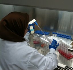 Ommoleila Molavi合作论文数Faculty of Pharmacy, Tabriz University of Medical Sciences15
 Montazer-Saheb Soheila合作论文数12
 Farahzadi Raheleh合作论文数5
 Mohammad Saeid Hejazi合作论文数3Afsaneh Lavasanifar合作论文数3Elnaz Mehdizadeh Aghdam合作论文数Faculty of Pharmacy, Tabriz University of Medical Sciences3
 Ali Shayanfar合作论文数Drug Applied Research Center and Faculty of Pharmacy, Tabriz University of Medical Sciences3
 Ezzatollah Fathi合作论文数Faculty of Veterinary Medicine, University of Tehran3Alireza Khodaei Ardakan合作论文数2Maryam Saadat合作论文数2
 Elham Ahmadian合作论文数Kidney Research Center, Tabriz University of Medical Sciences2
 Zakeri-Milani Parvin合作论文数Fac Pharm, Tabriz Univ Med Sci2
 Mohammad Reza Mofid合作论文数Fachbereich Chemie/Biochemie, Philipps-Universität Marburg2
 Vaez Haleh合作论文数Faculty of Pharmacy, Tabriz University of Medical Sciences2
 Valiollah Babaeipour合作论文数Department of Biochemical Engineering, Research Center of Biotechnology2
 Hadi Valizadeh合作论文数Faculty of Pharmacy and Drug Applied Research Center, Tabriz University of Medical Sciences2
 Javid Shahbazi Mojarrad合作论文数Department of Medicinal Chemistry, Tabriz University of Medical Sciences2
 Soodabeh Davaran合作论文数Faculty of Pharmacy, Tabriz University of Medical Sciences2Mohammad Mehdi Namvaran合作论文数1Mohammad Saied Hejazi合作论文数1Samira Khaledi合作论文数1Leila Haddad合作论文数1
 Behzad Baradaran合作论文数Immunology Research Center, Tabriz University of Medical Sciences1
 Muhammad Sarfraz合作论文数1
 Farshid Torkzaban合作论文数1
 Abdolhassan Kazemi合作论文数1Farshid Asiaee合作论文数1
 Elahe Pishgahzadeh合作论文数1Zahra Malekinejad合作论文数1
 Ashkan H Rad合作论文数1Ayse Baran合作论文数1
 Farnaz Khaleseh合作论文数Sch Pharm, Kermanshah Univ Med Sci1
 Yasin Bagheri合作论文数Kidney Research Center, Tabriz University of Medical Sciences1
 Aygun Nasibova合作论文数Department of Biophysics and Biochemistry, Baku State University1
 Shahnaz Sabetkam合作论文数1
 Mohmmad Mahmoudian合作论文数Faculty of Pharmacy, Tabriz University of Medical Sciences1
 Mehmet fırat Baran合作论文数Batman University, Batman University1
 Babak Elyasi Far合作论文数Dezful University of Medical Sciences1
 Amir Baghbanzadeh合作论文数Immunology Research Center, Tabriz University of Medical Sciences1
 Javad Ranjbari合作论文数1
 Hadi Hashemzadeh合作论文数Faculty of Bioscience, Tarbiat Modares University1
 Sajjad Janfaza合作论文数School of Engineering, University of British Columbia1
 Tooba Gholikhani合作论文数Student Research Committee, Tabriz University of Medical Sciences1Behnaz Valipour合作论文数Stem Cell Research Centre, Tabriz University of Medical Sciences;Student Research Committee, Tabriz University of Medical Sciences;Faculty of Medicine, Tabriz University of Medical Sciences;Student Research Committee, Tabriz University of Medical Sciences1
 Azita Dilmaghani合作论文数UR1290-BIOGER, INRA1
 Maleki-Dizaji Nasrin合作论文数Dept Pharmacol, Tabriz Univ Med Sci1
 Ardalan Mohammad Reza合作论文数Kidney Res Ctr, Tabriz Univ Med Sci1
 Mehran Mesgari Abbasi合作论文数Drug Applied Research Center, Tabriz University of Medical Sciences1Hoseini Vahid合作论文数1




