Nucleus segmentation is usually the first step in pathological image analysis tasks. Generalizable nucleus segmentation refers to the problem of training a segmentation model that is robust to domain gaps between the source and target domains. The domain gaps are usually believed to be caused by the varied image acquisition conditions, e.g., different scanners, tissues, or staining protocols. In this paper, we argue that domain gaps can also be caused by different foreground (nucleus)-background ratios, as this ratio significantly affects feature statistics that are critical to normalization layers. We propose a Distribution-Aware Re-Coloring (DARC) model that handles the above challenges from two perspectives. First, we introduce a re-coloring method that relieves dramatic image color variations between different domains. Second, we propose a new instance normalization method that is robust to the variation in foreground-background ratios. We evaluate the proposed methods on two H &E stained image datasets, named CoNSeP and CPM17, and two IHC stained image datasets, called DeepLIIF and BC-DeepLIIF. Extensive experimental results justify the effectiveness of our proposed DARC model. Codes are available at https://github.com/csccsccsccsc/DARC .
Nucleus segmentation is a challenging task due to the crowded distribution and blurry boundaries of nuclei. Recent approaches represent nuclei by means of polygons to differentiate between touching and overlapping nuclei and have accordingly achieved promising performance. Each polygon is represented by a set of centroid-to-boundary distances, which are in turn predicted by features of the centroid pixel for a single nucleus. However, using the centroid pixel alone does not provide sufficient contextual information for robust prediction and thus degrades the segmentation accuracy. To handle this problem, we propose a Context-aware Polygon Proposal Network (CPP-Net) for nucleus segmentation. First, we sample a point set rather than one single pixel within each cell for distance prediction. This strategy substantially enhances contextual information and thereby improves the robustness of the prediction. Second, we propose a Confidence-based Weighting Module, which adaptively fuses the predictions from the sampled point set. Third, we introduce a novel Shape-Aware Perceptual (SAP) loss that constrains the shape of the predicted polygons. Here, the SAP loss is based on an additional network that is pre-trained by means of mapping the centroid probability map and the pixel-to-boundary distance maps to a different nucleus representation. Extensive experiments justify the effectiveness of each component in the proposed CPP-Net. Finally, CPP-Net is found to achieve state-of-the-art performance on three publicly available databases, namely DSB2018, BBBC06, and PanNuke. Code of this paper is available at https://github.com/csccsccsccsc/cpp-net.
Nucleus segmentation is an important task in medical image analysis. However, machine learning models cannot perform well because there are large amount of clusters of crowded nuclei. To handle this problem, existing approaches typically resort to sophisticated hand-crafted post-processing strategies; therefore, they are vulnerable to the variation of post-processing hyper-parameters. Accordingly, in this paper, we devise a Boundary-assisted Region Proposal Network (BRP-Net) that achieves robust instance-level nucleus segmentation. First, we propose a novel Task-aware Feature Encoding (TAFE) network that efficiently extracts respective high-quality features for semantic segmentation and instance boundary detection tasks. This is achieved by carefully considering the correlation and differences between the two tasks. Second, coarse nucleus proposals are generated based on the predictions of the above two tasks. Third, these proposals are fed into instance segmentation networks for more accurate prediction. Experimental results demonstrate that the performance of BRP-Net is robust to the variation of post-processing hyper-parameters. Furthermore, BRP-Net achieves state-of-the-art performances on both the Kumar and CPM17 datasets. The code of BRP-Net will be released at https://github.com/csccsccsccsc/brpnet.
Brain tumor segmentation from Magnetic Resonance Imaging scans is vital for both the diagnosis and treatment of brain cancers. It is widely accepted that accurate segmentation depends on multi-level information. However, exiting deep architectures for brain tumor segmentation fail to explicitly encourage the models to learn high-quality hierarchical features. In this paper, we propose a series of approaches to enhance the quality of the learnt hierarchical features. Our contributions incorporate four aspects. First, we extend the popular DeepMedic model to Multi-Level DeepMedic to make use of multi-level information for more accurate segmentation. Second, we propose a novel dual-force training scheme to promote the quality of multi-level features learnt from deep models. It is a general training scheme and can be applied to many exiting architectures, e.g., DeepMedic and U-Net. Third, we design a label distribution-based loss function as an auxiliary classifier to encourage the high-level layers of deep models to learn more abstract information. Finally, we propose a novel Multi-Layer Perceptron-based post-processing approach to refine the prediction results of deep models. Extensive experiments are conducted on two most recent brain tumor segmentation datasets, i.e., BRATS 2017 and BRATS 2015 datasets. Results on the two databases indicate that the proposed approaches consistently promote the segmentation performance of the two popular deep models.
Thanks to the powerful representation learning ability, convolutional neural network has been an effective tool for the brain tumor segmentation task. In this work, we design multiple deep architectures of varied structures to learning contextual and attentive information, then ensemble the predictions of these models to obtain more robust segmentation results. In this way, the risk of overfitting in segmentation is reduced. Experimental results on validation dataset of BraTS 2018 challenge demonstrate that the proposed method can achieve good performance with average Dice scores of 0.8136, 0.9095 and 0.8651 for enhancing tumor, whole tumor and tumor core, respectively. The corresponding scores for BraTS 2018 testing set are 0.7775, 0.8842 and 0.7960, respectively, winning the third position in the BraTS 2018 competition among 64 participating teams.
Gliomas are the most common primary brain malignancies, with different degrees of aggressiveness, variable prognosis and various heterogeneous histologic sub-regions, i.e., peritumoral edematous/invaded tissue, necrotic core, active and non-enhancing core. This intrinsic heterogeneity is also portrayed in their radio-phenotype, as their sub-regions are depicted by varying intensity profiles disseminated across multi-parametric magnetic resonance imaging (mpMRI) scans, reflecting varying biological properties. Their heterogeneous shape, extent, and location are some of the factors that make these tumors difficult to resect, and in some cases inoperable. The amount of resected tumor is a factor also considered in longitudinal scans, when evaluating the apparent tumor for potential diagnosis of progression. Furthermore, there is mounting evidence that accurate segmentation of the various tumor sub-regions can offer the basis for quantitative image analysis towards prediction of patient overall survival. This study assesses the state-of-the-art machine learning (ML) methods used for brain tumor image analysis in mpMRI scans, during the last seven instances of the International Brain Tumor Segmentation (BraTS) challenge, i.e., 2012-2018. Specifically, we focus on i) evaluating segmentations of the various glioma sub-regions in pre-operative mpMRI scans, ii) assessing potential tumor progression by virtue of longitudinal growth of tumor sub-regions, beyond use of the RECIST/RANO criteria, and iii) predicting the overall survival from pre-operative mpMRI scans of patients that underwent gross total resection. Finally, we investigate the challenge of identifying the best ML algorithms for each of these tasks, considering that apart from being diverse on each instance of the challenge, the multi-institutional mpMRI BraTS dataset has also been a continuously evolving/growing dataset.
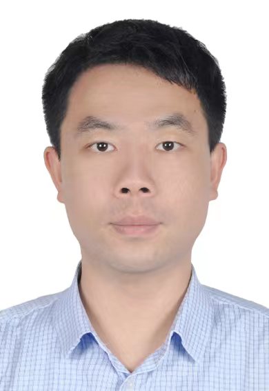 Changxing Ding (丁长兴)合作论文数School of Future Technology, South China University of Technology6
 Dacheng Tao (陶大程)合作论文数College of Computing & Data Science, Nanyang Technological University;Lee Kong Chian School of Medicine, Nanyang Technological University;DaXiao Robot5
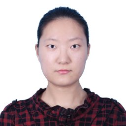 Chenhong Zhou合作论文数Department of Computer Science, Faculty of Science, Hong Kong Baptist University2
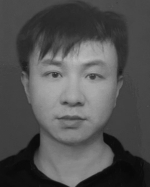 Liu Minfeng合作论文数2
 Aaron S. Jackson合作论文数1Mina Rezaei合作论文数1thomas mackie合作论文数1Hongdou Yao合作论文数1Haley Knapp合作论文数1Mehul Soni合作论文数1
 Irina Sanchez合作论文数1
 Jean Stawiaski合作论文数1Cheng-En Lee合作论文数1Bao Pham合作论文数1Kay Sun合作论文数1
 Jin Zhu合作论文数1
 Feng Shi (石峰)合作论文数School of Biomedical Engineering, ShanghaiTech University;Department of Research and Development, Shanghai United Imaging Intelligence Co1Konstantin Harmuth合作论文数1Mohammed Safwan合作论文数1
 Jonathan Ventura合作论文数Department of Computer Science & Software Engineering, California Polytechnic State University1
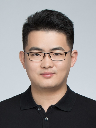 Hao Chen (陳浩)合作论文数Department of Computer Science and Engineering, School of Engineering, The Hong Kong University of Science and Technology;Division of Life Science, School of Science, The Hong Kong University of Science and Technology;Department of Chemical and Biological Engineering, School of Engineering, The Hong Kong University of Science and Technology1Eric Deutsch合作论文数1
 Sudeep Gupta合作论文数1Lutao Dai合作论文数1
 Steven Colleman合作论文数1Escudero Guilherme合作论文数1Thomas Batchelder合作论文数1King Nik合作论文数1Brad Paschke合作论文数1Vivekanandan Ajeet合作论文数1Lee Yuehchou合作论文数1Abhijit Amrutkar合作论文数1Emmett Sartor合作论文数1Anmol Popli合作论文数1
 Ou Yu-Nian合作论文数1Fan Zhou合作论文数1Wu Shaocheng合作论文数1
 Hongwei Bran Li (李宏伟)合作论文数Harvard Medical School;Massachusetts General Hospital1
 Wenqi Li (李文琦)合作论文数NVIDIA1Bharath Halandur Nagaraja合作论文数1
 Ramiro German Rodriguez Colmeiro合作论文数1Ayush Karnawat合作论文数1Issam Mahmoudi合作论文数1
 Willi Gierke合作论文数1
 Muneeza Azmat合作论文数1Catà Marcel合作论文数1Sara Sedlar合作论文数1Michael Pound合作论文数1Andrew French合作论文数1




