Predicting isocitrate dehydrogenase (IDH) mutations in gliomas using magnetic resonance imaging (MRI) is clinically important for treatment planning. This study compared two artificial intelligence (AI) models, GliomaDepth-IDH (ResNet34-based) and GliomaVista-IDH (Vision Transformer-based), with 18 physicians (eight neuroradiologists, five neurosurgeons, and five neurosurgery residents) in predicting IDH mutation status. On the Brain Tumor Segmentation Challenge dataset, the GliomaVista-IDH AI model achieved an area under the curve (AUC) value of 0.97, significantly outperforming all physician groups. However, external validation on a Japanese cohort revealed performance degradation: GliomaDepth-IDH declined to an AUC of 0.75 and GliomaVista-IDH to 0.82, with GliomaVista-IDH showing significant calibration issues (Brier score = 0.32). High-performing physicians achieved comparable results (AUC = 0.88) with superior calibration (Brier score = 0.19). Inter-rater reliability analysis revealed substantial variability across physician groups. These findings suggest that AI models can assist many physicians, while experienced practitioners remain competitive with better-calibrated predictions in challenging domains.
TLS in brain metastases of patients with NSCLC, and B and plasma cell cluster in the murine intracranial TME
The progression of isocitrate dehydrogenase-mutant glioma (IDH-G) from slow-growing tumor to fatal disease is associated with transcriptional and DNA methylation changes that remain poorly understood. Here, we profiled a longitudinal cohort of 36 IDH-G samples from 19 patients by joint-capture multi-omic single-nucleus DNA methylation, single-nucleus RNA sequencing and bulk exome sequencing. We show that IDH-G progression is associated with an increase in malignant stem-like states, decreased differentiation and methylation loss, which marks tumors with worse clinical outcome. Methylation loss was uniformly observed across malignant cells within individual tumors, suggesting that it may underlie rather than result from the increase in stem-like states. Analysis of cell-state heritability and plasticity using high-resolution phylogenetic trees links DNA methylation loss to alterations in glioma cell-state encoding and heritability. Our study offers insights into how DNA methylation loss reshapes cellular transitions and how it may mark clinically more aggressive tumors across IDH-G subsets.
Recent advances in molecular profiling have revealed that ependymomas are biologically distinct entities with unique molecular alterations and clinical behaviors. These findings have been incorporated into the 2021 WHO Classification of Tumors of the Central Nervous System, integrating anatomical location and molecular features into the diagnostic framework. This review summarizes the current molecular classification of ependymomas, role of DNA methylation profiling in diagnosis, and biological and clinical characteristics of the major molecular subgroups. Additionally, we discuss current treatment strategies, molecular risk stratification, and future directions in the management of ependymomas.
Glioblastoma (GBM) is the most aggressive primary brain tumor, with a poor prognosis despite advances in multimodal treatment. Current standard of care includes maximal safe resection and radiotherapy with concomitant and adjuvant temozolomide, which is called the Stupp regimen. Tumor-treating fields are incorporated into standard therapies and have demonstrated survival benefits in newly diagnosed GBM. Bevacizumab improves progression-free survival; however, its effect on overall survival remains limited. Recent advances in comprehensive molecular analyses have accelerated the development of novel therapies for GBM. Oncolytic virotherapy has emerged as a promising strategy for inducing direct tumor lysis and antitumor immunity. Immune checkpoint inhibitors, dendritic cell vaccines, CAR-T cell therapy, and gene therapy have been actively investigated. Precision oncology approaches based on genomic profiling, including basket and umbrella trials, are expanding the therapeutic possibilities for selected patients with actionable alterations.
Gender equity, work-life balance, and career development have become important concerns in the medical profession. However, empirical evidence on how neuro-oncology professionals experience working conditions, career barriers, and perceived inequity remains limited. We conducted a nationwide, cross-sectional, anonymous online survey of members of the Japan Society for Neuro-Oncology (JSNO) to evaluate working conditions, career-related concerns, job satisfaction, perceived workplace discrimination, and organizational challenges. Among 292 respondents (response rate, 35.9
Neuroendoscopic surgery, which allows tumor removal through a narrow surgical corridor, has been increasingly applied to intraventricular tumors. Although this approach is often regarded as minimally invasive, the restricted operative field and proximity to critical deep brain structures necessitate precise surgical manipulation. Periventricular vein injury can result in serious postoperative neurological deficits, underscoring the importance of meticulous dissection and hemostasis. During tumor removal, preservation of venous drainage should be prioritized. Aggressive resection should be avoided when strong adhesion to the veins is encountered. Gentle counterpressure using small cotton patties, combined with irrigation and suction, facilitated safe dissection and effective venous bleeding control. Hemostasis in neuroendoscopic surgery demands patience. The accurate identification of bleeding points is key. Blind coagulation in a blood-filled field should be avoided. Temporary packing with cotton patties can be effective without interrupting surgical workflow. The appropriate use of angled endoscopes and flexible transitions between the dry and wet fields is important for the reliable assessment of residual tumors and hemostasis. Furthermore, careful consideration of surgical instruments and timely conversion to microscopic surgery, when necessary, are critical for minimizing complications. Mastery of these principles can facilitate a safer neuroendoscopic management of intraventricular tumors.
 Nobuhito Saito合作论文数124Takayanagi Shunsaku合作论文数119
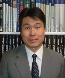 Akitake Mukasa合作论文数Department of Neurosurgery, University of Tokyo Hospital91
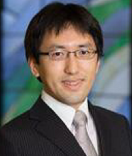 Hirokazu Takami (高見浩数)合作论文数Faculty of Medicine, The University of Tokyo;Department of Neurosurgery, The University of Tokyo Hospital80Yoshitaka Narita合作论文数73Nomura Masashi合作论文数62Ishida Joji合作论文数61
 Yoshihiro Otani合作论文数Department of Neurological Surgery, Okayama City Hospital48
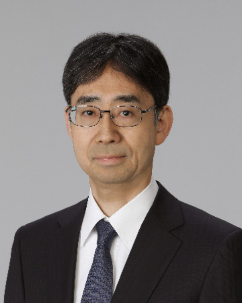 Motoo Nagane (永根基雄)合作论文数Department of Neurosurgery, Faculty of Medicine, Kyorin University46Ryo Nishikawa合作论文数45
 Koichi Ichimura合作论文数40
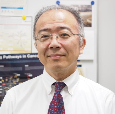 Hiroyuki Aburatani (油谷浩幸)合作论文数Laboratory for Systems Biology and Medicine, Research Center for Advanced Science and Technology, University of Tokyo36Kitagawa Yosuke合作论文数Department of Neurosurgery, Dokkyo Medical University35Taijun Hana合作论文数31
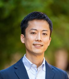 Nejo Takahide合作论文数30Higuchi Fumi合作论文数29
 Takayuki Terai合作论文数Faculty & Graduate School of Engineering, The University of Tokyo29
 Satoshi Takahashi合作论文数The Institute of Medical Science, The University of Tokyo28Tsukasa Koike合作论文数27Andrew S. Chi合作论文数Laura and Isaac Perlmutter Cancer Center, NYU Langone Health/Department of Neurosurgery, NYU Langone Health27Keisuke Ueki合作论文数27
 Hiroaki Wakimoto合作论文数Mass General Research Institute;Harvard Medical School;Massachusetts General Hospital26
 Tracy Batchelor合作论文数Harvard Medical School;Department of Neurology, Brigham and Women's Hospital;Cancer Center, Mass General Research Institute, Massachusetts General Hospital26
 Daniel Cahill合作论文数Stephen E. and Catherine Pappas Center for Neuro-Oncology, Massachusetts General Hospital, Harvard Medical School, Harvard University;Department of Neurosurgery, Massachusetts General Hospital, Harvard Medical School, Harvard University26
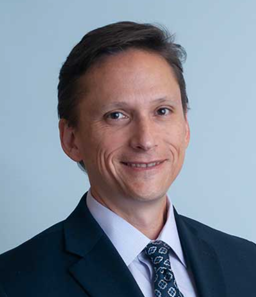 Anthony Iafrate合作论文数Department of Pathology, Massachusetts General Hospital Cancer Center;Harvard Medical School25Otani Ryohei合作论文数25
 Masahiro Shin合作论文数Department of Neurosurgery, The University of Tokyo Hospital25
 Yoshiki Arakawa合作论文数Cell Motility Laboratory, London Research Institute24
 Erika Yamazawa合作论文数22
 Kensuke Tateishi (立石健祐)合作论文数Department of Medical Life Science, Graduate School of Medical Life Science, Yokohama City University;Neurosurgical Oncology Laboratory, Yokohama City University22Masayuki Kanamori合作论文数Department of Neurosurgery, Tohoku University Graduate School of Medicine21
 Genta Nagae合作论文数Department of Gastroenterology, Jichi Medical School21Kaoru Tamura合作论文数20Ikemura Masako合作论文数20Shohei Nambu合作论文数19
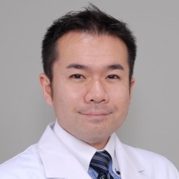 Masamichi Takahashi合作论文数National Cancer Center Hospital19
 Akio Sagara (相良明男)合作论文数Helical Fusion Co., Ltd.;Department of Quantum Science and Energy Engineering, Graduate School of Engineering, Tohoku University;SOKENDAI;National Institute for Fusion Science19
 Ryuta Saito (齋藤竜太)合作论文数Department of Neurosurgery, Tohoku University Hospital19Hiroshi Horiike合作论文数19
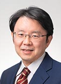 Mitsutoshi Nakada合作论文数Kanazawa University19Satomi Kaishi合作论文数18
 Koji Yoshimoto合作论文数18Lelic Nina合作论文数18
 William T Curry合作论文数18
 Tomonari Suzuki合作论文数17
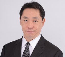 Hirofumi Nakatomi合作论文数Department of Neurosurgery, Fuji Brain Institute & Hospital17Kentaro Fujii合作论文数Synchrotron Radiation Research Center, Japan Atomic Energy Research Institute17Kazuhiko Kurozumi合作论文数Department of Neurological Surgery, Okayama University Graduate School of Medicine17




