Chemical electropolymerization can reliably produce two-dimensional (2D) and ultrathin conductive polymer films but offers limited control over nanomorphology and mechanical stiffness. To address this limitation, we investigate the use of DNA as a tethered dopant template, where the fixed charges of anionic DNA macromolecules grafted onto an electrode are used to dope and finely control electropolymerization in deionized water. The grafted layer adopts distinct conformations and stiffness depending on whether it is single- or double-stranded. Because the chemical composition of the dopant remains unaltered, variations in the resulting conductive polymer films arise solely from differences in the physical properties of the dopant layer. Our findings show that the nanomorphology of the polymer film directly reflects the morphology of the template, and that the dopant enhances mechanical stiffness with minimal impact on electrochemical performance. These results demonstrate that tethered dopant templating is a versatile platform for controlling the conductivity and electrochemical performance of molecularly engineered 2D polymer films, while dopant selection provides a powerful means for manipulating morphological and mechanical properties.
Development of PDMS-based microparticles for use in cell expansion applications requires a precise understanding of surface stability and its impact on long-term cellular performance. This study presents a systematic evaluation of plasma polymerised allylamine (ppAam) and acrylic acid (ppAac) coatings on PDMS discs as a precursor to PDMS microparticles. The study focuses on their characterisation and biological compatibility over a 30-day period. Surface characterisation was performed using X-ray Photoelectron Spectroscopy (XPS), which confirmed the successful presence of amine and carboxyl groups associated with allylamine and acrylic acid-coated discs, respectively. Water contact angle studies revealed decreased hydrophobic nature of the coated PDMS discs and the gradual recovery of hydrophobicity over time. When Sf9 insect cells are grown on functionalised surfaces, plasma polymerised discs outperformed uncoated discs; however, their metabolic activity declined slightly over a 30-day period. These results suggest that although the coatings are chemically stable for a month, their functionality is retained for about 14 days, suggesting that they should be utilised within 14 days. This work provides a framework for the synthesis and implementation of plasma-polymerised PDMS for advanced insect cell proliferation for future use in protein expression.
Despite significant advances in oncology, current cancer therapies remain constrained by toxicity, resistance, and limited selectivity. Endophytic fungi symbiotic microorganisms inhabiting plant tissues represent a sustainable and underexplored source of structurally diverse anticancer metabolites. These include alkaloids, terpenoids, polyketides, and peptides that disrupt microtubule dynamics, interfere with DNA replication, and induce mitochondrial-mediated apoptosis. They also modulate key oncogenic signalling pathways such as nuclear factor kappa B (NF-κB), signal transducer and activator of transcription 3 (STAT3), and phosphatidylinositol-3-kinase/protein kinase B (PI3K/Akt), thereby enhancing the efficacy of existing chemotherapies. Endophyte derived compounds further inhibit angiogenesis, suppress metastasis, and stimulate immune responses, offering multi-target mechanisms with reduced toxicity. This review examines strategies that enhance the discovery and yield of these bioactive metabolites, including One Strain Many Compounds (OSMAC), microbial co-culture, epigenetic activation, genome mining, and synthetic biology. A comparative assessment of endophyte-derived versus conventional anticancer agents highlights their potential for scalable, eco-sustainable production. Collectively, endophytic fungi are positioned as promising contributors to the next generation of accessible, cost-effective, and environmentally responsible anticancer therapies.
Growth factors (GFs) are the unique signaling molecules that enable constant communication and feedback between cells, modulating cell behavior and maintaining the structure and function of tissues. Traditional methods to detect and quantify GFs, such as ELISA, offer high specificity but are usually costly, present lower limits of detection, typically require sample processing, and are time-consuming. Electrochemical sensing emerges as a promising alternative due to their rapid responses and easy operation; however, they are prone to biofouling when challenged in complex biological environments. This study introduces an antifouling agent, lubricin (LUB), into an electrochemical immunosensor for the detection nerve growth factor (NGF). Through the combination of a specific NGF antibody with LUB, the developed sensor presents a great sensitivity, achieving a limit detection of 0.59 ng mL-1 for the target NGF. Our results show that the proposed sensing configuration can detect NGF directly in unprocessed and undiluted cell culture media with much faster analytical responses when compared to ELISA.
This study addresses the challenges of tracking cell-mediated biodegradation in cartilage tissue engineering, where hydrogels and scaffolds play a crucial role in providing structural support and promoting tissue regeneration. This research area has been rarely studied, offering potential insights into bridging the gap betweenin vitroandin vivoconditions for real-time monitoring of tissue regeneration alongside biodegradation. We developed dual-labeled hydrogel/scaffold composites for real-time monitoring of scaffold degradation in response to cell activity. Gelatin methacryloyl (GelMA) hydrogels are extensively explored for cartilage tissue engineering, albeit concerns remain regarding their mechanical properties under load-bearing conditions. To address this, a hydrogel/scaffold composite system was employed in this study, where a poly (ϵ-caprolactone) (PCL) hex prism edge structure acts as a scaffold to support the cell-laden GelMA hydrogel. Fluorophore labeling of GelMA and PCL facilitated non-invasive monitoring of the hydrogel/scaffold composite biodegradation under cell proliferation conditions. Initially, the behavior of fluorescent-tagged Hydrogel/Scaffold was examined under accelerated degradation conditions. Subsequently, human adipose-derived mesenchymal stem cells loaded into fluorescent-labeled hydrogel/scaffolds were evaluated for their biocompatibility potential and chondrogenesis. Results demonstrated a correlation between the loss of fluorescence from the hydrogel/scaffold degradation, accompanied by extracellular matrix accumulation. The fluorescently labeled hydrogel/scaffold holds promising application for cartilage tissue engineering, offering the capability to monitor biodegradation using high-throughput and contactless techniques.
Advanced tissue engineering (TE) strategies are vital to address challenging musculoskeletal conditions, such as volumetric muscle loss. These disorders impose a considerable economic burden and affect individuals' quality of life, highlighting the need for innovative treatments, such as TE, to address these challenges. Here, we examine how scaffold fibre orientation influences mechanical properties and cellular behaviour by utilising melt electrowriting (MEW) as a high-resolution 3D printing technique that combines aspects of electrospinning and melt based polymer deposition. In this work, we investigated the effects of fibre orientation in MEW scaffolds, and its effect on the scaffold mechanical properties as well as cell orientation and alignment. MEW scaffolds were mechanically characterised through uniaxial strain testing to determine critical parameters, including strain at failure, ultimate tensile strength, Young's modulus (E), fatigue rate, recovery time, and yield strain. These mechanical properties were analysed to define an optimal strain regime for transitioning from static to dynamic culture conditions under muscle-like cyclic loading, relevant to muscle's viscoelastic behaviour. In parallel, static cultures of primary human skeletal muscle myoblasts and normal human dermal fibroblasts (NHDFs) were grown on MEW scaffolds, with varying architectures, to study the effects of fibre aspect ratio on cell alignment. Cell alignment was visualised using DAPI/phalloidin staining and quantified with the ImageJ directionality plugin, enabling a systematic comparison of scaffold designs. This approach evaluates the potential of supportive scaffold architectures to promote aligned cell growth, offering insights into designing effective scaffolds for tissue regeneration.
Drug delivery platforms are frequently susceptible to nonspecific adsorption of biological materials upon contact with biological fluids that can interfere with the controlled release of drugs, affecting both the rate and amount of drug released. To improve the controlled release efficiency of drugs within biological environments, lubricin (also known as PRG4) antifouling coatings were applied to control the release of poly(3,4-ethylenedioxythiophene) (PEDOT) films doped with the model drug phenol red. Drug release experiments comparing uncoated and lubricin-coated PEDOT were performed in nonfouling PBS buffer to assess the impact of lubricin coating on the release of drug from the films, while identical experiments were performed in concentrated solutions of highly fouling proteins to investigate lubricin's capacity to mitigate the effects of surface biofouling on the drug release properties. These experiments revealed that lubricin coating did not create a physical or diffusional barrier to the release of phenol red from PEDOT under nonfouling conditions. Likewise, within a highly fouling protein solution, the lubricin-coated PEDOT films showed greatly enhanced phenol red release compared with uncoated films under passive conditions and active release under an applied negative potential. This greater phenol red release from the lubricin-coated PEDOT was attributed to lubricin's capacity to protect the surface from fouling by nonspecifically adsorbed proteins, which impede the release of phenol red. This study demonstrated that lubricin can effectively improve drug release from PEDOT films in highly fouling environments, resulting in more accurate and reliable dosing.
Stimuli-responsive materials (SRMs) are materials that change properties when exposed to external or internal stimuli. They respond to physiological changes within cells and tissues, as well as external triggers including light, magnetic fields, ultrasound, and electricity. In medicine, SRMs have diverse applications spanning drug delivery, tissue engineering, and diagnostics. They enable targeted drug release at specific times and locations, facilitate tissue generation and repair, and enhance disease detection capabilities. Beyond medical uses, SRMs are employed in smart coatings and artificial muscle systems. The breadth of biomedical applications for SRMs is extensive, generating substantial research into novel and innovative material development. Challenges in creating safe and efficient SRMs for medical treatments have driven innovative approaches in two key areas: functionalizing and modifying naturally occurring materials and developing new synthetic nanomaterials. The complexity of producing effective SRMs has necessitated creative solutions to overcome safety and efficiency barriers in medical applications. This ongoing research continues to expand the potential therapeutic uses of these responsive materials. This review examines literature focused on SRM development for external stimuli responses, particularly light, magnetic fields, ultrasound, and electricity, rather than covering the complete spectrum of stimuli-responsive applications.
Tissue engineering holds promise for the treatment of osteoarthritis (OA), where protective hydrogel scaffolds have been combined with mesenchymal stem cells (MSCs) to promote chondrogenesis. Quantification of chondrogenesis by MSCs in 3D culture requires the imaging and detection of deposited extracellular matrix (ECM) components like collagen and proteoglycans. ECM protein quantification should be performed in a non-destructive, label-free, and simple manner. Here, we demonstrate a nanoplasmonic colorimetric device for the imaging of collagen requiring only a simple optical microscope. MSCs were encapsulated in the hydrogel-forming peptide Fmoc-diphenylalanine (Fmoc-FF) with arginine glycine aspartic acid (RGD) added. We showed, by colorimetric histology, that increased concentrations of RGD resulted in a significant increase in collagen deposition after 21 days. Traditional techniques such as immunohistological staining were not able to detect any RGD dependent increases in ECM deposition. Through an in-depth biophysical analysis we were able to correlate elevated RGD with enhanced cell-viability, collagen deposition, and reduced hydrogel stability. In summary, plasmon-enhanced colorimetric histology provides a non-destructive, label-free means to image collagen without resorting to destructive sample processing and complex immunohistological staining. This approach holds broad potential for routine quantification of collagen-rich biomaterials, promising widespread applications across research and clinical settings.
Conductive hydrogels offer an exciting opportunity to combine the hydrophilicity, biocompatibility, and tuneable viscoelastic properties of hydrogels with the conductive properties of electroactive species and conducting polymers. Reported applications of conductive hydrogels include but are not limited to electroactive wound dressings, wearable electronics, stimuli-responsive drug delivery systems, and tissue-engineered implants. With the rise of electroactive materials in biomaterials research, the electrical and electrochemical measurement techniques used to characterise their conductive properties have also emerged. The vast range of novel materials and the wide scope for application-specific requirements may leave researchers unable to discern the appropriate techniques, measurement conditions, and analysis to apply within their research. This review concisely summarises the techniques utilised to characterise the electrical properties of conductive hydrogels, including four-point probe conductivity measurements, cyclic voltammetry, and electrochemical impedance spectroscopy. Furthermore, the limitations and practical considerations of each technique are detailed. Recommendations for optimal sample preparation and experimental parameters are made, referencing current literature where conductive hydrogels have been successfully characterised. The fundamental principles of conductivity, electrical percolation, and indirect electrical stimulation are also discussed, to provide researchers with a comprehensive resource to develop and translate conductive hydrogels within biomedical research. STATEMENT OF SIGNIFICANCE: Electrically conductive hydrogels have gained increasing attention in wearable electronics, drug-delivery systems, and tissue engineering research. In existing literature there is a lack of standardisation in the electrical and electrochemical characterisation of these materials, as well as a variation in reported experimental conditions and data interpretation. This review is significant in providing a concise and practical resource to guide researchers in conducting accurate and impactful characterisation of the electrical properties of conductive hydrogels. It will contribute to research as a unique guide for best practise, promoting the advancement and translation of conductive hydrogels and their ever-expanding applications.
Melanin, a natural biopolymer with a conjugated backbone, stands out as a promising candidate for bioelectronics owing to its redox activity, metal ion chelation, hydration-dependent electrical response, wet adhesion, potential electronic transport, anti-inflammatory properties, and excellent thermal stability. In this study, we explore the impact of hybridizing liquid crystalline graphene oxide (LCGO) into the conductive melanin-like polymer polydopamine (PDA) on the morphology, stability, and synergistic electrochemical performance. Our findings reveal that the gold electrode coated with PDA:LCGO composites exhibit more than two orders of magnitude lower impedance and more than 100 times higher charge storage capacity compared to both bare gold electrodes and those coated with pure PDA. Furthermore, we demonstrate that the morphology and the electrode performance could be tuned by adjusting the synthesis parameters in the PDA:LCGO electrochemical deposition. This PDA:LCGO composite, with its superior electrochemical performance, holds promise for diverse bioelectronics applications ranging from biosensors to implantable bionic interfaces.
To address the problems associated with pathogenic bacteria in healthcare settings, the development of novel antibacterial materials is of high priority. For such purposes, endophytic fungi - symbiotic microorganisms residing within healthy plant tissues - represent a promising yet largely unexplored source of antibacterial compounds. In this study, an antibacterial extract derived from an endophytic Alternaria fungus previously isolated from Eremophila longifolia was incorporated within gelatin methacryloyl (GelMA) to produce a novel antibacterial hydrogel. Whilst rheological and compression testing revealed the addition of the extract resulted in reduction in the crosslink density of the hydrogel, all GelMA-extract formulations produced a solid mechanical stable hydrogel. The GelMA hydrogel containing a range of extract concentrations demonstrated variable inhibition of bacterial (Staphylococcus aureus) growth, with a concentration of 10 mg mL-1 extract demonstrating complete inhibition over 24 h, while showing no toxicity toward brine shrimp nauplii, indicating good biocompatibility. The GelMA-extract demonstrated minimal rapid release from the hydrogel, followed by a slower release at longer times. As such, the developed hydrogel composite is promising for antibacterial applications in biomedical settings, while the results also highlight the potential for utilising endophytic extracts in the development of novel antibacterial materials.
Metal nanoparticles (MNPs) produced through biosynthesis approaches have shown favourable physical, chemical, and antimicrobial characteristics. The significance of biological agents in the synthesis of MNPs has been acknowledged as a promising alternative to conventional approaches such as physical and chemical methods, which are confronted with certain challenges. To meet these challenges, the use of endophytic fungi as nano-factories for the synthesis of MNPs has become increasingly popular worldwide in recent times. This review provides an overview of the synthesis of MNPs using endophytic fungi, the mechanisms involved, and their important biomedical applications. A special focus on different biomedical applications of MNPs mediated endophytic fungi involved their antibacterial, antifungal, antiviral, and anticancer applications and their potential as drug delivery agents. Furthermore, this review highlights the significance of the use of endophytic fungi for the green synthesis of MNPs and discusses the benefits, challenges, and prospects in this field.
Using a newly invented surface-tethered dopant approach for the electrochemical polymerization growth of conductive polymer films, nanocomposites are created consisting of two distinct phases of polypyrrole (PPy) and Poly(3,4-ethylene dioxythiophene) (PEDOT). The growth of the conductive nanocomposite films takes advantage of the unique way in which a conductive polymer film forms when grown using a surface-tethered dopant, starting out as a highly nanoporous film that gradually becomes more and more solid as additional polymer deposition occurs preferentially within the nanopores rather than at the films outer surface. Nanocomposite films can thus be fabricated simply and controllably by first growing one nanoporous conductive polymer film followed by the deposition of a second conductive polymer within the nanopores of the first. This study investigates the morphological and electrochemical properties of nanocomposite films grown using a surface-tethered dopant and how these properties are affected by simple changes in electrochemical polymerization parameters.
Oxygen plays a crucial role in human embryogenesis, homeostasis, and tissue regeneration. Emerging engineered regenerative solutions call for novel oxygen delivery systems. To become a reality, these systems must consider physiological processes, oxygen release mechanisms and the target application. In this review, we explore the biological relevance of oxygen at both a cellular and tissue level, and the importance of its controlled delivery via engineered biomaterials and devices. Recent advances and upcoming trends in the field are also discussed with a focus on tissue-engineered constructs that could meet metabolic demands to facilitate regeneration.
Nerve growth factor (NGF) plays a crucial role in cellular growth and neurodifferentiation. To achieve significant neuronal regeneration and repair using in vitro NGF delivery, spatiotemporal control that follows the natural neuronal processes must be developed. Notably, a challenge hindering this is the uncontrolled burst release from the growth factor delivery systems. The rapid depletion of NGF reduces treatment efficacy, leading to poor cellular response. To address this, we developed a highly controllable system using graphene oxygen (GO) and GelMA hydrogels modulated by electrical stimulation. Our system showed superior control over the release kinetics, reducing the burst up 30-fold. We demonstrate that the system is also able to sequester and retain NGF up to 10-times more efficiently than GelMA hydrogels alone. Our controlled release system enabled neurodifferentiation, as revealed by gene expression and immunostaining analysis. The increased retention and reduced burst release from our system show a promising pathway for nerve tissue engineering research toward effective regeneration.
The nondestructive localization and traceability of polymers by fluorescent tagging has become a valuable tool for biomedical applications. Integration of fluorescent molecule to the pristine polymers could modify polymers' degradation rate which is still unpredictable from a scaffold application standpoint. The current study focused to understand the material perspective of fluorescently tagged biodegradable polymers such as polycaprolactone (PCL) and poly (d,l-lactide-co-glycolide) (PLGA) with fluorescein amine isomer I (FITC). PCL-FITC and PLGA-FITC were characterized using FTIR for surface chemistry analysis and rheology for their mechanical properties. The grafted materials were utilized to form 3-dimentional scaffolds, and their degradation was monitored under accelerated degradation conditions triggered by pH. It was found that PCL and PCL-FITC had a very slow degradation rate, when compared to PLGA and PLGA-FITC. Both the FITC tagged materials displayed a faster degradation rate compared to their respective pristine material. Biocompatibility of the FITC conjugated polymers was tested using human-adipose derived stem cells (hADSCs) revealing that the sub products from the degradation of the polymers over 7 days did not negatively affect the cellular metabolic activity. This work highlights the significance of initial characterization of fluorescent modified polymers for future biomedical application.
This study investigates the electrochemical behavior of GelMA-based hydrogels and their interactions with PC12 neural cells under electrical stimulation in the presence of conducting substrates. Focusing on indium tin oxide (ITO), platinum, and gold mylar substrates supporting conductive scaffolds composed of hydrogel, graphene oxide, and gold nanorods, we explored how the substrate materials affect scaffold conductivity and cell viability. We examined the impact of an optimized electrical stimulation protocol on the PC12 cell viability. According to our findings, substrate selection significantly influences conductive hydrogel behavior, affecting cell viability and proliferation as a result. In particular, the ITO substrates were found to provide the best support for cell viability with an average of at least three times higher metabolic activity compared to platinum and gold mylar substrates over a 7 day stimulation period. The study offers new insights into substrate selection as a platform for neural cell stimulation and underscores the critical role of substrate materials in optimizing the efficacy of neural interfaces for biomedical applications. In addition to extending existing work, this study provides a robust platform for future explorations aimed at tailoring the full potential of tissue-engineered neural interfaces.
 Gordon Wallace合作论文数Intelligent Polymer Research Institute, University of Wollongong110
 Rob Kapsa合作论文数School of Engineering, RMIT University53
 Anita Quigley合作论文数Centre for Clinical Neurosciences and Neurological Research, St Vincent's Hospital (Melbourne),43
 Saimon Moraes Silva合作论文数Iverson Health Innovation Research Institute, Swinburne University of Technology32
 Michael J. Higgins合作论文数Department of Human Genetics, Roswell Park Cancer Institute21George Wren Greene合作论文数School of Dentistry, State University of New York at Buffalo21
 Joselito Razal合作论文数Institute for Frontier Materials Fibres and Textiles, Deakin University;ARC Research Hub for Future Fibres, Deakin University17
 Mark Cook合作论文数Division of Engineering and IT, The University of Melbourne13
 Lilith Caballero Aguilar合作论文数BioFab3D, St Vincents Hosp12
 Graeme M. Clark合作论文数Department of Otolaryngology, University of Melbourne, Parkville, Victoria 3052, Australia12
 Cathal O'Connell合作论文数Intelligent Polymer Research Institute, University of Wollongong11
 Zhilian Yue合作论文数Department of Chemical Engineering, University of Cambridge11
 Sina S. Jamali合作论文数Iran University of Science and Technology;Department of Electrical Engineering;Department of Electrical Engineering, Iran University of Science and Technology11
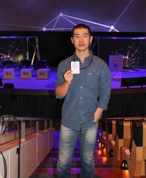 Mingyu Han合作论文数ARC Ctr Excellence Electromat Sci, Deakin Univ10
 Serena Duchi合作论文数Laboratory of Medical Genetics;Department of Internal Medicine;University of Bologna10Magdalena Kita合作论文数10
 Paul Stoddart合作论文数School of Engineering, Swinburne University of Technology9
 Pauline E Desroches合作论文数Aikenhead Ctr Med Discovery ACMD, St Vincents Hosp Melbourne8
 Maria Forsyth合作论文数Institute for Frontier Materials, Deakin University8
 Kerry J Gilmore合作论文数8Tharun Mysore合作论文数8Brianna C. Thompson合作论文数7
 Dorna Esrafilzadeh合作论文数Graduate School of Biomedical Engineering, Faculty of Engineering, The University of New South Wales7Jan Weber合作论文数6
 Matthew Jordan Russo合作论文数ARC Ctr Excellence Electromat Sci, Deakin Univ6
 Carmine Onofrillo合作论文数*Department of Experimental, Diagnostic and Specialty Medicine, Alma Mater Studiorum-Università di Bologna;Institut National de la Santé et de la Recherche Médicale, University Grenoble Alpes;University Grenoble Alpes, Alma Mater Studiorum-Università di Bologna6Orawan Ngamna合作论文数6
 Peter C. Innis合作论文数Intelligent Polymer Research Institute, University of Wollongong6
 Alexandre Xavier Mendes合作论文数5M Kita合作论文数5
 Claudia Di Bella合作论文数Department of Surgery, Melbourne Medical School, Faculty of Medicine, Dentistry and Health Sciences, The University of Melbourne5
 Mirren Charnley合作论文数Ctr Microphoton, Swinburne Univ Technol5
 Jalili Rouhollah合作论文数Intelligent Polymer Research Institute, University of Wollongong5
 del Rosal Blanca合作论文数Fluorescence Imaging Grp, Univ Autonoma Madrid5
 Jun Chen合作论文数Intelligent Polymer Research Institute, Australian Institute for Innovative Materials, University of Wollongong5
 Amy Halliday合作论文数5
 Rosanne M Guijt合作论文数University of Tasmania, University of Tasmania5Tallman Dennis合作论文数North Dakota State University5Elise M. Stewart合作论文数4
 Adriana Nascimento合作论文数4Bc Kim合作论文数4Yu Chen合作论文数4
 David Nisbet合作论文数John Curtin School of Medical Research, College of Health and Medicine, The Australian National University4
 Stephen O'Leary合作论文数Department of Otolaryngology,, The University of Melbourne4
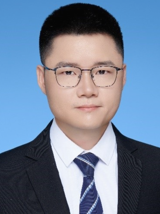 Xiao Liu (刘肖)合作论文数School of Biological Science and Medical Engineering, Beihang University4Romeo Tony C合作论文数4
 Andrew I. Minett合作论文数Intelligent Polymer Research Institute, University of Wollongong4
 Sally L Mcarthur合作论文数Cooperative Research Centre for Eye Research and Technology, University of New South Wales4
 Kara Vine-Perrow合作论文数School of Chemistry and Molecular Bioscience, Faculty of Science, Medicine and Health, University of Wollongong;Targeted Cancer Therapeutics Research Laboratory, Illawarra Health and Medical Research Institute, University of Wollongong4




