Background Remission From Stage D Heart Failure (RESTAGE-HF) was a multicenter study demonstrating that a selected patient population with left ventricular assist devices (LVADs) could achieve functional recovery and subsequent durable pump explantation. The purpose of our study was to describe the impact of surgical approach to deactivation in this study cohort. Methods The study prospectively enrolled 40 patients with HeartMate II LVAD. Overall, 19 (52.8%) of 36 evaluable patients reached the predefined criteria and underwent LVAD deactivation. Three approaches were used: complete explantation (n = 8), partial explantation (n = 10), and percutaneous decommissioning (n = 1). The primary outcome was survival free from transplantation or LVAD reimplantation. Results Patients with partial explantation had a longer duration of heart failure and a larger posterior wall thickness before ventricular assist device implantation. There were no differences in other preimplantation parameters. Median follow-up was 7.9 years. Median time to deactivation was similar between approaches. Post-LVAD survival free from LVAD reimplantation or transplantation was 90% at 1 year and remained stable after 4 years at 74% for the entire cohort. No significant intergroup differences in 7-year survival were observed (87.5% vs 40%; P = .06). Conclusions This analysis represents the longest follow-up of post-LVAD patients stratified by surgical approach and suggests that select patients can achieve sustained myocardial recovery after LVAD deactivation.
BACKGROUND:Limited studies have evaluated lung ultrasound (LUS) in ambulatory heart failure (HF). A six-zone LUS assesses B-lines, a marker of congestion. The Butterfly IQ+ probe features an automated B-line counter (ABLC), eliminating manual counting. We evaluated LUS quality by novice HF providers after training, compared expert manual counts to ABLC, and explored associations between LUS and clinical HF metrics. METHODS:Three novice providers underwent 2 h of didactics and 30 proctored exams. Image quality was independently reviewed by two LUS experts. B-lines were counted manually by experts and ABLC. We assessed associations between LUS and four clinical metrics: provider-assessed volume status, > 30% NT-proBNP increase, > 5 lb weight gain, and PAD above goal (CardioMEMS). RESULTS:Seventy-five subjects were enrolled. Overall, LUS quality was excellent, with 88% good quality. Surprisingly, agreement between expert B-line counts was moderate (Gwet's AC1: 0.49, 95% CI: 0.27 to 0.71) while the accuracy of experts as compared to ABLC was modest (Expert 1 = 61.2%, Expert 2 = 40.3%). Experts correctly identified 93% of positive studies but only 19% of negative studies versus ABLC. Provider volume assessments substantially agreed with LUS (Gwet's AC1: 0.76, 95% CI: 0.61 to 0.91), but providers identified only half of positive LUS cases, suggesting utility in detecting mild volume overload. Only volume overload correlated with positive LUS. Only 25% of subjects had a CardioMEMS. CONCLUSION:Novice providers can perform high-quality LUS after brief training. ABLC reduces B-line counting variability. LUS detects mild pulmonary congestion undetectable by clinical exam, potentially preventing worsening in HF patients.
Purpose: Detecting volume overload in the outpatient setting is key to preventing heart failure (HF) hospitalizations. Lung ultrasound (LUS) can be used to assess pulmonary congestion and guide therapy by identifying B-lines. While prior studies have focused on the inpatient setting, the primary aim of this study was to assess the quality of LUS images performed by outpatient nurse practitioners (NPs) after a formal period of training. We further explored the relationship between clinical markers of HF and B-lines assessed with a novel automated B-line counter.
OBJECTIVES:Heart failure exacerbations are a common cause of hospitalizations with a high readmission rate. There are few validated predictors of readmission after treatment for acute decompensated heart failure (ADHF). Lung ultrasound (LUS) is sensitive and specific in the assessment of pulmonary congestion; however, it is not frequently utilized to assess for congestion before discharge. This study assessed the association between number of B-lines, on LUS, at patient discharge and risk of 30-day readmission in patients hospitalized for acute decompensated heart failure (ADHF).METHODS:This was a single-center prospective study of adults admitted to a quaternary care center with a diagnosis of ADHF. At the time of discharge, the patient received an 8-zone LUS exam to evaluate for the presence of B-lines. A zone was considered positive if ≥3 B-lines was present. We assessed the risk of 30-day readmission associated with the number of lung zones positive for B-lines using a log-binomial regression model.RESULTS:Based on data from 200 patients, the risk of 30-day readmission in patients with 2-3 positive lung zones was 1.25 times higher (95% CI: 1.08-1.45), and in patients with 4-8 positive lung zones was 1.50 times higher (95% CI: 1.23-1.82, compared with patients with 0-1 positive zones, after adjusting for discharge blood urea nitrogen, creatinine, and hemoglobin.CONCLUSION:Among patients admitted with ADHF, the presence of B-lines at discharge was associated with a significantly increased risk of 30-day readmission, with greater number of lung zones positive for B-lines corresponding to higher risk.
BackgroundExpert opinion and professional society statements have called for multi-tier care systems for the management of cardiogenic shock (CS). However, little is known about how to pragmatically define centers with different levels of care (LOC) for CS.MethodsEleven of 23 hospitals within our healthcare system sharing a common electronic health record were classified as different LOC according to their highest mechanical circulatory support (MCS) capabilities: Level 1 (L-1)—durable left ventricular assist device, Level 1A (L-1A)—extracorporeal membrane oxygenation, Level 2 (L-2)—intra-aortic balloon pump and percutaneous ventricular assist device; and Level 3 (L-3)—no MCS. All adult patients treated for CS (International Classification of Diseases, ICD-10 code R57.0) between 2016 and 2022 were included. Etiologies of CS were identified using associated diagnostic codes. Management strategies and outcomes across LOC were compared.ResultsHigher LOC centers had higher volumes: L-1 (n = 1): 2,831 patients, L-1A (n = 4): 3,452, L-2 (n = 1): 340, and L-3 (n = 5): 780. Emergency room admissions were more common in lower LOC (96% at L-3 vs. 46% L-1; p < 0.001), while hospital transfers were predominant at higher LOC (40% at L-1 vs. 2.7% at L-3; p < 0.001). Men comprised 61% of the cohort. Patients were younger in the higher LOC [69 (60–78) years at L-1 vs. 77 (67–85) years at L-3; p < 0.001]. Patients with acute myocardial infarction (AMI)-CS and acute heart failure (AHF)-CS were concentrated in higher LOC centers while other etiologies of CS were more common in L-2 and L-3 (p < 0.001). Cardiac arrest on admission was more prevalent in lower LOC centers (L-1: 2.8% vs. L-3: 12.1%; p < 0.001). Patients with AMI-CS received more percutaneous coronary intervention in lower LOC (51% L-2 vs. 29% L-1; p < 0.01) but more coronary arterial bypass graft surgery at higher LOC (L-1: 42% vs. L-1A: 23%; p < 0.001). MCS use was consistent across levels for AMI-CS but was more frequent in higher LOC for AHF-CS patients (L-1: 28% vs. L-2: 10%; p < 0.001). Despite increasing in-hospital mortality with decreasing LOC, no significant difference was seen after multivariable adjustment.ConclusionThis is the first report describing a pragmatic classification of LOC for CS which, based on MCS capabilities, can discriminate between centers with distinct demographics, practice patterns, and outcomes. This classification may serve as the basis for future research and the creation of CS systems of care.
Introduction: Mechanical unloading (MU) with Intra-aortic balloon pump (IABP) or Impella is commonly used to prevent left ventricular (LV) distension in VA-ECMO patients, but its impact on shock severity is not well described. Methods: Patients treated with VA-ECMO in our hospital between 2016-2022 for cardiogenic shock (CS) due to LV failure not related to cardiac surgery were included if no MU was used or it was instituted within 24h of ECMO cannulation. Patients were grouped according to MU: ECMO alone, ECMO + IABP or ECMO + Impella. Linear mixed effects models adjusting for age, sex, hypertension, acute myocardial infarction (AMI) etiology of shock and pre-ECMO cardiac arrest (CA) were used to assess the impact of MU on hemodynamic and perfusion markers. R version 4 was used for analysis. Results: Among 68 patients included, 18 received ECMO alone, 17 ECMO + IABP, and 33 ECMO + Impella. Age and CA were similar across groups. The Impella group had a higher proportion of men and patients with AMI. Pulmonary artery diastolic pressure was unchanged in all groups. Compared to ECMO alone, MU did not enhance lactate clearance (interaction: IABP p=0.7, Impella p=0.22), but it did improve the vasopressor-inotropic score (IABP p=0.03, Impella p=0.01), serum creatinine (IABP p=0.03, Impella p=0.04), and alanine transaminase (IABP only p=0.02). Survival to decannulation was similar in patients with ECMO alone (50%), Impella (64%) and IABP (65%) (p=0.68). Conclusions: In adults treated with VA-ECMO for CS unrelated to cardiac surgery, LV MU can lead to improvements in CS severity.
PurposeVaccination against COVID 19 has been shown to improve outcomes after transplant. There are prior reports of vaccines leading to sensitization in transplant recipients, however, little is known about the development of de novo (dn) anti-HLA antibodies (abs) after COVID-19 vaccination (COVAX). The purpose of this study was to prospectively survey immunological markers in heart transplant recipients after receiving their 3rd dose of COVAX.MethodsThirty-one subjects (mean 30 months post-transplant) were evaluated 1, 3and 5 months after COVAX. Single antigen bead assay for anti-HLA abs (thresholdMFI >1000), COVID-19 spike abs (threshold ≥250 U/mL) and donor/recipientcell free DNA testing (threshold ratio >.12) were performed.Results(Table 1, Fig 1): Over 5 months, 32% of subjects developed dn anti-HLA abs and 19%developed dn donor specific abs (DSA). Antibodies developed by 1 month with titers decreasing by 5 months. All sensitized subjects developed Class II antibodies, with 60% having concomitant Class I. One of four subjects with pre COVAX DSAs had a significant rise after COVAX. No significant cellular or antibody mediated rejection was seen during follow up, but 32% of subjects demonstrated a new rise in cell free DNA. We did not see a temporal relationship between intercurrent COVID infection and dn DSA during study follow up.ConclusionAfter the 3rd COVAX dose, one third of subjects developed dn anti-HLA abs and one fifth dn DSAs. These preliminary findings warrant further study, especially as additional boosters are recommended. Vaccination against COVID 19 has been shown to improve outcomes after transplant. There are prior reports of vaccines leading to sensitization in transplant recipients, however, little is known about the development of de novo (dn) anti-HLA antibodies (abs) after COVID-19 vaccination (COVAX). The purpose of this study was to prospectively survey immunological markers in heart transplant recipients after receiving their 3rd dose of COVAX. Thirty-one subjects (mean 30 months post-transplant) were evaluated 1, 3and 5 months after COVAX. Single antigen bead assay for anti-HLA abs (thresholdMFI >1000), COVID-19 spike abs (threshold ≥250 U/mL) and donor/recipientcell free DNA testing (threshold ratio >.12) were performed. (Table 1, Fig 1): Over 5 months, 32% of subjects developed dn anti-HLA abs and 19%developed dn donor specific abs (DSA). Antibodies developed by 1 month with titers decreasing by 5 months. All sensitized subjects developed Class II antibodies, with 60% having concomitant Class I. One of four subjects with pre COVAX DSAs had a significant rise after COVAX. No significant cellular or antibody mediated rejection was seen during follow up, but 32% of subjects demonstrated a new rise in cell free DNA. We did not see a temporal relationship between intercurrent COVID infection and dn DSA during study follow up. After the 3rd COVAX dose, one third of subjects developed dn anti-HLA abs and one fifth dn DSAs. These preliminary findings warrant further study, especially as additional boosters are recommended.
Objectives- Heart failure exacerbations are a common cause of hospitalizations with a high readmission rate. There are few validated predictors of readmission after treatment for acute decompensated heart failure (ADHF). Lung ultrasound (LUS) is sensitive and specific in the assessment of pulmonary congestion; however, it is not frequently utilized to assess for congestion before discharge. This study assessed the association between number of B-lines, on LUS, at patient discharge and risk of 30-day readmission in patients hospitalized for acute decompensated heart failure (ADHF). Methods_This was a single-center prospective study of adults admitted to a quaternary care center with a diagnosis of ADHF. At the time of discharge, the patient received an 8-zone LUS exam to evaluate for the presence of B-lines. A zone was considered positive if >= 3 B-lines was present. We assessed the risk of 30-day readmission associated with the number of lung zones positive for B-lines using a log-binomial regression model. Results- Based on data from 200 patients, the risk of 30-day readmission in patients with 2-3 positive lung zones was 1.25 times higher (95% CI: 1.08-1.45), and in patients with 4-8 positive lung zones was 1.50 times higher (95% CI: 1.23-1.82, compared with patients with 0-1 positive zones, after adjusting for discharge blood urea nitrogen, creatinine, and hemoglobin. Conclusion- Among patients admitted with ADHF, the presence of B-lines at discharge was associated with a significantly increased risk of 30-day readmission, with greater number of lung zones positive for B-lines corresponding to higher risk.
To the Editor: Immunogenicity sought through vaccination carries a risk of stimulating new antibody production to the donor organ after transplant and prior reports have described development of de novo anti-HLA antibodies following influenza vaccination.1 Development of de novo donor-specific antibodies (DSA) after cardiac transplant is associated with worse outcomes.2 We sought to prospectively survey immunological markers after the third COVID-19 vaccination (COVAX) dose in cardiac transplant recipients (the fourth COVAX dose was not available at the time of this study). Current guidelines recommend a fourth dose with the updated (bivalent messenger RNA) vaccine, with the option for additional updated doses at the clinician’s discretion.3 From August 2021 to June 2022, 31 participants were prospectively studied prior to and at 1, 3, and 5 mo after the third COVAX dose (details of specific COVAX regimens are included in Table S1, SDC,https://links.lww.com/TP/C907). At each time point, Luminex single antigen bead assay for anti-HLA antibodies, spike antibodies to COVID-19, and donor/recipient cell-free DNA (cfDNA) (AlloSure, Brisbane, CA) were performed. We defined the presence of de novo antibody as an antibody with mean fluorescent intensity (MFI) >1000 during follow-up, that was either not detectable or present with MFI ≤1000 before the third COVAX dose. Anti-HLA antibodies were classified as DSA or non-DSA. A new rise in cfDNA above the threshold ratio of <0.12 was reported. Endomyocardial biopsy was performed according to the standard programmatic schedule or if a patient demonstrated clinical evidence of possible rejection. Rejection was reported if there was International Society for Heart and Lung Transplantation grading ≥1R/2 acute cellular rejection or any antibody-mediated rejection. Thirty-one participants with an average age of 61 ± 9 y ranging from 4 to 34 mo (interquartile range: 9–14) posttransplant were enrolled before their third COVAX dose. Data showing spike antibody response to the third COVAX dose are included in Figure S1 (SDC, https://links.lww.com/TP/C907). Six participants had a history of COVID infection before enrollment. All 31 participants were receiving tacrolimus, 18 were managed on mycophenolate, 10 on everolimus, 15 on prednisone, and the 3 remaining participants were managed on tacrolimus monotherapy (malignancy and recurrent infections). There were no significant changes in immunosuppression during the 5-mo follow-up period. Ten participants (32%) developed de novo anti-HLA antibodies (100% class II antibodies, 60% both class I/class II) with 6 (19%) participants developing de novo DSA (100% class II, 17% both class I/class II) over the 5-mo follow-up. Table 1 shows the proportion of patients with antibodies at each time point. Overall, the proportion of participants demonstrating de novo anti-HLA antibodies rose over time; however, de novo DSA fell below threshold in 1 participant by month 5. De novo anti-HLA antibody levels were modest, with all below 3000 MFI (with 1 exception). Neither the development of COVID threshold spike titers (in response to the third COVAX) nor differences in immunosuppression impacted the development of de novo anti-HLA antibodies. Figure 1 shows the individual trends in MFI in the 6 participants who developed de novo DSA. We did not see a temporal relationship between intercurrent COVID infection and development of de novo DSA. Only 1 of 4 participants with preexisting DSAs developed a significant rise (>1000 MFI) after the third COVAX. TABLE 1. - Development of anti-HLA antibodies and AlloSure post third COVAX dose Rate of de novo DSAa Rate of de novo NDSAa Any de novo anti-HLA AlloSure >0.12b 1 mo 14.3% (4/28) 17.9% (5/28) 25.0% (7/28) 23.3% (7/30) 3 mo 14.8% (4/27) 14.8% (4/27) 22.2% (6/27) 12.9% (4/31) 5 mo 20.0% (5/25) 24.0% (6/25) 32.0% (8/25) 16.1% (5/31) aDe novo DSA > MFI 1000, de novo NDSA > MFI 1000.bNew rise in AlloSure above 0.12.COVAX, COVID-19 vaccination; DSA, donor-specific anti-HLA antibodies; MFI, mean fluorescent intensity; NDSA, non–donor-specific anti-HLA antibodies. FIGURE 1.: De novo DSA pre and post third COVAX dose. The figure shows trend in DSA MFI in individual participants pre and post the third dose of COVAX. Six participants are represented. Dotted black line shows MFI threshold of 1000, which was used as the threshold for de novo DSA development in this study. Development of COVID infection after the third dose of COVAX is marked. Dotted lines mark participants who missed 1 surveillance time point and whose MFI rose out of range. COVAX, COVID-19 vaccination; DSA, donor-specific antibodies; MFI, mean fluorescent intensity.Overall 10 (32%) participants had a new rise in cfDNA, Table 1 shows the proportion of these participants at each time point (individual trends included in Figure S2, SDC,https://links.lww.com/TP/C907). Only the minority (3/10) of patients with rise in cfDNA, demonstrated development of de novo DSA. Notably, only 1 of 7 participants who developed intercurrent COVID had a subsequent rise in cfDNA. There were no clinical events that prompted cardiac biopsy during follow up (cfDNA surveillance was not part of our clinical protocol at the time and did not prompt biopsy), and no rejection was seen on surveillance biopsy. McCune et al demonstrated de novo DSAs in 7% (3/43) of heart recipients after the second COVAX dose with similar rates of participants showing increase in MFI of preexisting DSAs as seen in our study.4 Only 1 other study (in kidney transplant) has looked at immunogenicity after the third COVAX dose and in contrast showed no de novo DSA development over 1-mo follow-up.5 Differences in heart and kidney immunosuppression protocols (specifically the use of induction) may explain these results. The primary limitations of this study are its small sample size and the lack of a control group who did not receive COVAX. Although we did not demonstrate rejection, we cannot exclude asymptomatic rejection nor can we exclude the possibility that participants received influenza vaccinations by outside providers during follow-up. In conclusion, we report the frequency of de novo anti-HLA antibody production after the third dose of COVAX in a cohort of heart transplant recipients. We feel these data suggest an anti-HLA response in a portion of transplant recipients after COVAX. It is important to emphasize that there are no data from this (or other studies) to suggest that this immune response to COVAX is associated with worse outcomes. These preliminary findings might suggest the importance of surveillance of immunological markers after COVID-19 vaccination, especially as patients receive additional recommended doses.
Background Previous registry data have suggested important sex-based differences in management and clinical outcomes for patients with acute myocardial infarction related cardiogenic shock (AMI-CS). Methods: We classified 11 hospitals in a metropolitan health network as different levels of care (LOC) for CS based on their highest mechanical circulatory support (MCS) capability; L1: Durable LVAD, L1A: ECMO, L2: Impella. We identified all patients ≥ 18 years of age treated for AMI-CS (ICD-10 diagnosis codes) from 2016 to 2022. Sex differences in management and hospital outcomes are reported. Descriptive statistics and logistic regression were performed using PRISM version 9. Results: In our cohort (n=1,956), women were a minority in L1 (30%) and L1A (31%) and L2 (41%) and were older with decreasing LOC, L1: 72y. (61-81), L1A: 74y. (64-82) and L2: 81y. (72-88), p<0.01 for trend.Across LOC, left heart catheterization (LHC) and percutaneous coronary interventions (PCI) were used similarly in women and men, while coronary artery bypass grafting (CABG) in surgical centers (L1 and L1A) and MCS in PCI centers (except L1A) were used more frequently in men (Figure). After adjustment for LOC and other relevant covariates, female sex (aOR: 1.35, 95%CI: 1.09-1.67; p=0.007), age (aOR: 1.04, 95%CI: 1.03-1.05, p<0.001) and admission cardiac arrest (aOR: 5.36, 95%CI: 3.63-8.54, p<0.001) were associated with higher hospital mortality. Conclusion: In a metropolitan health system, despite similar rates of LHC and PCI across different LOC, women with AMI-CS received less CABG and MCS than men and had higher adjusted hospital mortality.
Project Leaders: Diyar Saeed, MD, PhD; David Feldman, MD, PhD and David D'Alessandro, MD Task Force 1 Co-Chairs: Guy MacGowan, MD and Palak Shah, MD, MS Contributing Writers: Manreet Kanwar, MD, Antonio Loforte, MD, Feras Khaliel, MD, Salpy V. Pamboukian, MD, MSPH, Jeffrey J. Teuteberg, MD, Jaime-Juergen Eulert-Grehn, MD and Doug Horstmanshof, MD Task Force 2 Co-Chairs: Christopher Hayward, MD and David Feldman, MD, PhD Contributing Writers: Ryan Tedford, MD, Louis Benson-Louis IV, MD, Jaime Hernandez-Montfort, MD, Erin Coglianese, MD, Samer Najjar, MD, Aleem Siddique, MD, David Schibilsky, MD, Juliane Vierecke, MD Task Force 3 Co-Chairs: Simon Maltais, MD, PhD and Diyar Saeed, MD, PhD Contributing Writers: Sern Lim, MD; Hiroo Takayama, MD, PhD; Scott Silvestry, MD; Jiri Maly, MD; Jan Schmitto, MD, PhD; Benjamin Sun, MD; Daniel Zimpfer, MD, PhD, Jens Garbade, MD, PhD; Keyur B. Shah, MD; Marzia Leache, MD Task Force 4 Co-Chairs: Jennifer Cowger, MD, MS and Laurens F. Tops, MD, PhD Contributing Writers: Elena Sandoval, MD, Gabriel Sayer, MD, Saima Aslam, MD, Jerry D. Estep, MD, Holger W. Buchholz, MD, Ranjit John, MD, Thomas Schlöglhofer, MSc, Ivan Netuka, MD, Snehal R Patel, MD, Jennifer Cook, MD, Rebecca Cogswell, MD, Jaime-Jürgen Eulert-Grehn, MD, and Kristin Sandau, PhD, RN Task Force 5 Co-Chairs: Paul Mohacsi, MD and Ulrich Jorde, MD Contributing Writers: Yogita Rochlani, MD, Sarah E. Schroeder, RN, Dawn Christensen, RN, Thomas Schlöglhofer, MSc, Agnieszka Ciarka, MD, Sarah Schettle, PA, Sean Pinney, MD, Melana Yuzefpolskaya, MD, Nir Uriel, MD, Kristin Sandau, RN, Tonya Elliot, RN, MSN, Stuart Russell,MD, Jennifer Cowger, MD, Desiree Robson, RN, Maya Guglin, MD, Jose Gonzalez Costello, MD and Evgenij Potapov, MD Task Force 6 Co-Chairs: Martin Schweiger, MD, Elizabeth Blume, MD, David Feldman, MD, PhD and Stephan Schueler, MD, PhD Contributing Writers: Christina Vander Pluym, MD, Pirooz Eghtesady, MD, PhD, Jill Gelow, MD, De Rita Fabrizio, MD, Ari Cedars, MD Task Force 7 Co-Chairs: Emma Birks MD and Simon Maybaum, MD Contributing Writers: Gene Kim, MD, Edo Birati, MD, Van-Khue Ton, MD, PhD, Mark S Slaughter, MD and Stavros G Drakos, MD, PhD Task Force 8 Co-Chairs: Aly El Banayosy, MD; Jamila Kremer, MD and Marvin Slepian, MD Contributing Writers: Paulino Alvarez, MD, Francisco Arabia, MD, Jack Copeland, MD, Sachin Kumar MD, Jacob Lavee, MD and Pascal Leprince, MD Task Force 9 Co-Chairs: Sarah E. Schroeder ACNP-BC, MSN RN; Mandeep Mehra MD and Jennifer A. Cowger, MD. Contributing writer: David D'Alessandro, MD Expert Reviewers: Francis Pagani, MD, PhD21; Lynne Warner Stevenson, MD22; James Kirklin, MD23; Finn Gustafsson, MD24; Maria G Crespo-Leiro, MD25 and Vivek Rao, MD, PhD26 In 2013, the International Society for Heart and Lung Transplantation (ISHLT) published the first official guidelines for implantable mechanical circulatory support (MCS) as commissioned by its Board of Directory. Considering the substantial growth and technological advancement in the MCS field, much of the content of the 2013 report is no longer clinically relevant and new information is needed. In response to this and at the request of the Board of Directors to keep ISHLT guidelines appropriately updated, the MCS Council approved and commissioned the development of a focused update. The 2013 MCS guidelines were organized into individual Task Forces covering preoperative, intraoperative, and postoperative management of MCS patients. These guidelines exclusively pertain to patients treated with implantable left ventricular assist devices (LVADs). In addition to updating and augmenting this content, the 2023 Guidelines update includes 4 additional Task Forces resulting in the most comprehensive resource guiding the management of patients with durable mechanical circulatory support (DMCS). As the field of MCS has evolved, these guidelines now pertain to all configurations of DMCS including single and biventricular support. During the development of this document some notable changes occurred that are relevant to the field. Most significantly, the HeartMate III was introduced into practice; and following a successful clinical trial, it was approved for use in both the USA and Europe. As a result, the HeartMate II pump was rapidly phased out of clinical practice. More recently, Medtronic discontinued new implants of the HVAD. As there are a significant number of HVAD and HMII supported patients still in clinical practice, these guidelines remain pertinent and continue to guide the management of patients supported with these pumps. Eight years after the original guidelines were published, the Centers for Medicare and Medicaid Services redefined the categories for the approval of LVADs replacing the traditional bridge-to-transplant and Destination Therapy terminology. The traditional terminology remains widely used worldwide and as such continues to be used in these updated guidelines. Also, of note, these guidelines are intended to specifically guide the management of DMCS patients. Notably absent are utilization and management guidelines for temporary mechanical support, as these guidelines are forthcoming. The terminology used in this guidelines-update is important and should be considered by the reader. As the new guidelines include additional implantable devices, we have substituted DMCS for implantable MCS and LVAD throughout the document. This term is used when a statement or recommendation is applicable broadly to all durable heart pumps and configurations. More specific terms such as LVAD, BIVAD, or TAH are used when a statement or recommendation is specific to a device or configuration. Writers were encouraged to use DMCS whenever possible. Each Task Force was extensively reviewed by the Writing Committee, Co-Chairs, and by outside reviewers who were identified by the manuscripts leads. Every effort was made to avoid guideline recommendations which are not generally practiced in most medical centers or were otherwise controversial or unsettled in 2023. The 2023 MCS Update is comprised of 9 individual Task Forces. These include:Task Force 1: Selection of candidates for DMCS and risk management before implantation for fixed comorbidities.Task Force 2: Patient optimization, consent, and appropriate timing for MCS: Modifiable risk management before implantation.Task Force 3: Intraoperative and immediate postoperative managementTask Force 4: Inpatient management of patients with DMCS.Task Force 5: Outpatient management of the mechanical circulatory support device recipient.Task Force 6: VAD in adults with congenital heart disease.Task Force 7: Evaluation for recovery.Task Force 8: Section on biventricular assist devices and total artificial heart specifications.Task Force 9: Section on center quality metrics, outcomes, volume, and staffing. The contributing writers represent an international and multidisciplinary community, reflecting the membership of the International Society of Heart and Lung Transplantation and respecting its commitment to Gender, Geography and Generation. Task force leaders were chosen for their expertise and contributions to the MCS field, and the writing groups were selected to include junior and senior members from a range of specialties depending on the focus of the section. Task Force leaders were instructed to follow general guidelines conventions as outlined in Table 1. Following each task force is a table summarizing the 2013 recommendations on the left (not present with the new sections) and the 2023 updates on the right. While certain content was moved to better organize the material, the authors made every attempt to make the changes obvious, with side-by-side comparisons. Omitted recommendations from 2013 are simply not included in the 2023 updates. In this update, we have reviewed the prior recommendations and made the following determination: Unchanged: Either reproduced verbatim or slightly modified if the change in wording did not alter clinical practice. Modified: Used when a prior recommendation was substantially changed or altered in a way which could lead to a change in clinical practice. This could include expanding the scope of a prior recommendation, a change in the classification or the level of evidence supporting a recommendation. New: Used when adding a recommendation which did not previously exists. As with the original 2013 MCS guidelines, the authors have made every attempt to provide the best level of evidence, as a basis for these recommendations. Despite these efforts, a large portion of these recommendations continue to be based on consensus or expert opinion. While this document is comprehensive and designed to stand-alone, it references other ISHLT documents that are summarized and referenced within. The 2023 MCD Guidelines Update represents a tremendous amount of work done over several years during tumultuous and challenging times in our medical communities. The Task Force leaders were responsible for the comprehensiveness and quality of their section's content. During the editing phase, some of the content was moved between Task Forces to more sensibly organize the material. Due to the amount of time required to complete this document, additional updates were required to adjust for significant developments in the field. We applaud and congratulate the contributing writers and our reviewers for this momentous contribution to our MCS field. Diyar Saeed, MD, PhD; David Feldman, MD; and David D'Alessandro, MD Table 1Class IStrongly supported by evidence or consensus opinion. Such a treatment is strongly recommendedClass IIaEvidence or consensus opinion mostly in favor. Such a treatment is reasonable to consider.Class IIbEvidence or consensus opinion conflicting or less well established. Such a treatment may be reasonable to consider.Class IIIEvidence or consensus opinion is against as the treatment is not effective or harmful. Such a treatment should be avoided.Level of evidence AData derived from multiple randomized clinical trials or meta-analyses.Level of evidence BData derived from a single randomized clinical trial or nonrandomized studies.Level of evidence CConsensus opinion or case reports. Clinical evidence lacking. Open table in a new tab Selection of candidates for DMCS and risk management before implantation for fixed comorbidities Chairs: Guy MacGowan, MDa and Palak Shah, MD, MSb Contributing Writers: Manreet Kanwar, MDc, Antonio Loforte, MDd, Feras Khaliel, MDe, Salpy V. Pamboukian, MDf, MSPH, Jeffrey J. Teuteberg, MDg, Jaime-Juergen Eulert-Grehn, MDh, and Doug Horstmanshof, MDi aNewcastle Upon Tyne Hospitals, and Newcastle University, Newcastle upon Tyne, UK bInova Heart and Vascular Institute, Falls Church, Virginia cAllegheny General Hospital, Pittsburgh, Pennsylvania dBologna University, Cardiothorac, Transplant and Vasc Surg Department, Bologna, Italy eKing Faisal Specialist Hospital & Research Center, Riyadh, Saudi Arabia fUAB, Birmingham, Alabama gStanford University Medical Center, Stanford, California hDeutsches Herzzentrum, Berlin, Germany iIntegris Baptist Medical Center, Oklahoma City, Oklahoma Expert Reviewers Finn Gustafsson, MD, Copenhagen University Hospital, Copenhagen, Denmark Francis Pagani, MD, University of Michigan Health, Ann Arbor, Michigan Lynne Warner Stevenson, Vanderbilt University, Medical Center, Nashville, Tennessee Two major indications for durable mechanical circulatory support (DMCS) are accepted by regulatory bodies and payors both in the United States of America (USA) (1-4) and European countries (5, 6): bridge to cardiac transplantation (BTT) or permanent therapy for end-stage refractory heart failure, referred to as destination therapy (DT). As of October 10, 2018, the Interagency Registry for Mechanically Assisted Circulatory Support (INTERMACS) database has acquired data from 152 of 163 hospitals (93%) implanting durable Food and Drug Association (FDA)-approved devices in USA between 2006 and 2017 (1-4). According to the most recent North-American reports, more than 25,000 patients have received MCS therapy, of whom over 18,000 underwent continuous-flow (CF) LVAD device implantation (1-4). The intention to treat at the time of implant category has evolved over time. Before the approval of continuous flow devices, approximately 200 implants per year were entered into the INTERMACS database. Only a small fraction of these implants were for DT. After approval of continuous flow devices for BTT, pulsatile technology was quickly supplanted by continuous flow pumps, and the volume of implants recorded in INTERMACS tripled. The volume of implants again grew dramatically after the approval of a continuous flow device for DT, and the DT indication accounted for roughly one-third of all new implants (1-6). Despite the majority of patients being implanted as BTT, only about half of these patients are actually listed for transplantation at the time of DMCS. While transplantation may be the ultimate intention for those not listed, these patients are often not initially eligible for transplantation for a variety of reasons. Implants under these circumstances are often colloquially referred to as “bridge to candidacy” (BTC), as in the United States the FDA does not recognize BTC as an approved indication similar to many European countries (1-6). In some patients, contraindications to transplant such as pulmonary hypertension, renal impairment, or obesity may improve after a period of DMCS such that transplant candidacy may be reconsidered. Conversely, these same contraindications may persist, or the patient may experience an adverse event during support that makes them ineligible for transplant. To illustrate this point, as many as 17% of DT recipients eventually undergo heart transplant, whereas many BTT patients, particularly those implanted as BTC, are no longer eligible for transplant after a period of support (1-6). The frequency of CF LVAD implants for the DT indication increased with time (1-6). In patients who underwent centrifugal flow CF LVAD implant, the DT indication increased from 0% in 2012 to 27% in 2017, reflecting the impact of FDA approval of newer generation pumps for DT support. Between 2014 and 2020, the DT indication increased from 46% to 73% of patients in the United States who underwent axial flow CF LVAD support (7), whereas BTT frequencies declined (1-4). Over a mean support duration of 20 months, according to the most recent INTERMACS analysis (2008-2017), 1-year survival has reached 83%, and median survival has now surpassed 5 years with CF LVADs (1-4) (7) with similar results according to recent European data (5, 6). Bridge to recovery may also be a goal of DMCS therapy in some patients (8-10). Clinical practice has demonstrated several examples of reverse myocardial remodeling in a variety of clinical conditions either occurring spontaneously (e.g., nonischemic cardiomyopathy, myocarditis, treatable forms of inflammatory cardiomyopathies and recent onset disease) or facilitated through intervention (e.g., treatment of tachycardia-induced cardiomyopathy, pharmacological therapy, or cardiac resynchronization therapy) (1, 5, 8-10). LVADs provide significant volume and pressure unloading of the left ventricle and increased cardiac output, which allows reversal of the compensatory responses of the overloaded myocardium. As a result, some patients placed on long-term, DMCS demonstrate improvement of cardiac function, permitting weaning from the MCS device (Myocardial recovery with DMCS is focus of Task Force 7). Device explantation for myocardial recovery occurs in only 1% to 3% of all implants, though the proportion of patients achieving responder status, defined as a left ventricular internal diastolic diameter ≤6.0 cm and a left ventricular ejection fraction ≥40%, with mechanical unloading is 10% to 12% (8-12). In a recent prospective, multicenter nonrandomized study in patients with LVADs due to nonischemic cardiomyopathy (aged between 18 and 59 and with duration of heart failure less than or equal to 5 years) 40% achieved the primary end-point of alive free from mechanical support/heart transplantation 1-year post-LVAD explant (9). Due to limited organ availability and changing prioritization schema for organ allocation, patients receiving DMCS are being supported for longer periods of time. In addition, the initial intent of DMCS implant may not be the ultimate therapy the patient receives. Listed patients become ineligible for transplant and initially ineligible patients becoming transplant candidates (13). In recognition of this, the field has evolved to use the terms short-term (e.g., bridge-to-recovery and bridge-to-transplant) or long-term (e.g., destination therapy) support. In the United States specifically, the Centers for Medicare and Medicaid Services (CMS) made a National Coverage Decision (NCD) to formally recognize the terms short-term and long-term (14). In addition, being listed for transplant is no longer a critical step in the decision-making framework in many regions across the globe and many regulatory bodies do not make the distinction between BTT and DT. Class I 1. Patients with advanced heart failure symptoms (New York Heart Association functional class IIIB-IV) refractory to maximal medical management, inotrope dependent or on temporary circulatory support, should be considered for durable mechanical circulatory (DMCS) support for short-term support as bridge to transplantation or bridge to candidacy. Level of Evidence: A. 2. Patients with advanced heart failure symptoms (New York Heart Association functional class IIIB-IV) refractory to maximal medical management, inotrope dependent or on temporary circulatory support, should be considered for DMCS for long-term support if transplant is unlikely to occur in the short-term, if a period of support will improve transplant candidacy, or as destination therapy for patients who are ineligible for transplant. Level of Evidence: A. Class IIA 1. Patients with dilated cardiomyopathy, particularly of recent onset and nonischemic etiology refractory to maximal medical therapy, should be considered for DMCS as bridge-to-recovery. Pharmacological treatment should be with maximally tolerated neurohormonal modulation, and surveillance for recovery of left ventricular function should be undertaken. Level of Evidence: B The treatment of advanced heart failure has been furthered by the addition of new medications, monitoring devices, and interventions, all of which have resulted in improved outcomes in selected populations that may delay the individual need for DMCS. Despite these advances, heart failure with reduced ejection fraction (HFrEF) remains a progressive disease. Patients who develop symptoms of heart failure despite ongoing optimal management will experience deterioration in their quality of life and progressive risk for mortality. There is no single “best” prognostic marker or risk score that allows for early identification of patients who are in imminent need for DMCS or transplant therapy, which can result in referral for advanced therapies very late in the disease process, after the development of the sequela of long-term HF (sarcopenia, malnutrition, organ failure, fixed pulmonary vascular resistance) or frank cardiogenic shock that can reduce the probability of success with MCS. Due to these complexities, patients with advanced HFrEF should be regularly assessed by a dedicated advanced heart failure team for optimization of therapy, regular comprehensive risk assessment, and early facilitation of shared decision making to define goals of care as well as education regarding therapeutic options, including MCS and transplant when appropriate. Another important role of the advanced HF team is to reduce the probability of patients under management deteriorating to the point of severe cardiogenic shock (INTERMACS profile 1 and 2) before consideration of MCS therapy. Before initiation of the evaluation for DMCS, reversible factors for HFrEF need to be evaluated and treated (e.g., valvular disease, coronary ischemia, arrhythmias, cardiotoxic agents). Guideline-directed medical and device therapy for HfrEF should be optimized including, but not limited to beta-blockers, angiotensin receptor/neprilysin inhibitors, mineralicorticoid receptor antagonists, sodium-glucose cotransporter-2 (SGLT2) inhibitors, and cardiac resynchronization therapy. Class I 1. All potential DMCS patients should be managed by an advanced heart failure team for optimization of therapies, risk assessment, and shared decision making. Level of Evidence: C. 2. All patients should have any reversible causes of heart failure addressed before consideration for DMCS. Level of Evidence: C. 3. All patients referred for DMCS should have their transplant candidacy assessed before implant. Level of Evidence: C. INTERMACS Profiles are used to delineate HF severity and associated risk in patients with NYHA IIIB to IV symptoms being considered for DMCS (15-17). The ROADMAP study assessed outcomes in INTERMACS profile 4 to 7 patients compared to medical therapy, demonstrating higher survival with improved functional status, improved quality of life, and reduced depression despite a greater rate of major adverse events with LVAD in the first year of support (18). A further analysis of data from this trial suggested benefit was seen in INTERMACS profile 4, but not 5 to 7 patients (19), and there is insufficient evidence from recent clinical trials to support routine implantation in class 5 to 7 patients (13). Class I 1. All patients being considered for DMCS should have their NYHA class assessed. Level of Evidence: C. 2. All patients being assessed for DMCS should have their INTERMACS profile determined. Level of Evidence: C. Class IIa 1. Long-term DMCS for patients who are in acute cardiogenic shock should be reserved for the following: a. Patients whose ventricular function is either deemed unrecoverable or unlikely to recover without long-term device support. b. Patients who are deemed too ill to maintain normal hemodynamics and vital organ function with temporary MCSDs or who cannot be weaned from temporary MCSDs or inotropic support. c. Patients with the capacity for meaningful recovery of end-organ function and quality of life. d. Patients without irreversible end-organ damage. Level of Evidence: C. 2. Patients who are inotrope dependent should be considered for DMCS, as they represent a group with high mortality with ongoing medical management. Level of Evidence: B. 3. Patients with end-stage systolic heart failure who do not fall into recommendations 1 and 2 above should undergo routine risk stratification at regular intervals to determine the need for and optimal timing of DMCS. This determination may be aided by risk assessment calculators and cardiopulmonary stress testing. Level of Evidence: C. 4. Heart failure patients who are at high-risk for 1-year mortality using prognostic models should be referred to advanced therapy including heart transplant, or DMCS (BTT or DT) as appropriate. Level of Evidence: C. DMCS should be considered in patients whose ventricular function is unlikely to recover or who are too ill to maintain normal hemodynamics and vital organ function without MCS. Ideally, patients who develop markers of increasing risk for HF mortality should be managed in partnership with an advanced heart failure program, with the purpose of early referral being partnered management, regular risk assessment, patient education and ongoing evaluation of the need for advanced therapies. Several risk scores and tests are available for risk stratification of HFrEF patients and include the Seattle Heart Failure Model, the Heart Failure Survival Score, and cardiopulmonary stress testing. A tool that can be used to trigger referral of a HF patient to an advanced heart failure program includes the I NEED HELP acronym (20) (Table 1). Seattle Heart Failure Model. No change Heart Failure Survival Score (HFSS). No change Role of cardiopulmonary stress testing. No change Need for inotropes. No change Prediction of survival post-MCS While risk-stratification models (21-26) have demonstrated an ability to define groups of patients at elevated risk for adverse outcomes, they have had limited application in actual decision making due to their limited application to an individual patient and dependence on small data sets. Newer models of predicting outcomes after LVAD implantation based on Bayesian network (BN) algorithms are demonstrating promise by drawing on the >400 preimplant variables available in the INTERMACS data set and the advantages of Bayesian analytics which allows for dynamic incorporation of multiple variables (27). Risk scores and BN models do not take into account patient-specific characteristics, clinical management practices and pump-patient interactions after implant. Consistent variables predictive of mortality include older age, renal and hepatic function, previous cardiac operations, lower INTERMACS profile, preoperative ventilator dependence, ischemic etiology of heart disease, and frailty. Overall, prediction of mortality after DMCS implantation remains challenging on an individual basis and ongoing efforts to refine these models remains critical to aid MCS teams in guiding patients through what can be difficult decisions where the preexisting bias tends to be in favor of accepting risk given even modest chances of success. Class I 1. INTERMACS profile 1 to 3 patients benefit in terms of survival from implantation of a LVAD Level of Evidence: A. Class IIb 1. INTERMACS profile 4 may benefit in terms of survival from implantation of a LVAD. Level of Evidence: B. Class IIa 1. Patients being considered for DMCS who have a history of coronary artery bypass grafting should have appropriate imaging to assess the location and course of the bypass grafts to guide the surgical approach. Level of Evidence: C. Class IIb 1. If possible, permanent DMCS should be delayed in the setting of an acute infarct (at least 5 days). Level of Evidence: C. Evaluation of MCS candidate with congenital heart disease: Topic moved to TF 6 Valvular disease: Topic moved to TF 3 Infective endocarditis: Topic moved to TF 3 Intracardiac shunts: Topic moved to TF 3 Intracardiac thrombus: Topic moved to TF 3 Class I 1. Atrial flutter or fibrillation is not a contraindication to DMCS. Level of Evidence: C. Class IIb 1. Patients with medically refractory atrial tachyarrhythmias may benefit from ablation of the arrhythmia or AV node (with subsequent ICD/pacemaker placement) before LVAD implantation. Level of Evidence: C. Class IIa 1. Patients with treatment refractory recurrent sustained ventricular tachycardia or ventricular fibrillation in the presence of untreatable arrhythmogenic pathologic substrate (e.g., giant cell myocarditis, scar, sarcoidosis), a biventricular support or a TAH is preferred over isolated LV support. Level of Evidence: C. Class IIa 1. All patients with known atherosclerotic vascular disease or significant risk factors for its development should be screened for peripheral vascular disease before DMCS. Level of Evidence: C. 2. Imaging to assess intrathoracic atherosclerotic burden should be considered. Level of Evidence: C. Class IIb 1. DMCS may be reasonable in select patients with manageable peripheral vascular disease. Level of Evidence: C. Class III 1. Consideration of DMCS in the setting of irreversible multiorgan failure is not recommended. Level of Evidence: C. Improvement in renal function after LVAD has been documented (28), however INTERMACS data have shown that preimplant renal dysfunction predicts higher mortality after LVAD implant. The progressive reduction in survival with higher grades of renal dysfunction supports consideration of LVAD implant before cardiorenal syndrome is advanced. For patients with severe renal dysfunction and other major comorbidities, initial support with a temporary device while awaiting organ recovery before implanting a durable pump could be considered (29, 30). Class IIb 1. For patients with severe renal dysfunction, initial support with a temporary device to assess for potential of renal recovery before implanting DMCS can be considered. Level of Evidence: B. Class I 1. All patients being considered for DMCS should have an invasive hemodynamic assessment of pulmonary vascular resistance. Level of Evidence: C. Pulmonary assessment: Modified from Task Force 2 Chest Imaging—It is advisable to obtain a preoperative chest radiograph in patients with undergoing thoracic surgery, to allow for a baseline image for any postoperative comparisons (31). Characterization of cardiac and extra-cardiac structures with computed tomography (CT) or magnetic resonance imaging (MRI) allows for identification of previous grafts, chest irregularities, aortic anatomy, diaphragmatic abnormalities, etc. and hence aids in determining practical surgical feasibility (32, 33). Assessment of oxygenation and hypercapnia—An arterial blood gas (ABG) analysis is rarely needed as part of preoperative assessment but might be useful in patients with resting SpO2 <93%, an abnormal serum bicarbonate, and severe abnormalities on PFTs (e.g., FEV1 <1 L or <50% predicted) (34, 35). A significantly abnormal ABG should lead to a reassessment of the indication for the proposed procedure and aggressive preoperative preparation. Current data do not support the routine use of preoperative ABG analyses to stratify risk for postoperative pulmonary complications. Pulmonary function testing (PFTs)—Few studies have compared the incremental value obtained by spirometry with the risk estimate based on clinical evaluation. The direct impact of spirometry finding on predicting rates of prolonged mechanical ventilation, postoperative pneumonia, prolonged intensive
Purpose Some transplant patients mount an insufficient response to standard COVID-19 vaccine (COVAX) dosing and a booster dose has now been approved. Vaccination carries the theoretical risk of stimulating antibody production to the donor. There are no prior studies surveying de novo donor specific HLA antibodies (dnDSA) after COVAX. HLA-DQ is the most common dnDSA post cardiac transplant and is associated with worse outcomes. We reviewed the development of dnDSA to HLA-DQ in cardiac transplant patients after COVAX. Methods DSA testing (luminex single antigen bead) was performed routinely at the time of surveillance endomyocardial biopsy. We retrospectively recorded any dnDSA to HLA-DQ with MFI >1000 after COVAX. We further identified a historical cohort (pre vaccination) of 32 patients who had DSA testing a minimum of 5 months after transplant. Results 32 adult patients completed COVAX (16 Pfizer-BioNTech, 8 Moderna, 8 Johnson and Johnson) 3-79 mths after transplantation. 16 patients had DSA testing 1-5 mths post COVAX (4-84 mths post transplant) (Table 1). Three patients (18.8 %) demonstrated dnDSA to HLA-DQ (MFI 6360-25,824) 10-20 weeks post COVAX and all had episodes of high grade acute cellular rejection. One additional patient had rejection post COVAX without dnDSA. All 3 patients with dnDSA had robust response to the vaccine with antibodies to spike protein RBD of > 200 U/ml (Elecsys®). Eight vaccinated patients without dnDSA had anti-RBD testing, half had titres > 200 U/ml. In the historical pre COVAX cohort, 3 patients (9 %) demonstrated dnDSA (2022-25480 MFI), one of these had cellular rejection and one developed antibody mediated rejection. Conclusion One fifth of cardiac transplant recipients developed dnDSA in the weeks following COVAX, all associated with rejection. While no definitive conclusions may be drawn, we believe these data suggest the need for immunological surveillance after COVAX. This may be even more important after a booster dose.
Purpose Several studies have reported that peak oxygen consumption (pVO2) is diminished after continuous flow (CF) LVAD support and recent data shows that pVO2 post LVAD predicts outcomes. It has been suggested that augmentation of native cardiac output (NCO) and not LVAD flow supports exercise. Using simultaneous metabolic and hemodynamic measures, we further studied the determinants of maximal exercise with the Heartmate 3 (HM3) LVAD. Methods Subjects underwent maximal upright bicycle exercise testing utilizing a 10-15 W ramp protocol with hemodynamic measurements from a right heart and radial arterial catheter, and CPET testing with a metabolic cart (Metalyzer 3B, Cortex, Germany). Mid-point hemodynamics were acquired to plot pressure/flow relationships. NCO was calculated by subtracting LVAD flow from Fick cardiac output (FCO). Arterial lactate and regional tissue oxygenation (rSO2) with the NIRS SenSmart™ were also measured. Results Six HM3 subjects (43-76 yrs) were studied at least 12 weeks post implant, all were on at least 2 neurohormonal agents. All patients performed maximal exercise (RER 1.10-1.34). Most did not demonstrate chronotropic incompetence; achieving median 80.8 (63.5- 90.9) % of predicted heart rate. We saw robust oxygen extraction, with median peak A-VO2 difference 14.2 (10.5-15.8) mL/100mL with no change in rSO2 and median peak lactate 6.5 (3.5-9.9) mmol/L. However, 5/6 subjects achieved pVO2 ≤14 ml/kg/min. During exercise, there was minimal augmentation of LVAD flow (0.4-1.6 L/min) while NCO increased by 2.0-7.1 L/min. Mean pulmonary artery (mPA)/FCO relationships were available for 5 subjects (Fig.1), demonstrating abnormal rate of rise of mPA in all patients (slope 3.7-7.0, normal < 3). Conclusion Neither chronotropic response nor peripheral extraction appeared to limit exercise. Steep rise in mPA suggests either impaired pulmonary vasoreactivity and or cardiac reserve may be the key determinants of exercise capacity during HM3 support. Further study will clarify these factors.
Abstract Aims Venoarterial extracorporeal membrane oxygenation (VA‐ECMO) is increasingly being used to support patients in cardiogenic shock (CS). Early determination of disposition is paramount, as longer durations of support have been associated with worse outcomes. We describe a stepwise, bedside weaning protocol to assess cardiopulmonary recovery during VA‐ECMO. Methods and results Over 1 year, we considered all patients on VA‐ECMO for CS for the Weaning Protocol (WP) at our centre. During the WP, patients had invasive haemodynamic monitoring, echocardiography, and blood gas analysis while flow was reduced in 1 LPM decrements. Ultimately, the circuit was clamped for 30 min, and final measures were taken. Patients were described as having durable recovery (DR) if they were free of pharmacological and mechanical support at 30 days post‐decannulation. Over 12 months, 34 patients had VA‐ECMO for CS. Fourteen patients were eligible for the WP at 4–12 days. Ten patients tolerated full flow reduction and were successfully decannulated. Twenty‐four per cent of the entire cohort demonstrated DR with no adverse events during the WP. Patients with DR had significantly higher ejection fraction, cardiac index, and smaller left ventricular size at lowest flow during the WP. Conclusions We describe a safe, stepwise, bedside weaning protocol to assess cardiac recovery during VA‐ECMO. Early identification of patients more likely to recover may improve outcomes during ECMO support.
PurposeThe RESTAGE-HF multicenter study demonstrated high rates of myocardial recovery by implementing protocolized HF pharmacological therapy, LVAD optimization and regular cardiac function monitoring. The purpose of this study was to identify the pre-implant and early post-implant predictors of myocardial recovery sufficient to allow LVAD explantation.MethodsWe studied 36 HeartMate II LVAD patients with advanced non-ischemic cardiomyopathy, cardiomegaly, age <60yo and chronic HF history <5 years who completed the RESTAGE-HF study protocol. LVAD explantation took place when patients met the following criteria with the LVAD reduced to minimum speed: LVEF >45%, left ventricular end diastolic diameter (LVEDD) <60mm, end systolic diameter <50mm, PCWP ≤15mmHg, cardiac index >2.4 L/min/m2. Univariate and stepwise multivariable logistic regression were used to determine independent effects of pre-implant and early post-implant clinical, laboratory, echocardiographic and hemodynamic parameters on resultant myocardial recovery sufficient for explantation.ResultsNineteen patients (53%) met the reverse remodeling criteria and underwent LVAD explantation. On both multivariable and univariate analysis ethnicity, post LVAD pre-discharge on pump LVEDD <6.0cm and (an unloaded) change in LVEDD after LVAD implantation >0.75cm were associated with subsequent explantation. Additionally pre-LVAD PA Systolic Pressure <55mmHg and PA Diastolic Pressure <27mmHg and post LVAD pre-discharge on pump LVEF >33% and LVESD <4.7cm were also associated with explantation by univariate analysis (Table 1).ConclusionIn this multi-center study ethnicity and a good initial response to unloading (LVEDD on pump pre discharge <6.0cm and reduction in LVEDD >0.75cm) were identified as predictors of myocardial recovery sufficient for subsequent LVAD removal. Larger scale prospective studies are required to validate these findings that could inform patient management. The RESTAGE-HF multicenter study demonstrated high rates of myocardial recovery by implementing protocolized HF pharmacological therapy, LVAD optimization and regular cardiac function monitoring. The purpose of this study was to identify the pre-implant and early post-implant predictors of myocardial recovery sufficient to allow LVAD explantation. We studied 36 HeartMate II LVAD patients with advanced non-ischemic cardiomyopathy, cardiomegaly, age <60yo and chronic HF history <5 years who completed the RESTAGE-HF study protocol. LVAD explantation took place when patients met the following criteria with the LVAD reduced to minimum speed: LVEF >45%, left ventricular end diastolic diameter (LVEDD) <60mm, end systolic diameter <50mm, PCWP ≤15mmHg, cardiac index >2.4 L/min/m2. Univariate and stepwise multivariable logistic regression were used to determine independent effects of pre-implant and early post-implant clinical, laboratory, echocardiographic and hemodynamic parameters on resultant myocardial recovery sufficient for explantation. Nineteen patients (53%) met the reverse remodeling criteria and underwent LVAD explantation. On both multivariable and univariate analysis ethnicity, post LVAD pre-discharge on pump LVEDD <6.0cm and (an unloaded) change in LVEDD after LVAD implantation >0.75cm were associated with subsequent explantation. Additionally pre-LVAD PA Systolic Pressure <55mmHg and PA Diastolic Pressure <27mmHg and post LVAD pre-discharge on pump LVEF >33% and LVESD <4.7cm were also associated with explantation by univariate analysis (Table 1). In this multi-center study ethnicity and a good initial response to unloading (LVEDD on pump pre discharge <6.0cm and reduction in LVEDD >0.75cm) were identified as predictors of myocardial recovery sufficient for subsequent LVAD removal. Larger scale prospective studies are required to validate these findings that could inform patient management.
Introduction Mitral regurgitation (MR) often co-exists with severe left ventricular (LV) systolic dysfunction and there is controversy regarding the need for corrective surgery at the time of left ventricular assist device (LVAD) implantation as it often improves with mechanical LV unloading. We describe three patients with significant MR after LVAD implant in which transcatheter mitral valve repair (TMVR) [MitraClip] was utilized. To our knowledge, this is the largest case series of TMVR post-LVAD implant reported. Case Report Patient #1 is a 40-year-old male with severe LV dysfunction due to a non-ischemic cardiomyopathy (CM) who presented in cardiogenic shock (CS) and underwent centrifugal-flow LVAD [Heartmate 3]. MR was severe due to bileaflet tethering at time of implant and improved to mild to moderate immediately after. With medical and device optimization, EF improved to 35-40% however he had right ventricular (RV) dysfunction with recurrence of severe MR secondary to bileaflet prolapse. Given NYHA III symptoms, he underwent TMVR with improvement in MR to mild to moderate and improvement in his symptoms to NYHA class II. Patient #2 is a 27-year-old female who presented with acute severe LV dysfunction with moderate to severe functional MR due to viral myocarditis. She required extracorporeal membrane oxygenation and initially improved but decompensated after removal of support and subsequently underwent implantation of Heartmate 3. Her LV function recovered and she was scheduled for LVAD explantation but pre-operative tranesophageal echocardiogram revealed moderate to severe MR secondary to bileaflet prolapse so the procedure was deferred. She successfully underwent TMVR with improvement to mild to moderate MR and is planned for LVAD explant in the future. She had unchanged NYHA I symptoms before and after TMVR. Patient #3 is a 76-year-old male who underwent an axial-flow LVAD [Heartmate 2] seven years prior for ischemic CM. He reported worsening dyspnea despite device and medication optimization and was found to have moderate to severe MR (secondary to leaflet tethering). He underwent a successful TMVR with reduction in MR to mild to moderate, as well as improvement of NYHA class III to II. Summary Persistence of MR after LVAD implantation can be associated with worse outcomes. We describe three patients who underwent successful TMVR to reduce morbidity.
PurposeRESTAGE-HF is a multi-center prospective study performed in 6 US centers which showed a protocol of optimized LVAD unloading, combined with standardized specific pharmacological therapy to induce reverse remodeling and regular testing of myocardial function resulted in a high incidence of LVAD explantation. The purpose of this study was to investigate the long-term outcomes after explantation from the RESTAGE-HF study including durability and long term myocardial structure and function.MethodsForty patients with chronic advanced HF due to non-ischemic cardiomyopathy, requiring LVAD implantation as BTT or DT were enrolled. Prior to implant age was 35.1±10.8 years, 27 (67.5%) were male, HF duration was 20.8±20.6 months, 95% required inotropic and 20% temporary mechanical support, ejection fraction (EF) was 14.5±5.3%, end-diastolic diameter (EDD) 7.33±0.89cm and end-systolic diameter (ESD) was 6.74±0.88cm. LVAD Explant criteria (with LVAD reduced to no net flow) were EF>45%, EDD<60mm, ESD <50mm, PCWP≤15mmHg and cardiac index>2.4 L/min/m2.ResultsOverall 19 (52.3%) of 36 evaluable patients receiving the protocol reaching the explant criteria were explanted (4 enrolled patients not undergoing the protocol due to complications unrelated to the study were considered non evaluable). Of all the enrolled patients 40% (16/40) achieved the primary endpoint (reaching explant criteria within 18 months and remaining free from transplant/VAD/death at 12 months), p<0.0001. Long term follow-up showed post-explantation survival free from transplant or VAD reimplantation to be 90% at 1-year and 74% at 3 and 5 years. At 4.7 ± 0.9 (range 3.7-6.4) years follow up mean LVEDD was 55.4±5.3mm, ESD was 41.0±8.4mm and was EF 45.3±11% for the 14 ongoing explanted patients. The timecourse of LVEDD and LVEF are shown in Fig 1.ConclusionIn this multicenter prospective study, this strategy resulted in sustained durable recovery from chronic HF in the majority of explanted patients. RESTAGE-HF is a multi-center prospective study performed in 6 US centers which showed a protocol of optimized LVAD unloading, combined with standardized specific pharmacological therapy to induce reverse remodeling and regular testing of myocardial function resulted in a high incidence of LVAD explantation. The purpose of this study was to investigate the long-term outcomes after explantation from the RESTAGE-HF study including durability and long term myocardial structure and function. Forty patients with chronic advanced HF due to non-ischemic cardiomyopathy, requiring LVAD implantation as BTT or DT were enrolled. Prior to implant age was 35.1±10.8 years, 27 (67.5%) were male, HF duration was 20.8±20.6 months, 95% required inotropic and 20% temporary mechanical support, ejection fraction (EF) was 14.5±5.3%, end-diastolic diameter (EDD) 7.33±0.89cm and end-systolic diameter (ESD) was 6.74±0.88cm. LVAD Explant criteria (with LVAD reduced to no net flow) were EF>45%, EDD<60mm, ESD <50mm, PCWP≤15mmHg and cardiac index>2.4 L/min/m2. Overall 19 (52.3%) of 36 evaluable patients receiving the protocol reaching the explant criteria were explanted (4 enrolled patients not undergoing the protocol due to complications unrelated to the study were considered non evaluable). Of all the enrolled patients 40% (16/40) achieved the primary endpoint (reaching explant criteria within 18 months and remaining free from transplant/VAD/death at 12 months), p<0.0001. Long term follow-up showed post-explantation survival free from transplant or VAD reimplantation to be 90% at 1-year and 74% at 3 and 5 years. At 4.7 ± 0.9 (range 3.7-6.4) years follow up mean LVEDD was 55.4±5.3mm, ESD was 41.0±8.4mm and was EF 45.3±11% for the 14 ongoing explanted patients. The timecourse of LVEDD and LVEF are shown in Fig 1. In this multicenter prospective study, this strategy resulted in sustained durable recovery from chronic HF in the majority of explanted patients.
 Daniel J. Goldstein合作论文数28Donna M Mancini合作论文数Icahn School of Medicine at Mount Sinai13D MANCINI合作论文数12Gerin R. Stevens合作论文数11Steve Xydas合作论文数10
 Kenneth B. Margulies合作论文数Margulies Laboratory, Perelman School of Medicine, University of Pennsylvania10
 Omar Saeed合作论文数10Y. Naka合作论文数9
 Sandhya Murthy合作论文数9
 Randall Starling合作论文数Kaufman Center for Heart Failure, Cleveland Clinic9E. Shane合作论文数8Ravi Vuthoori合作论文数7David T Majure合作论文数7
 Isaac George合作论文数Department of Surgery;Division of Cardiothoracic;Columbia University;College of Physicians7Cassandra Heaney合作论文数6
 Christopher Petrilli合作论文数6Marie Galvao合作论文数6M Oz合作论文数6
 Lima Brian合作论文数Department of Cardiac Surgery, Baylor University Medical Center6David J. Farrar合作论文数6Vicki Addesso合作论文数6Philip Formica合作论文数5Harold Fernandez合作论文数5Auris Browne合作论文数5G. Stevens合作论文数5
 Keith David Aaronson合作论文数Division of Cardiovascular Medicine, Department of Internal Medicine, Medical School, University of Michigan;Samuel and Jean Frankel Cardiovascular Center, University of Michigan;Center for Circulatory Support, University of Michigan5
 Jesus Eduardo Rame合作论文数5Frazier O H合作论文数5Patel Snehal R合作论文数5Shin Jooyoung J合作论文数5Bhatia Vivek合作论文数5
 Mehmet Oz合作论文数Columbia University5Marco R. Ditullio合作论文数5Susan McRee合作论文数5
 Brian Lowe合作论文数Department of Internal Medicine, College of Medicine, University of Nebraska Medical Center5
 Josef Stehlik合作论文数School of Medicine, University of Utah5B. Lima合作论文数4David D'Alessandro合作论文数4H. Fernandez合作论文数4Ricardo Bello合作论文数4Pauline Edwards合作论文数4
 Jaimin R Trivedi合作论文数Division of Thoracic and Cardiovascular Surgery, University of Louisville4
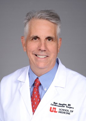 Mark S. Slaughter合作论文数Department of Cardiovascular and Thoracic Surgery, School of Medical, University of Louisville4
 Srinivas Murali合作论文数Department of Biomedical Engineering,Carnegie Mellon University4Dan Tzivoni合作论文数4
 Craig H. Selzman合作论文数Division of Cardiothoracic Surgery, University of Utah4
 Ronald Zolty合作论文数4
 Snehal G. Patel合作论文数David H. Koch Center for Cancer Care, Memorial Sloan Kettering Cancer Center4Gayathri Kamalakkannan合作论文数4




