Background Radiation Oncology (RO) clerkship grades and letters of recommendation (LoR) are non-standardized and a poorly defined landscape, thus diminishing the ability to evaluate residency candidates. Other specialties have implemented a standardized letter of evaluation (SLOE) that objectively compares applicants via standardized categories across participating institutions. We sought to design a radiation oncology SLOE (RO-SLOE) by surveying stakeholders on key performance metrics for inclusion. In parallel, we collected current grading practices. This information will inform a RO-SLOE that should be both feasible and fair for cross institution comparison during the RO-residency application cycle. Methods Anonymous REDCap surveys were distributed via email to rotating RO MSs from two institutions (n=43) and all program directors (PDs) of ACGME-accredited RO departments (n=88). Respondents answered questions on the perceived utility of RO-SLOE, 13 key proposed inclusion areas, and current evaluation practices. Likert-type scores (1, strongly agree; 5, strongly disagree) are reported as the median and interquartile range (IQR). Results 25 PDs (28%) and 18 (42%) MSs completed the survey (response rate 43/131, 33%). Respondents (n=43) agreed with the value of RO-SLOE for grading (2, IQR1-3) and residency selection (2, IQR1-3) as well as 12 of 13 of the proposed criteria to be included within the RO-SLOE. There were mixed perceptions among stakeholders regarding the inclusion of the students' ability to contribute to DEI (3, IQR1-3). The disclosure of the relationship between the evaluator and the student (2, IQR2-2) and median score awarded by the evaluator (2, IQR2-3), likewise, the students' work ethic and preparedness (1, IQR1-1) as well as the ability to communicate empathically with patients (1, IQR1-2) should be included. There were no standardized grading criteria for rotating MSs, but the most common was "honors/high pass/pass/fail (H/HP/P/F)" (83%, 20/24). Several PDs reported assigning 90-100% rotators with honors (35%, 7/20). No PD reported a threshold for honors. While the perceived value of LoR within residency selection (3, IQR2-4) was indifferent, there was consensus on the limited utility of clerkship and away rotation grades for discrimination of residency candidates (4, IQR2.5-4). Conclusion Currently within RO MS rotations, there is heterogeneity in evaluations and suggestions of grade inflation particularly in the "H/HP/P/F" systems. This may explain why many PDs question the utility of RO rotation grades during residency selection. Key stakeholders are receptive to adopting an RO-SLOE for use in MS grading and residency applications, and there is alignment on core elements for inclusion.
Purpose: Geographic and program signals allow applicants to denote preferred regions and institutions for residency training. For the 2025 Match, program signals were introduced for radiation oncology (RO). We describe the experiences of RO applicants and program directors (PDs) with these novel program signals and geographic signals. Methods and Materials: Anonymous REDCap surveys were distributed to RO applicants and PDs after Match Day 2025. Consented participants answered questions on their perspectives and behaviors surrounding signals. Results: All surveyed applicants (71, 100%) used the newly offered program signals. Many applicants (46, 65%) and PDs (25, 63%) were aware of the RO professional society guidelines regarding signaling home and away rotations. Most informed applicants did not signal their home (16/23, 70%) or all away (21/37, 57%) rotations, despite awareness of guidelines. Likewise, informed PDs advised internally (17/24, 71%) and externally rotating applicants to not signal (14/24, 58%), also against guideline recommendations. Geographic signals continued to be widely used by RO applicants (59/71, 83%), and most PDs reported using geographic signals during the selection process (30/40, 75%). Interview invitation (P = .16) and matching (P = .26) to a program within a division of a geographic signal did not differ by applicant type. Conclusions: Program signals were universally used by surveyed RO applicants for the Match 2025, with most employing all tokens. Surveyed RO PDs reported signals influenced stages of residency recruitment, including interview invitations and ranking. Our data show that program and geographic signals may influence interview invites, but their impact on final match outcomes remains unclear. There was notably poor alignment of applicant and RO PD behaviors with the best practices regarding program signals as described by governing bodies, which should be addressed in future match cycles.
Purpose: Postmastectomy radiation therapy is known to increase risk of complications in the reconstruction setting. We aim to identify the variables associated with reconstruction failure and other major complications. Methods and Materials: A prospectively collected institutional database was queried for patients with up to stage IIIC breast cancer treated from 2000 to 2017, undergoing mastectomy, immediate implant or autologous tissue reconstruction, and radiation to the recon-structed breast within 1 year of surgery. Reconstruction failure was defined as complication requiring surgical revision or implant removal. Additional major complications were defined as any infection, contracture, necrosis, or fibrosis. Covariates of interest included age, body mass index, smoking status, stage, hormone receptor and HER2 status, systemic therapy timing, radiation technique, nodal irradiation, and interval between surgery and start of postmastectomy radiation therapy. Differences in complication rates were assessed with x2 or Fisher exact tests. Competing risk regression was used to estimate hazard ratios; covariates were included one at a time to avoid over adjustment. Results: A total of 206 reconstructed breasts in 202 patients resulted from our initial query, with 139 treated with intensity-modulated radiation therapy (IMRT) and 67 treated with conventional radiation therapy (CRT). Median follow-up was 45 months (range, 4-210 months); patient cohorts were generally similar. Eight patients were excluded from toxicity analysis for insufficient follow-up (<2 years). Overall, reconstruction failure and major complication rates were significantly lower in the IMRT group. Reconstruction failure rates were 3.0% for IMRT versus 16.4% for CRT (P = .002), and major complication rates were 6.8% for IMRT versus 24.6% for CRT (P < .001). On univariate analysis, CRT was significantly predictive of implant failure (hazard ratio, 5.54; P = .003) and increased complication rates (hazard ratio, 3.83; P = .001). Significance persisted on multivariable analysis. Survival outcomes were similar, with no difference in 2 year overall survival (P = .12) and local recurrence (P = .41). Conclusions: Using IMRT may improve reconstruction outcomes over CRT, with significantly lower reconstruction failure and com-plication rates without compromising local control or survival.(c) 2022 American Society for Radiation Oncology. Published by Elsevier Inc. All rights reserved.
Supplemental Tables 1-3. Supplementary Table 1: Correlations in the vandetanib/RT/TMZ arm between best radiographic responses with pre-treatment and on-treatment changes in blood biomarkers. Supplementary Table 2: Correlations in the vandetanib/RT/TMZ arm between overall survival with pre-treatment and on-treatment changes in blood biomarkers. Supplementary Table 3: Comparison of PFS or OS by log-rank p-value based on tissue biomarker analysis
The NCCN Guidelines for Central Nervous System (CNS) Cancers focus on management of the following adult CNS cancers: glioma (WHO grade 1, WHO grade 2-3 oligodendroglioma [1p19q codeleted, IDH-mutant], WHO grade 2-4 IDH-mutant astrocytoma, WHOgrade 4 glioblastoma), intracranial and spinal ependymomas, medulloblastoma, limited and extensive brain metastases, leptomeningeal metastases, non-AIDS-related primary CNS lymphomas, metastatic spine tumors, meningiomas, and primary spinal cord tumors. The information contained in the algorithms and principles of management sections in the NCCN Guidelines for CNS Cancers are designed to help clinicians navigate through the complex management of patients with CNS tumors. Several important principles guide surgical management and treatment with radiotherapy and systemic therapy for adults with brain tumors. The NCCN CNS Cancers Panel meets at least annually to review comments from reviewers within their institutions, examine relevant new data from publications and abstracts, and reevaluate and update their recommendations. These NCCN Guidelines Insights summarize the panel's most recent recommendations regarding molecular profiling of gliomas.
The NCCN Guidelines for Central Nervous System (CNS) Cancers focus on management of the following adult CNS cancers: glioma (WHO grade 1, WHO grade 2-3 oligodendroglioma [1p19q codeleted, IDH-mutant], WHO grade 2-4 IDH-mutant astrocytoma, WHO grade 4 glioblastoma), intracranial and spinal ependymomas, medulloblastoma, limited and extensive brain metastases, leptomeningeal metastases, non-AIDS-related primary CNS lymphomas, metastatic spine tumors, meningiomas, and primary spinal cord tumors. The information contained in the algorithms and principles of management sections in the NCCN Guidelines for CNS Cancers are designed to help clinicians navigate through the complex management of patients with CNS tumors. Several important principles guide surgical management and treatment with radiotherapy and systemic therapy for adults with brain tumors. The NCCN CNS Cancers Panel meets at least annually to review comments from reviewers within their institutions, examine relevant new data from publications and abstracts, and reevaluate and update their recommendations. These NCCN Guidelines Insights summarize the panel's most recent recommendations regarding molecular profiling of gliomas.
Introduction: Patients with brain metastases (BrMs) arising from EGFR and ALK driven non-small cell lung cancer (NSCLC) have favorable prognoses and evolving treatment options. We evaluated multicenter outcomes for stereotactic radiosurgery (SRS) to multiple (>= 4) BrMs, where randomized data remain limited. Methods: Data were collected retrospectively from 5 academic centers on EGFR and ALK NSCLC who received SRS to >= 4 BrMs with their first SRS treatment between 2008 and 2018. Analyzed endpoints included overall survival (OS), freedom from CNS progression (FFCNSP), and freedom from whole-brain radiotherapy (FFWBRT).Results: Eighty-nine patients (50 EGFR, 39 ALK) received a total of 159 SRS treatments to 1,080 BrMs, with a median follow up of 51.3 months. The median number of BrMs treated with SRS treatment-1 was 6 (range 4-26) and median for all treatments was 9 (range 4-47). Sixteen patients (18 %) had received WBRT prior to SRS treatment-1. The median OS was 24.2, 21.2, and 33.2 months for all patients, EGFR, and ALK subsets, respec-tively. After multivariable adjustment, only receipt of a next-generation tyrosine kinase inhibitor was associated with OS (HR 0.40, p = 0.005). No differences in OS were observed based on number of BrMs treated. The median FFCNSP was 9.4, 11.6, and 7.5 months, for all patients, EGFR, and ALK subsets, respectively. After multivariable adjustment, the number of BrMs (continuous) treated during treatment-1 was the only negative prognostic factor associated with FFCNSP (HR 1.071, p = 0.045). The 5-year FFWBRT was 73.6 %.Conclusions: This multicenter analysis over a >10-year period demonstrated favorable OS, FFCNSP, and FFWBRT, in patients with EGFR and ALK driven NSCLC receiving SRS to >= 4 BrMs. These data support SRS as an option in the upfront and salvage setting for higher burden CNS disease in this population.
Purpose/Objective(s)Hypofractionated external beam radiotherapy with 10 to 30 fractions (fx) and stereotactic body radiation therapy (SBRT) for prostate cancer (PC) have similar rates of cancer control and toxicity at 5 years as compared to conventional treatment. There is a lack of a radiobiological modeling from lab experiments to guide this transition. By assuming a unified multi-activation (UMA) model of lab-measured radiosensitivity (RS) of the cell lines to the entire dose range, we explored the clinical dose-response, modeling tumor-control probability (TCP) and normal-tissue complication probability (NTCP).Materials/MethodsPublished cell survival curves (CSC) with experimental error bars for PC of 5 DU145 CSCs, 3 CP3 CSCs, 6 LNCaP + 2 LnCaP with BCL2 overexpressed or down-regulated CSCs, 3 wild-type (WT)-22RV1 CSCs, 3 post-radiation radioresistant (RR)-22RV1 CSCs, 2 hypoxia-22RV1 CSCs, 3 urothelial HCV-29 CSCs and 4 large intestine (LI) or small intestine (SI) stem cell and organoid CSCs were fitted with a UMA model by using a logit transformation and c2-regression to obtain the model parameters of g and n and their deviations. Cell survival at any fractional dose (=total dose/fx) and TCP and NTCP for the total number of cancer cells or normal tissue cells corresponding to a grade of toxicity were analytically determined from the parameters.ResultsAll of the CSCs had been excellently fitted with R2 > 0.95 and Q-factor > 0.2 excepting for two in-vivo LI/SI crypt CSCs each with an outline point. We identified radiobiological parameters and predicted TCP/NTCP curves and listed the data for the 8Gyx5 SBRT of PC in Table 1 that are comparable with the results listed in the HyTEC papers. The 0% TCP to RR or hypoxia PC and 100% NTCP to SI are similar to conventional EBRT. The Dg reflects the major uncertainties in the lab-measured RS. D50, the total dose at 50% TCP or NTCP, correlated with RS and number of cells. Cell heterogeneity greatly changed RS and shapes of TCP and NTCP curves.ConclusionThese findings may be useful for the design and evaluation of new clinical treatment schemes in PC translating lab work into clinical application.
PURPOSE:Management paradigms now allow for systemic targeted drugs before central nervous system (CNS)-directed radiation therapy (RT) in selected asymptomatic patients with non-small cell lung cancer (NSCLC) and brain metastases (BM). We aimed to quantify how novel targeted agents with improved CNS activity, such as second-generation anaplastic lymphoma kinase (ALK) inhibitors (eg, alectinib), might affect the role of CNS-directed RT.METHODS AND MATERIALS:This retrospective, observational, real-world, patterns-of-care study used a nationwide, electronic, health record-derived, de-identified, longitudinal database. A random sample of patients with ALK+ advanced NSCLC and BM on first-line ALK-inhibitor monotherapy between January 1, 2014 and August 31, 2019 were included. Using an index date of the first instance of BM, the outcome was brain-directed local treatment within 4 months. Trends over time were reported and tested using multivariable modified Poisson regression with robust error variance, including an indicator during or after 2017 (when alectinib was approved).RESULTS:Of the 352 included patients, 146 had BM. In addition, 104 patients received CNS-directed local therapy, and 42 did not. The majority of patients (89.4%) were treated with RT alone. Of those receiving RT, stereotactic radiosurgery monotherapy was the most common (53%), followed by whole brain RT alone (39%). On multivariable analysis, patients who had their first BM during or after 2017 had a decreased rate of receiving local BM treatment versus those before 2017 with an adjusted incidence rate ratio of 0.63 (95% confidence interval [CI], 0.41-0.95; P = .026). We found no change in the proportion of BM treated with whole brain RT during or after 2017 versus before (adjusted incidence rate ratio: 0.70; 95% CI: 0.24-2.06; P = .517).CONCLUSIONS:We found decreasing use of CNS-directed RT in patients with NSCLC with new BM on first-line ALK inhibitors. Clinical outcomes for these patients require continued investigation, because physicians may become increasingly comfortable deferring upfront local therapy for BM in lieu of novel targeted agents with improved CNS activity.
PURPOSE:This guideline provides evidence-based recommendations for adults with isocitrate dehydrogenase (IDH)-mutant grade 2 and grade 3 diffuse glioma, as classified in the 2021 World Health Organization (WHO) Classification of Tumours. It includes indications for radiation therapy (RT), advanced RT techniques, and clinical management of adverse effects. METHODS:The American Society for Radiation Oncology convened a multidisciplinary task force to address 4 key questions focused on the RT management of patients with IDH-mutant grade 2 and grade 3 diffuse glioma. Recommendations were based on a systematic literature review and created using a predefined consensus-building methodology and system for grading evidence quality and recommendation strength. RESULTS:A strong recommendation for close surveillance alone was made for patients with oligodendroglioma, IDH-mutant, 1p/19q codeleted, WHO grade 2 after gross total resection without high-risk features. For oligodendroglioma, WHO grade 2 with any high-risk features, adjuvant RT was conditionally recommended. However, adjuvant RT was strongly recommended for oligodendroglioma, WHO grade 3. A conditional recommendation for close surveillance alone was made for astrocytoma, IDH-mutant, WHO grade 2 after gross total resection without high-risk features. Adjuvant RT was conditionally recommended for astrocytoma, WHO grade 2, with any high-risk features and strongly recommended for astrocytoma, WHO grade 3. Dose recommendations varied based on histology and grade. Given known adverse long-term effects of RT, consideration for advanced techniques such as intensity modulated radiation therapy/volumetric modulated arc therapy or proton therapy were given as strong and conditional recommendations, respectively. Finally, based on expert opinion, the guideline recommends assessment, surveillance, and management for toxicity management. CONCLUSIONS:Based on published data, the American Society for Radiation Oncology task force has proposed recommendations to inform the management of adults with IDH-mutant grade 2 and grade 3 diffuse glioma as defined by WHO 2021 classification, based on the highest quality published data, and best translated by our task force of subject matter experts.
Radiation oncology is a highly multidisciplinary medical specialty, drawing significantly from three scientific disciplines—medicine, physics, and biology. As a result, discussion of controversies or changes in practice within radiation oncology involves input from all three disciplines. For this reason, significant effort has been expended recently to foster collaborative multidisciplinary research in radiation oncology, with substantial demonstrated benefit.1, 2 In light of these results, we have adopted this “team-science” approach to the traditional debates featured in this journal. This article is part of a series of special debates entitled “three discipline collaborative radiation therapy (3DCRT)”, in which each debate team has included three multidisciplinary team members, with the hope that this format would be both engaging for the readership and foster further collaboration in the science and clinical practice of radiation oncology. All 3DCRT debates thus far have included a radiation oncologist, medical physicist, and radiobiologist on each team. For this debate, we break that trend and include a patient representative along with a radiation oncologist and medical physicist on each team. We hope this patient perspective adds a valuable new aspect to our debate format and encourages the continued inclusion of patient perspectives in future clinical discussions. Medical physicists have historically contributed to patient care in radiation oncology primarily through the implementation and oversight of technology and comprehensive quality and safety programs.3, 4 However, with the introduction of more robust equipment and widespread automation, we anticipate that the time required for these tasks will decrease accordingly.5, 6 In addition, our dynamic healthcare environment continuously pressures the medical profession to redefine its contribution and value. So where can a more unfettered medical physicist provide “top of the license” contributions to the quality of patient care? One recent effort has been to cultivate increased engagement of the medical physicist with the patient for the intended result of maximizing the patient's understanding of their treatment and improving the overall healthcare experience. But does the implementation of a direct, patient-facing role for the physicist result in a substantial improvement in the patient experience and/or the quality of care? And if so, does this outweigh the value of other possible technical contributions to which physicists could re-allocate their time? Quality and safety initiatives will presumably grow as health care organizations and accreditors work to implement meaningful patient safety programs with defined executive responsibilities and accountability to specific outcomes. Does direct patient care by the physicist take time away from the physicist's contributions to these quality and safety initiatives, or is this patient interaction a valuable and critical component of such initiatives? In other words, where does the medical physicist most enhance the quality of care in radiation oncology—with the patient's technology, or with the patient? This is the subject of this month's three discipline collaborative radiation therapy debate. Arguing for the proposition will be Dr. Todd Atwood, Dr. Krisha Howell, and Mr. Charles Pearson. Dr. Atwood is an Associate Professor and Senior Associate Division Director of Transformational Clinical Physics at UC San Diego. As a native of North Carolina, Todd attended the University of North Carolina at Chapel Hill, before receiving his MS and PhD degrees from Wake Forest University. After completing a medical physics residency at Stanford University, he began to focus on maximizing the impact medical physicists have on patient care. Dr. Howell is currently an Associate Professor in Gynecologic and Sarcoma Radiation Oncology and acting Clinical Director at Fox Chase Cancer Center/Temple University. Originally from Southeastern Michigan, she received her Medical Doctorate at Wayne State University School of Medicine. She completed a residency in Radiation Oncology at the Medical University of Charleston. Additionally, she received brachytherapy fellowship training at Princess Margaret Hospital. Charles Pearson is the patient partner for the proposition. Mr. Pearson received his B.A. degree in economics from Seattle University and his M.P.H. degree in hospital administration from UC Berkeley. Prior to his management consulting career, he spent 7 years in line executive positions in both academic and non-academic (300–500+ bed) acute care hospitals. During his 40 years of directly providing management consulting services to hospitals and healthcare systems, he contracted with approximately 200 clients in 39 states. Arguing against the proposition will be Dr. Narottam Lamichhane, Dr. Stephanie Weiss, and Ms. Louise Bird. Dr. Lamichhane is an assistant professor and medical physicist in the Department of Radiation Oncology at the University of Maryland School of Medicine. He completed his therapeutic medical physics residency from the University of Miami Miller School of Medicine. His training and research interests focus on treatment planning, quality assurance, imaging, and experimental therapeutics. Dr. Weiss is a professor in the Department of Radiation Oncology, Chief of the Division of Neurologic Oncology, and Director of the Radiation Oncology Residency and Fellowship Training Program at Fox Chase Cancer Center/Temple University. She completed her residency training at Johns Hopkins Hospital and has also served as an attending physician for Brigham and Women's Hospital/Dana Farber Cancer Institute and on the faculty at Harvard Medical School. Louise Bird is the patient partner against the proposition. She lives and works in rural Saskatchewan, Canada, and is a breast cancer survivor of 18 years. She participates in many different Provincial and Pan Canadian initiatives, including serving as co-chair of the Patient and Family Advisory Council for the Saskatchewan Cancer Agency. For more about her patient story, see https://cancerfoundationsask.ca/patient/louise-bird/. As the field of radiation oncology has evolved, so has the role of the medical physicist. While the primary function of the medical physicist in radiation oncology has always centered around the design and delivery of safe and efficacious therapy, the day-to-day responsibilities of medical physicists have consistently adapted to provide patients with the highest level of care. To assure the continued value of the medical physicist in the changing healthcare landscape, the American Association of Physicists in Medicine (AAPM) created a new initiative, called “Medical Physics 3.0” (https://www.aapm.org/MedPhys30/), which aims to “redefine and reinvigorate the role of physics in modern medicine.”7 When evaluating the current needs of radiation oncology patients, one desire stands out—patients want to be more involved in their care. Research has shown that radiation oncology patients want comprehensive and detailed information about their disease and treatment procedures8; however, this is not always easily achieved. From the perspective of the patient, radiation oncology is often viewed as a complex and overwhelming medical specialty. After receiving a cancer diagnosis, patients are quickly introduced to an array of complicated imaging and treatment modalities, often with little understanding of the role they play in their care. In addition, patients also frequently face concerns and misconceptions about the use of radiation and how it can safely and effectively treat their disease. These circumstances commonly result in patients looking for answers and information about the technical aspects of their care online, where even the most reliable sources have been shown to be nonspecific or too complicated for the general public.9, 10 Unfortunately, all of these factors have the potential to negatively influence the patient experience. More importantly, the combination of these factors can create anxiety and patient-related distress, which has been shown to negatively influence outcomes following radiation therapy.11 Ensuring that all patients have the information they need to understand and feel comfortable with their care is a necessity for the field of radiation oncology. Medical physicists are ideally positioned to help address some of these concerns by leading efforts to demystify the radiation therapy process for patients. Using their comprehensive knowledge of the technology involved in radiation oncology and the specifics of each patient's treatment plan, medical physicists could ensure that all patient questions and concerns related to the technical aspects of their care are adequately addressed. Additionally, research has shown that education assists with patient enlistment in their own care, which can lead to improved adherence to treatment regimens.12 Traditionally, medical physicists have had some patient contact, but these interactions have typically been limited to brief clinical encounters or meetings with technologically savvy and inquisitive patients. Recently, more comprehensive patient-facing roles have been explored to evaluate the potential of further integrating medical physicists into direct patient care. One example is including the medical physicist in the initial radiation oncology consult to facilitate a collaborative approach to patient care at the beginning of treatment.13 This process introduces the patient to both the medical and technical experts on the care team and creates an opportunity for the radiation oncologist and medical physicist to transfer knowledge at an early stage in the treatment planning process. More extensive direct patient interactions by the medical physicist have also been studied. As part of the Physics Direct Patient Care protocol, medical physicists established independent professional relationships with patients to oversee and communicate all of the technical aspects related to the patient's care.14 After attending a dedicated patient communication training program, medical physicists routinely met with patients for two physicist–patient consults to describe the role of a medical physicist, explain the treatment planning and delivery process, review the patient's treatment plan, and answer all technical questions. The results from this trial indicated that physicist–patient consults were associated with statistically significant decreases in patient anxiety and increases in patient satisfaction. In addition to improving the patient experience, patient-facing roles for medical physicists would also strengthen clinical collaborations with radiation oncologists. Effective communication and teamwork have traditionally been assumed to be skills of expert individual practitioners, and formal training and assessment in these areas has been largely absent. By expanding the direct patient care team to include medical physicists, opportunities for shared decision-making would arise and communications bridging the technical and medical aspects of patient care would increase. This approach works to create a well-understood plan of care, which greatly reduces the chances of errors becoming consequential and injuring patients, and expresses a culture of strong, clear, and visible attention to safety.15, 16 The field of radiation oncology is interdisciplinary and requires a lot of teamwork. In the midst of this teamwork, the physicist plays a vital role in maintaining patient safety and quality of care. This delicate balance of teamwork in radiation oncology requires each division to prioritize and focus on their expertise. The smooth workflow of the radiation therapy department is facilitated by each team member carrying out their required work with diligence. A safety gate of this entire workflow is the division of physics, and a major focus of routine radiation oncology physics work is chart review. The process of chart review occurs within various steps of a clinical physics workflow such as pretreatment initial chart review, weekly chart review, and end of treatment chart review. The initial chart review is one of the most effective ways of diagnosing pretreatment errors and ensuring compliance with the prescription.17 Since the largest number of errors occurs during the planning and the pretreatment processes, chart review represents an opportunity to improve the quality assurance of the entire workflow.18 Similarly, the weekly chart review also plays a significant role in providing quality control during the course of patient treatment to catch or rule out any gross errors. In the current state of the radiation therapy workflow, the treating radiation oncologist, nurse, and clinical care team perform the direct patient interaction. The motivation behind the physicist being involved in direct patient care is noteworthy. The responsibilities of clinical physicists are evolving in the current era. However, adding direct patient care as another responsibility of a clinical physicist also comes with many challenges. For a radiation oncology department, and specifically for the division of physics, the allotment of staff is based on various factors within the department and guidance from professional societies. As such, the number of physicists required for a radiation therapy department is guided by the number of treatments, radiation oncologists, machines, special procedures, and many other clinical factors. The addition of direct patient interaction will add extra responsibilities to the established physics workflow that may not only detract from completing existing responsibilities, but also lead to miscalculation in terms of allocation of medical physicist FTE in a given department, hospital, or hospital system. As such, this will require restructuring the standard physics workflow within the department and may require additional physics resources that may not always be feasible. Most importantly, and realistically, additional responsibilities without additional staffing would likely result in a reduction in the amount of time available for traditional physics work such as chart review. This may cause a strain on the physics team that leads to potential errors and the compromise of patient safety. The American Society for Radiation Oncology published details surrounding implementation of best clinical practices in its “safety is no accident” report, and emphasizes the open communication between different divisions within the department.19 In our interpretation, clear communication between physician and physicist requires effective information sharing without impinging on the predefined responsibilities of an individual division. This allows physicists to be involved in the clinical decision-making and still focus on the patient safety that will ultimately ensure the highest quality clinical care for patients. Furthermore, the involvement of the physicist in patient care does not necessarily have to involve direct interaction. In light of the current common telehealth practices, the introduction of the care team with live video or a recorded video may provide the patient the assurance they need. Additionally, during the development of a patient's individualized treatment, the information on various treatment techniques as well as the physics behind the treatment modalities and peer review process can be shared with patients either through brochures or as a recorded video, thus assuring patients that safe and effective protocols are being practiced. Finally, a detailed discharge summary, drafted and reviewed by the care team, will be provided to the patient and to their primary care provider, which provides information about the radiotherapy treatment. Our colleagues argue that expanding the role of the medical physicist would detract from quality and safety and, moreover, contradict radiation oncology staffing models. Although we respect their concern for patient safety, we believe it is misplaced. More engaged patient-facing roles and responsibilities would strengthen quality and safety efforts, while optimizing patient care and education by leveraging the medical physicist's unique strengths. As our colleagues mentioned, chart review is an integral, but time consuming, part of a quality and safety program. However, the time required for these tasks is decreasing as software improves and automated tools for clinical decision support are introduced.5, 20 As this trend continues, we anticipate medical physicists will have the freedom to break from rote tasks and better populate their workload with “top of license” activities. We see these activities defined as cultivating direct patient care roles to educate patients on the technical aspects of their treatment, to enlist patients in their own care, and to further engage in the technical decisions of a patient's own treatment plan design. These roles could lead to improved treatment adherence,12 result in shared technical decision making with the radiation oncologist, and foster increased communication among experts on the care team. All of these factors have the potential to bring about dramatic improvements in quality and safety.16 Furthermore, we challenge our colleagues’ concerns for medical physicist involvement in “patient care” as a misconception. The physicist as an active participant in patient care is securely set within the current purview of the medical physicist. Our colleagues’ misgiving may result from, as it stands, the lack of public transparency to the physicist's role in that care. We wish to increase this transparency and reinforce that, in the radiation treatment paradigm, highest quality care of the patient is not just the responsibility of the physician, but also an outcome for which the physicist shares accountability. The value a medical physicist has in patient care is evident in expectations built into the current patient relations and clinic flow. It, however, is not often executed in a manner to build the patient's education and trust. Yet, these elements are highly crucial. In a review of more than 8,000 patient satisfaction surveys, albeit missing a medical physicist component question, patient satisfaction was greatest with regard to their perceived provider relationships.21 Beyond that, there exists a uniqueness to the relationship between a medical physicist and patient, some of which cannot be supplemented. We need to look no further than the standard procedure for HDR remote afterloader major medical emergencies. As per safety protocol, a solo physicist initially enters the treatment room in a timely manner to attempt retraction of the source.22 In such an emergency scenario, it is highly plausible that the physicist and patient would occupy the same physical space alone in a high-pressure, time-sensitive moment. If the patient was not familiar with whom this individual was or what tasks they are authorized to perform, it could add anxiety, increase time, and decrease patient cooperation while attempting to extract the source to limit radiation exposure. To summarize, although direct patient interactions represent a new role for medical physicists, the evolution of the profession should be driven by the needs of radiation oncology patients, not confined by historical responsibilities. Current data suggests patients want extensive information about their disease and treatment procedures8 and that physicist-patient consults have the ability to address these needs, with low patient anxiety and high patient satisfaction.14 In addition, recent studies have shown that a comprehensive patient communication training program for medical physicists can be created by tailoring the accepted medical school curriculum,23 and that this type of program has the ability to increase the participants’ level of confidence across multiple communication categories.24 More broadly, the development of direct patient care roles for medical physicists has the potential to facilitate professional growth within the field of radiation oncology as a whole. As medical physicists take on new responsibilities, radiation oncologists will have more time for clinical tasks, such as multi-disciplinary clinics and tumor boards, which will help cultivate larger roles in oncologic management for all cancer patients.25, 26 We agree with the opposing team that the scope of medical physicists has evolved over the years. We also agree that re-fitting the vocation within the ever-changing dynamics of radiation oncology is the right approach for medical physicists. However, modern needs are not best served by direct patient care by medical physicists. Indeed, medicine has never involved patient interaction by all players. Clinically trained pathologists and radiologists best serve patients solely through direct peer-to-peer interaction. So too do medical physicists. The collaborative division of expertise in a functioning department of radiation oncology is akin to the clinical division of expertise that is enhanced by bringing these experts together in multi-disciplinary conferences. This promotes seamless throughput of patient care without compromising patient safety. We agree that patient awareness of the type of treatment and the methodology of treatment they are receiving is of utmost importance. However, this is not necessarily best achieved with direct physicist–patient interaction. Patient-related information sharing can be accomplished by electronic means, or printed materials provided by the patient's established clinical care team. This method is not only more efficient and cost-effective but potentially less overwhelming for the patient. We agree with the opposing team that medical physicists should use their technological expertise to increase the visibility of medical physics. However, providing each patient with a consultation by a physicist will require significant departmental resources, add an unnecessary burden to the patient in terms of increased time with the ever-expanding team, and may even depersonalize the experience. Is it not possible that the physician and the physicist may articulate the different rationale for particular recommendations or details of a treatment plan? Any discrepancy or perceived discrepancy has the potential to add stress and cause additional anxiety. Finally, even if the benefits were agreed to outweigh the risks (which we do not at this time concede), not every radiation oncology department will have sufficient resources to accommodate such costly time commitment from the physicist. Therefore, the inclusion of direct patient care duties must be evaluated with a view of the cost–benefit analysis impact on the medical physicist and the radiation oncology department.13 The opposing team brings up a good point regarding the new initiative by AAPM called “MedPhys 3.0” (https://www.aapm.org/medphys30/), and we support this initiative to redefine and reinvigorate the practice of medical physics. One of the initiatives of MedPhys 3.0 is to promote new physics contributions in all areas of medicine including domains beyond radiation medicine. The foundation of medicine is its underlying research. Medical physics research plays an important role in shaping the field of radiation oncology. Thus, we believe that the MedPhys 3.0 initiative may be best achieved by extending medical physics research into contemporary fields of medicine in lieu of directing physics efforts in direct patient interaction. None. All authors were responsible for preparation of arguments, and writing and reviewing the manuscript. The authors declare no conflict of interest.
Meningioma is the most common benign brain tumor of adults. For 50 years, neurosurgery was the standard of care and has provided the basis for our evidence for the regions at risk, behavior, and patterns of relapse for this tumor with a long but persistent natural history. As its role in treatment of grade I lesions continues to evolve, it is increasingly clear that radiotherapy plays an important part in durable local control and long-term patient outcomes. However, there is considerable variation in radiotherapeutic approach, particularly as pertains to defining the region at risk (i.e., GTV and CTV), technique (radiosurgery vs. conventional fractionation), and indications for and timing of treatment (i.e., up front or at recurrence). This chapter provides guidance to the radiotherapeutic approach of benign meningioma based on evidence from multidisciplinary sources, including recommendations for margins based on control by extent of resection and pathologic analysis of subclinical tumor extension after dural stripping, and on timing of intervention based on histologic behavior of recurrent tumor with and without radiotherapy up front.
PurposeApplication of linear‐quadratic (LQ) model to large fractional dose treatments is inconsistent with observed cell survival curves having a straight portion at high doses. We have proposed a unified multi‐activation (UMA) model to fit cell survival curves over the entire dose range that allows us to calculate EQD2 for hypofractionated SBRT, SRT, SRS, and HDRB.MethodsA unified formula of cell survival using only the extrapolation number of n and the dose slope of Do was derived. Coefficient of determination, R2, relative residuals, r, and relative experimental errors, e, normalized to survival fraction at each dose point, were calculated to quantify the goodness in modeling of a survival curve. Analytical solutions for α and β, the coefficients respectively describe the linear and quadratic parts of the survival curve, as well as the α/β ratio for the LQ model and EQD2 at any fractional doses were derived for tumor cells undertaking any fractionated radiation therapy.ResultsOur proposed model fits survival curves of in‐vivo and in‐vitro tumor cells with R2 > 0.97 and r < e. The predicted α, β, and α/β ratio are significantly different from their values in the LQ model. Average EQD2 of 20‐Gy SRS of glioblastomas and melanomas metastatic to the brain, 10‐Gy × 5 SBRT of the lung cancer, and 7‐Gy × 5 HDRB of endometrial and cervical carcinomas are 36.7 (24.3–48.5), 114.1 (86.6–173.1),, and 45.5 (35–52.6) Gy, different from the LQ model estimates of 50.0, 90.0, and 49.6 Gy, respectively.ConclusionOur UMA model validated through many tumor cell lines can fit cell survival curves over the entire dose range within their experimental errors. The unified formula theoretically indicates a common mechanism of cell inactivation and can estimate EQD2 at all dose levels.
Refer the patient 1 Nguyen C. Sirineni G. Diabetes insipidus: To treat or not to treat?. Int J Radiat Oncol Biol Phys. 2021; 109: 651-652 Abstract Full Text Full Text PDF Scopus (3) Google Scholar to a high-volume surgical center. If biopsy remains unsafe, rule out sarcoid by assessing cerebrospinal fluid (CSF) angiotensin-converting enzyme. A steroid trial will lead to a radiographic response. Metastatic cancer should be ruled out by adding abdominal and pelvic scans and repeat brain magnetic resonance imaging (MRI) to assess for progression or new lesions. Rathke’s cleft cysts do not enhance, and cyst drainage is typically successful. Diabetes Insipidus: To Treat or Not to Treat?International Journal of Radiation Oncology, Biology, PhysicsVol. 109Issue 3PreviewA 35-year-old woman with no significant history presented with polyuria and polydipsia after the birth of her second child. The patient was evaluated by endocrinology, and after appropriate laboratory tests, she was diagnosed with central diabetes insipidus (DI). Full-Text PDF
Leptomeningeal disease in patients with melanoma historically portends a grim prognosis, with median survival measured in weeks to months. The advent of effective immunotherapy and targeted agents may modify the outcome of such patients. This case report describes a 43-year-old patient diagnosed with stage IIIa BRAF-positive cutaneous melanoma in 2012 who subsequently developed leptomeningeal involvement as her sole site of melanotic metastasis. She received multiple systemic therapies and radiotherapy and survived 2.5 years after her diagnosis with central nervous system involvement. This case report highlights the importance of a multidisciplinary team and the advent of effective agents, which offers the potential for significantly improved outcomes for patients with metastatic melanoma involving the central nervous system.
 Brian M. Alexander合作论文数Dana-Farber Cancer Institute;Harvard Medical School;GCAR;Roche/Genentech Pharma R&D32
 Patrick Y. Wen合作论文数Department of Neurology, Harvard Medical School;Center For Neuro-Oncology, Dana-Farber Cancer Institute21
 Shelly B Hayes合作论文数17
 Jan Drappatz合作论文数Center for Neuro-Oncology, Department of Medical Oncology, Dana-Farber/Brigham and Women’s Cancer Center, 44 Binney St, Boston, MA 02115, USA14
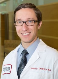 Churilla Thomas M合作论文数12
 Eudocia Q. Lee合作论文数Division of Cancer Neurology, Department of Neurology, Brigham and Women's Hospital, Harvard Medical School;Center for Neuro-Oncology, Dana-Farber Cancer Institute12
 Ayal A. Aizer合作论文数Harvard Medical School;Department of Radiation Oncology, Brigham and Women's Hospital12
 Yanqun Dong合作论文数11
 Nils D. Arvold合作论文数Harvard Radiation Oncology Program, Harvard Medical School11Kaley Thomas J合作论文数11
 Fred Hacker合作论文数10
 Penny Anderson合作论文数10
 Elizabeth Handorf合作论文数Fox Chase Cancer Center, Temple University Health System, Inc.9
 Paul J Kelly合作论文数8Laurence Collette合作论文数8
 Elizabeth Claus合作论文数Department of Biostatistics, School of Public Health, Yale University;Department of Neurosurgery, Brigham and Women’s Hospital;Harvard Medical School8
 Riccardo Soffietti合作论文数8M. Kocher合作论文数7Pinnell Nancy E合作论文数7
 Joshua E. Meyer合作论文数Department of Radiation Oncology, Fox Chase Cancer Center;Quantigic® Genomics7
 Schiff, David合作论文数School of Medicine, University of Virginia7
 Alona Muzikansky合作论文数Biostatistics Center, Massachusetts General Hospital7
 Tracy Batchelor合作论文数Harvard Medical School;Department of Neurology, Brigham and Women's Hospital;Cancer Center, Mass General Research Institute, Massachusetts General Hospital7Richard J. Bleicher合作论文数Mercy Hospital of Pittsburgh, Mercy Cancer Institute7
 Ian F. Parney合作论文数Cancer Center, Mayo Clinic;Department of Neurosurgery, Mayo Clinic7
 Andrew D. Norden合作论文数Boston Medical Center;OncoHealth7Lode Swinnen合作论文数6
 Larry Junck合作论文数Department of Neurology, Medical School, University of Michigan6
 Joachim Baehring合作论文数Department of Neurology, Yale School of Medicine, Yale University6
 Seema Nagpal合作论文数Neurosurgery Department, Stanford University6Galloway Thomas J合作论文数Department of Radiation Oncology, Fox Chase Cancer Center6
 Maciej M. Mrugala合作论文数Department of Neurology, Mayo Clinic6Shonka Nicole A合作论文数University of Nebraska Medical Center6Rusthoven Chad G合作论文数Department of Radiation Oncology, University of Colorado School of Medicine6Portnow Jana合作论文数6
 Keith L. Ligon合作论文数Center for Patient Derived Models, Dana-Farber Cancer Institute;Division of Neuropathology, Brigham and Women's Hospital;Medical School, Harvard University;Travera;Broad Institute, Massachusetts Institute of Technology;Boston Children's Hospital6
 Eric T Wong合作论文数Division of Hematology/Oncology, Department of Medicine, Rhode Island Hospital;Department of Neurology, Warren Alpert Medical School, Brown University;Lifespan Cancer Institute6
 Louis Nabors合作论文数Department of Neurology, School of Medicine, The University of Alabama at Birmingham;Department of Cell, Developmental and Integrative Biology, The University of Alabama at Birmingham6Charlie Ma合作论文数6
 Jona Hattangadi-Gluth合作论文数Radiation Oncology Division, Department of Radiation Medicine & Applied Sciences, School of Medicine, University of California, San Diego6Nicholas Butowski合作论文数6
 Mark A Hallman合作论文数6Mary Anne Bergman合作论文数5
 Katherine Barnett Peters合作论文数Department of Neurology, Duke University School of Medicine;Duke Cancer Institute5Talha Shaikh合作论文数5
 Tom Mikkelsen合作论文数Hermelin Brain Center, Henry Ford Hospital;Department of Neurology, Henry Ford Hospital;Ontario Brain Institute5
 Nancy U. Lin合作论文数Department of Medical Oncology, Dana-Farber Cancer Institute;Harvard Medical School5Tianyu Li合作论文数5
 Ian Dunn合作论文数The Roslin Institute, The University of Edinburgh/The Royal (Dick) School of Veterinary Studies, The University of Edinburgh5




