Purpose This study aimed to evaluate the protective effects of melatonin on colon tissue integrity in rats exposed to low- and high-dose rate radiotherapy. Materials and methods Forty rats were randomly divided into: Group1 (Control) received no treatment. Group2 was exposed to a single dose of 8 Gy radiotherapy at a low-dose rate (LDR) of 400 MU/min. Group3 (LDR+Melatonin) received of 50 mg/kg melatonin, followed by 8 Gy LDR radiotherapy (400 MU/min) to the abdominopelvic region 15 min later. Group4 was treated with 8 Gy radiotherapy at a high-dose rate (HDR) of 1400 MU/min. Group5 (HDR+Melatonin) received 50 mg/kg melatonin 15 min before 8 Gy HDR radiotherapy (1400 MU/min). Colon tissues were harvested 48 h after radiotherapy for analyses. Results Radiotherapy induced damage to colon tissues, characterized by disruption of crypt architecture and depletion of goblet cells. Melatonin treatment effectively preserved these structures, exhibiting a protective effect (p < 0.001). The Bax/Bcl-2 ratio, a key marker of apoptosis, was significantly reduced in melatonin-treated groups compared to radiotherapy-only groups (p < 0.001), suggesting a reduction in apoptotic cell death. Furthermore, melatonin administration resulted in a substantial increase in total antioxidant status (T-AS) and a significant decrease in total oxidative status (T-OS) and oxidative stress index (O-SI) levels (p < 0.001). Conclusions Melatonin significantly mitigated radiation-induced oxidative stress and apoptosis in healthy colon tissue, as evidenced by the reduction in the Bax/Bcl-2 ratio. Melatonin may serve as an effective radioprotective agent, preserving the integrity of colon tissue during both low- and high-dose rate abdominopelvic radiotherapy.
PURPOSE:Radiotherapy of abdominal and thoracic malignancies often results in unintended exposure of healthy gastrointestinal tissues, leading to oxidative stress, inflammation, and DNA damage in the gastric mucosa. Melatonin, a potent endogenous antioxidant with well-established anti-inflammatory and free radical-scavenging properties, has been proposed as a promising radioprotective agent. This study aimed to investigate the dose-rate-dependent radioprotective effects of melatonin on gastric tissue exposed to low- and high-dose rate radiotherapy. METHODS:Rats were randomly divided into control (n = 8), low-dose rate radiotherapy (LwDRR; n = 8; 8 Gy at 400 MU/min), high-dose rate radiotherapy (HgDRR; n = 8; 8 Gy at 1400 MU/min), and corresponding melatonin-treated groups (LwDRR + Mel; n = 8 and HgDRR + Mel; n = 8). Melatonin (50 mg/kg) was administered intraperitoneally 15 min before irradiation. Animals were sacrificed 48 h after treatment. Gastric tissues were evaluated histopathologically using H&E and PAS staining, oxidative DNA damage was assessed via 8-OHdG immunolocalization, and biochemical markers including TNF-α, IL-10, TAS, TOS, and OSI were analyzed. RESULTS:Both radiotherapy protocols significantly induced gastric tissue injury, characterized by histopathological alterations, increased oxidative DNA damage, and elevated inflammatory and oxidative stress markers. 8-OHdG expression and TNF-α, TOS, and OSI levels were significantly increased in irradiated groups, whereas TAS levels were reduced, particularly under high-dose rate irradiation. Melatonin administration markedly attenuated these alterations by reducing histopathological damage, decreasing 8-OHdG expression and oxidative stress markers, and increasing anti-inflammatory IL-10 levels. No significant differences were observed between low- and high-dose rate radiotherapy in any measured parameter. CONCLUSION:Melatonin effectively mitigates radiation-induced gastric injury by modulating oxidative stress, inflammatory responses, and oxidative DNA damage. Collectively, these results suggest that melatonin may serve as a promising adjunctive radioprotective agent for preserving healthy gastrointestinal tissues during radiotherapy.
PURPOSE:This study investigated the radioprotective effect of melatonin (Mel) on small intestinal damage in healthy rats exposed to Flattening Filter (FF) and Flattening Filter Free (FFF) X-rays during radiotherapy, with a comparative evaluation of the biological effects of FF and FFF irradiation on intestinal tissue injury. MATERIALS AND METHODS:Forty male Sprague-Dawley rats (12 weeks, 250±20 g) were randomly assigned to five groups. Group 1 (Control) received no treatment. Group 2 (FF) was exposed to 8 Gy radiotherapy at 4 Gy/min to the abdominopelvic region. Group 3 (FF+Mel) received melatonin 15 minutes before the same irradiation. Group 4 (FFF) was treated with 8 Gy at 14 Gy/min, and Group 5 (FFF+Mel) received melatonin before this exposure. Intestinal tissues were collected 48 h after the experimental procedures and evaluated histopathologically using hematoxylin-eosin (H&E) and periodic acid-Schiff (PAS) staining, immunofluorescently by 8-hydroxy-2'-deoxyguanosine (8-OHdG) antibody labeling, and biochemically by measuring serum 8-OHdG and malondialdehyde (MDA) levels. RESULTS:Significant histopathological differences were observed between the control (G1) and irradiated groups (FF (G2); FFF (G4)), including mucosal disruption, crypt loss/dilatation, goblet cell reduction, and hemorrhage. In addition, 8-OHdG expression was significantly higher in the FFF group (G4) compared to the FFF+Mel group (G5) (p<0.05). Biochemically, both 8-OHdG and MDA levels were elevated in irradiated groups (G2, G4) compared with controls (p<0.05). Notably, melatonin administration significantly reduced oxidative stress markers, with differences between FF (G2) and FF+Mel (G3), as well as FFF+Mel (G5) (p<0.05). CONCLUSION:Melatonin significantly reduced radiotherapy-induced oxidative stress and apoptosis in healthy small intestinal tissue histopathologically and biochemically. These findings suggest that melatonin may act as an effective radioprotective agent by preserving small intestinal integrity during abdominopelvic radiotherapy.
This study aimed to investigate the histopathological and molecular effects of melatonin on ovarian follicle reserve, SMAD7 immunolocalization, and SMAD7-related miRNAs (miR-182, miR-21, miR-92) in a rat model of adnexal torsion-detorsion (TD). Twenty-eight adult female rats were divided into four groups (n = 7). Group 1 (Control) underwent no procedure, while Group 2 (TD) received 2-h torsion followed by 2-h detorsion. Group 3 (TD + Solvent) and Group 4 (TD + Melatonin) were administered ethanol or melatonin intraperitoneally 15 min before detorsion, respectively. Ovarian tissues and serum samples were analyzed histopathologically, biochemically, and molecularly. In Group 2, follicular atrophy, cystic degeneration, and increased connective tissue and vascular congestion were observed, whereas Group 4 exhibited reduced cystic follicles and preserved follicular architecture. SMAD7 expression decreased in Group 2 and increased in Group 4. Although not reaching statistical significance, TOS tended to increase and TAS tended to decrease in Group 2, whereas the opposite trend was observed in Group 4. miR-21, miR-182, and miR-92 expression levels significantly increased in Groups 2 and 3 compared with the control but remained comparable to the control in Group 4. These findings indicate that melatonin contributes to the attenuation of acute structural damage in ovarian follicles, modulation of oxidative stress, and regulation of SMAD7-related miRNAs in the TD model. Melatonin may play a protective role against torsion-detorsion-induced acute ovarian injury, showing a strong association with the modulation of oxidative stress, SMAD7 immunoreactivity, and specific miRNA expression levels.
Ischemia and reperfusion (Is-Rp) of the kidney constitutes the leading cause of acute kidney injury (AKIn). This is an evolving process that is frequently linked to the failure of several organs. The distant organ injuries such as acute liver and lung injuries are the side effects of AKIn. In the current study, bilateral renal Is-Rp was induced in rats, and the impact of curcumin and melatonin on the lung and liver was investigated. Thirty-five adult rats, aged 10-12 weeks, were randomly assigned to five groups: the Control Group (Group 1), which underwent no surgical procedure. All other groups underwent 45 min of kidney ischemia, followed by 2 h of reperfusion. Group 2 was not administered any substance. Prior to reperfusion, the following treatments were applied: Group 3 received Is-Rp combined with melatonin (MEL), Group 4 received Is-Rp with curcumin (CUR), and Group 5 received Is-Rp with both MEL and CUR. Histological analysis was performed using Hematoxylin and Eosin (H&E), Periodic Acid-Schiff (PAS), Toluidine Blue (TB), and Masson's Trichrome (TRI) staining techniques. The preparations were evaluated for changes, and severity was graded on a scale of 0 (none), 1 (mild), 2 (moderate), and 3 (severe). The scores were aggregated and presented as liver and lung injury indices for each rat. The liver tissues of the rats were assessed for leukocyte accumulation, fibrotic alterations, sinusoidal expansion, and vascular congestion. Simultaneously, the lung tissues were evaluated for alveolar collapse, intra-alveolar bleeding, mononuclear leukocyte presence within the alveoli, intravascular leukocyte accumulation, and necrotic changes. Statistically significant differences were observed among the groups in lung injury scores, liver injury scores, and mast cell counts (p < 0.000∗∗). The use of melatonin-curcumin together reduces distant organ damage caused by renal Is-Rp injury. The concurrent use of these two antioxidants exhibits a more effective therapeutic effect than their individual application.
Aim This study aimed to evaluate the protective effects of oleuropein, a compound recognised for its antioxidant and anti-apoptotic properties, against renal ischemia/reperfusion injury (IRI) in rats. Materials and Methods Oleuropein, purified from Olea europaea L. (olive tree) by chromatographic methods, was injected intraperitoneally 48 h, 24 h, and 30 min prior to ischemia. Forty male Sprague-Dawley rats were randomly divided into five groups: Control, IRI, and three treatment groups subjected to IRI and treated with oleuropein at 10, 50, and 100 mg/kg, respectively. In all IRI groups, 45 min of bilateral renal ischemia was followed by 24 h of reperfusion. Biochemical analyses included total antioxidant status (TAS), total oxidant status (TOS), superoxide dismutase (SOD), and paraoxonase-1 (PON-1) levels in serum and kidney tissues. Renal function was assessed by serum creatinine, urea, and uric acid levels. Apoptotic markers, including cytochrome c and caspase-3, were evaluated by Western blotting and immunofluorescence. Results In the IRI group, TAS and SOD levels significantly decreased, whereas TOS and PON-1 increased (p<0.05). Oleuropein administration improved these parameters in a dose-dependent manner. At 50 mg/kg, oxidative stress markers and renal function tests approached control values (p<0.001). Apoptotic markers were elevated in the IRI group but markedly reduced after oleuropein treatment. Conclusion Oleuropein protected kidney tissue against IRI by reducing oxidative stress and apoptosis. These results suggest that oleuropein may serve as a potential therapeutic agent against renal IRI.
Radiation-induced nephropathy is a major concern, particularly for patients undergoing radiotherapy. This study investigates melatonin’s (MEL) protective effects against kidney damage induced by Flattening Filter (low dose rate-LDR) and Flattening Filter Free (high dose rate-HDR) X-ray by focusing on apoptosis and SIRT1 modulation in rats. Forty rats were divided into five groups (n = 8): Group 1 (control), Group 2 (FF-LDR, 400MU/min), Group 3 (MEL + FF-LDR), Group 4 (FFF-HDR, 1400MU/min), and Group 5 (MEL + FFF-HDR). Groups 3 and 5 received 10 mg/kg MEL intraperitoneally 15 min before an 8 Gy abdominal irradiation. Rats were sacrificed 48 h post-radiotherapy for histopathological and immunohistochemical analyses of the kidney. The FFF-HDR group showed fewer atypical glomeruli and lost brush borders, and epithelial connections compared to the FF-LDR group. The MEL + FFF-HDR group had significantly fewer atypical glomeruli and brush border losses than the MEL + FF-LDR group. Additionally, while SIRT1 expression increased in both the FF and FFF groups following MEL treatment, this increase was significant only in the MEL + FFF-HDR group compared to the control group. MEL pretreatment effectively decreased cellular apoptosis in both MEL + FFF-HDR and MEL + FF-LDR groups. Melatonin has been proven to be an effective radioprotective agent against renal nephropathy and cellular apoptosis induced by FF and FFF beams. Additionally, pre-treatment with MEL has been shown to enhance SIRT1 expression, thereby protecting the kidneys from acute radiation damage, particularly from high-dose rate radiation.
The objective of this study to examine the effects of curcumin and gallic acid use against oxidative stress damage in the autologous intraperitoneal ovarian transplantation model created in rats on ovarian follicle reserve, ovarian surface epithelium, and oxidant-antioxidant systems. 42 adult female Sprague Dawley rats (n=7) were allocated into 6 groups. Group 1 served as the control. In Group 2, rats underwent ovarian transplantation (TR) to their peritoneal walls. Group 3 received corn oil (CO) (0.5ml/day) one day before and 14 days after transplantation. Group 4 was administered curcumin (CUR) (100mg/kg/day), Group 5 received gallic acid (GA) (20mg/kg/day), and Group 6 was treated with a combination of curcumin and gallic acid via oral gavage after transplantation. Rats were sacrificed on the 14th postoperative day, and blood along with ovaries were collected for analysis. The removed ovaries were analyzed at light microscopic, fluorescence microscopic, and biochemical levels. In Group 2 and Group 3, while serum and tissue Total Oxidant Levels (TOS) and Oxidative Stress Index (OSI) increased, serum Total Antioxidant Levels (TAS) decreased statistically significantly (p˂0.05) compared to the other groups (Groups 1, 4, 5, and 6). The ovarian follicle reserve was preserved and the changes in the ovarian surface epithelium and histopathological findings were reduced in the antioxidant-treated groups (Groups 4, 5, and 6). In addition, immunofluorescence examination revealed that the expression of Cytochrome C and Caspase 3 was stronger and Ki-67 was weaker in Groups 2 and 3, in comparison to the groups that were given antioxidants. It can be said that curcumin and gallic acid have a histological and biochemical protective effect against ischemia-reperfusion injury due to ovarian transplantation, and this effect is stronger when these two antioxidants are applied together compared to individual use.
Objective This study aimed to explore the radioprotective effect of melatonin against acute testicular injury caused by low- and high-dose-rate radiotherapy in rats. Materials and methods A total of 40 12-week-old adult male rats were divided into 5 groups (n = 8). Group 1 comprised control rats who were not subjected to any procedures. A single dose of 8 Gy (Gy) radiotherapy was administered to the abdominopelvic regions of Group 2 (low-dose rate radiotherapy (LDRR) group) and Group 4 (high-dose rate radiotherapy (HDRR) group) rats at dose rates of 400 MU/min and 1400 MU/min, respectively. A single dose of 8 Gy radiotherapy was administered to Group 3 (low-dose rate radiotherapy + melatonin (LDRR + MEL) group) and Group 5 (high-dose rate radiotherapy + melatonin (HDRR + MEL) group) rats at dose rates of 400 MU/min and 1400 MU/min, respectively. Following this, 50 mg/kg/intraperitoneal melatonin was given to the rats 15 min before radiotherapy. The subjects were sacrificed 48 h after radiotherapy. Histological, immunohistochemical, immunofluorescence, and biochemical analyses of testicular tissue samples were performed. Results As a result of comparing the FF and FFF radiotherapy groups with the control group, a statistically significant difference was observed in histopathological, biochemical, immunohistochemical and immunofluorescent parameters (p < 0.001). An improvement in these parameters was observed in the groups where melatonin was applied along with radiotherapy (p < 0.001). Additionally, no statistically significant difference was found between FF and FFF dose rates (p > 0.05). Conclusion No significant difference was observed between these low- and high-dose rates of radiotherapy in terms of testicular damage and the effect of melatonin. Further, melatonin may be useful in preventing testicular damage caused by low- and high-dose rate radiotherapy.
OBJECTIVES:Recent studies have shown that oxidative stress, inflammation, and apoptosis play substantial roles in the pathogenesis of tissue damage and distant organ damage during ovarian transplantation. Because of their potential antioxidant and anti-inflammatory properties, investigating natural compounds like curcumin and gallic acid is crucial. This study investigated the effects of curcumin, gallic acid, and their combination on liver and kidney tissues in a rat model of ovarian transplantation. MATERIALS AND METHODS:For this study, we used 42 adult female Sprague-Dawley rats aged 11 to 12 weeks, which we randomly divided into 6 groups. After the transplant procedures, we performed histopathological analysis, biochemical examinations, and statistical analysis. RESULTS:Significant damage was shown in the liver and kidney tissues of the ovarian transplant only and transplant plus corn oil groups compared with the control group, as evidenced by histopathological evaluation, increased BAX/BCL-2 intensity indicating apoptotic activity, and elevated interleukin 6 levels. However, treatment with curcumin, gallic acid, or their combination attenuated these adverse effects, suggesting potential protective effects against transplant-induced injury. CONCLUSIONS:Our findings highlight the therapeutic potential of these compounds in mitigating tissue damage and inflammation associated with ovarian transplant.
Background: To compare the changes caused by low and high dose rate radiotherapy in the bladder tissue of rats and to examine the effect of melatonin on the bladder tissue.Materials and Methods: 40 adult rats were randomly divided into five groups with 8 rats in each group.Radiotherapy and melatonin treatment were not applied to group 1 (G1) control group rats.A single dose of 8 Gy and 400 MU/min radiotherapy was applied to G2 and G3 group rats.A single dose of 8 Gy and 1400 MU/min radiotherapy was applied to G4 and G5 group rats.G3 and G5 group (treated groups) rats were given 50 mg/kg melatonin intraperitoneally 15 minutes before the radiotherapy application.Rats were sacrificed under anesthesia and bladder tissues were removed.Histopathological examination was performed on the samples stained with hematoxylin eosin and toluidine blue.Results: Number of mast cells was increased in G2 and G4 (p<0.01).In addition, edema and vascular congestion were observed in these groups.In G3 and G5, acute phase markers decreased compared to radiotherapy received groups. Conclusion:Bladder tissue degradation was observed in G2 and G4 compared to the control group.However, there was no difference in bladder tissue between the groups given two different doses.The acute phase markers of edema, number of mast cells, inflammatory cell infiltration, and vascular congestion in the bladder tissue of the groups administered melatonin decreased.
Melatonin is a hormone that exhibits many bioactivities such as antioxidant, anti-inflammatory, anti-carcinogenic, anti-diabetic, neuroprotective, and anti-aging activity. In addition, melatonin has been used to strengthen the effect of drugs or agents used for treatment in many different disease models such as cancer, tumor, bacterial infection, autoimmune diseases, and gastrointestinal diseases. According to recent studies, curcumin is used as a pigment and powerful antioxidant and anti-inflammatory agent with therapeutic potential against various cancers. Melatonin and curcumin have been used in different models of disease or injury: such as Alzheimer's disease treatment, testicular tissue preservation, bladder cancer treatment, gastric mucosal damage, and prevention of nephrotoxicity. This review covers published literature studies on the effects of comparison or combined use of melatonin and curcumin as anticancer and antioxidant agents and it reveals whether melatonin potentiates the effect of curcumin when used together.
The objective of this study was to assess the impact of Vitamin E (Vit E) and Vitamin C (Vit C) on markers of the oxidant-antioxidant system, ovarian follicle reserves, and the surface epithelium in autologous intraperitoneal ovarian transplantation conducted in rats. The study aimed to investigate how these antioxidants influence various aspects related to transplantation outcomes, including oxidative stress markers, the preservation of follicle reserves, and the condition of the surface epithelium. A total of 20 adult female Wistar Albino rats were included in the study and randomly assigned to four different groups. Group 1, consisting of 5 rats, served as the control group and underwent a surgical procedure where their abdomens were opened and closed without any further intervention. Group 2, also consisting of 5 rats, underwent ovarian transplantation. In Group 3, comprising 5 rats, an intraperitoneal (IP) administration of 20 mg/kg body weight (b.w.) of Vitamin E (Vit E) was given 15 min prior to ovarian transplantation. Lastly, in Group 4, which included 5 rats, an IP administration of 50 mg/kg body weight (b.w.) of Vitamin C (Vit C) was given 15 min before ovarian transplantation. Vaginal cytology was performed in order to monitor the estrus phase in the rats. Biochemically, tissue and serum malondialdehyde (MDA) levels and erythrocyte superoxide dismutase (SOD) levels were measured. Histopathologically, the number of dysplastic changes in the ovarian surface epithelium and primordial, primary, secondary, Graaffian, and atretic follicles were examined. Dysplastic changes in the surface epithelium of Group 2 were found to be significantly higher than in Group 1 and 4 (p < 0.02). In Group 2, the ovarian follicle reserves (primordial, primary, secondary, and Graaffian follicles) were significantly lower than in other groups (p < 0.02). In addition, a significant decrease in SOD levels was found in Group 2 compared to other groups (p < 0.02). The study showed that Vit E and Vit C in autologous intraperitoneal ovarian transplantation preserved the ovarian follicle reserve. Vit C was found to be more effective than Vit E.
Polycystic ovary syndrome (PCOS) is a debilitating endocrine and metabolic disorder that affects a large proportion of women in their reproductive years. It differs by a range of symptoms including polycystic ovary appearance, hyperandrogenism, chronic anovulation, insulin resistance, and obesity. Despite years of research, the exact cause of PCOS remains elusive, but recent studies have suggested that epigenetic mechanisms may play a significant role in its pathogenesis. Of particular interest are micro-RNAs (miRNAs), short non-coding RNAs that are transcribed from DNA but not translated into protein. Recent research has demonstrated that abnormal expression of miRNAs is present in PCOS and may contribute to the development and progression of the disease. This review aims to provide an in-depth analysis of the current knowledge and challenges related to abnormal miRNA expression in PCOS, shedding light on a potential avenue for targeted therapies and improved management of this debilitating condition. The review summarizes the findings from various studies that have investigated the role of miRNAs in PCOS. It discusses the specific miRNAs that have been found to be dysregulated in PCOS and their potential impact on the pathophysiology of the disease. The review also highlights the challenges associated with studying miRNAs, including the complexity of their regulation and the need for standardized methodologies for miRNA profiling. Based on the available evidence, abnormal expression of miRNAs appears to be a significant contributor to the development and progression of PCOS. Targeting these dysregulated miRNAs could offer new therapeutic strategies for the management of PCOS. Biomarkers and gene therapies associated with miRNA may improve the accuracy and effectiveness of PCOS diagnosis and treatment. However, further research is needed to fully understand the functional roles of specific miRNAs and their potential as diagnostic or therapeutic targets.
Background: To investigate the effects of tannic acid (TA) use on ovarian folliculogenesis, p53 expression, and serum luteinizing hormone (LH), follicle-stimulating hormone (FSH), and testosterone (T) levels in rats with polycystic ovary syndrome (PCOS) model. Materials and Methods:A total of 18 Wistar female rats were used and divided into 3 experimental groups.Group 1 (G1, control), group 2 (G2, PCOS), group 3 (G3, PCOS+TA).Rats were induced with letrozole for 21 days to form a PCOS model.After 21 days, TA (40 mg/kg) was given by gavage for 10 days and the rats were sacrificed on the 10 th day.PCOS formation was evaluated by daily estrous cycle follow-up.Hematoxylin & eosin and p53 immunohistochemical staining was performed on ovaries.Serum FSH, LH, and T levels were determined by ELISA.Data were analyzed with the One-Way ANOVA test and Kruskal-Wallis H test. P<0.05 was considered statistically significant. Results:The number of cystic follicles was significantly increased in the PCOS group compared to the TA treatment group (p<0.05).The number of primary follicles was significantly increased in the TA treatment group (p<0.001).No significant change was observed in the number of primordial, secondary, and Graaf follicles between the experimental groups.A significant increase in LH and T was observed in the PCOS group (p<0.05).The increase in LH has significantly decreased TA administered rats (p<0.05).Although it was not significant, serum FSH level was increased in the PCOS+TA group.No immunoreactivity was detected in p53 staining in experimental groups. Conclusion:TA can decrease cystic follicle formation and increase primary follicle formation in PCOS.Also, it can regulate the hormonal expression of serum LH, FSH, and T in PCOS-modeled rats.
Endometriosis is a common gynecological hurting disorder in which tissue is similar to the tissue that normally lines the inner layer of the uterus. It often causes fertility problems. Unfortunately, effective treatments are limited. Therefore it's important to explore an imperative and easily accessible treatment to alleviate the probable pathologies and preserve fertility in endometriosis. Consequently, we aimed to investigate the effects of metformin, letrozole, and atorvastatin on inflammation and apoptosis in experimentally induced ovarian and peritoneal endometriosis in rat models. In the present study, 35 rats were randomly divided into five groups. Group 1: sham-operated control group. Group 2: untreated endometriosis group. Group 3: given 100 mg/kg/day of oral metformin. Group 4: given 0.1 mg/kg/day of oral letrozole. Group 5: given 2.5 mg/kg/day of oral atorvastatin. At the end of the 28 days, we examined Ki67, Bax and Bcl-2 immunoexpressions in ovarian and peritoneal tissues, and IL-6, IL-8, and TNF-α levels were evaluated from the peritoneal fluid. All medical treatment groups showed a significant decrease in Ki67 expression. A significant increase in Bax expression was also observed in all samples from all medical treatment groups (other than the untreated endometriosis groups). Further, a significant decrease in Bcl-2 expression was found in all medical treatment groups. IL-6, IL-8, and TNF-α levels were significantly lower in all medical treatment groups than in the endometriosis groups. In conclusion; Metformin, letrozole, and atorvastatin showed apoptosis induction and anti-inflammatory effects on both ovarian and peritoneal endometriosis in experimental models.
This study was aimed at investigating the effects of melatonin, oxytetracycline and N-acetylcysteine on the ovarian follicle reserves and surface epithelium in autologous intraperitoneal ovarian transplantation in rats. Thirty adult female Wistar Albino were selected and randomly divided into six groups (n = 5). Group 1, which was the control group, only had their abdomens opened and closed while Group 2 underwent ovarian transplantation. Group 3, 4, 5 and 6 received 20 μg/kg/IM melatonin, 10 mg/kg/IM oxytetracycline, 150 mg/kg/IP N-Asetil sistein (NAC) and 1% ethanol respectively 15 min before the ovarian transplantation. Vaginal cytology was performed to monitor the estrus phase and the follicle reserve and changes in the surface epithelium were histopathologically evaluated during the preparations. Moreover, cellular apoptosis in tissues was evaluated with immunofluorescence staining of Bcl-2 and Bax. The Bax/Bcl-2 ratio was then calculated as the mean fluorescence intensity (MFI) of Bax and Bcl-2 MFI. Dysplastic change was found only significantly higher in the transplantation group (G2) (p < 0.01). Histopathologically, it was found that the follicle reserve was preserved significantly in the oxytetracycline and melatonin treated group (G3, G4) (p < 0.01). It was also observed that the oxytetracycline treated group (G4) were able to show better preventive effects against dysplastic changes of the surface epithelium. Moreover, the melatonin treated group depicted a low Bax/Bcl-2 ratio compared to the group that only underwent transplantation (G2) (p < 0.01). This study indicated that oxytetracycline and melatonin might be more effective than N-acetylcysteine in protecting against oxidative stress during ovarian transplantation.
 Tansel Sapmaz合作论文数Faculty of Medicine, Health Sciences University15
 Kubra Sevgin合作论文数8
 Muhammetnur Tekayev合作论文数University of Health Sciences, Hamidiye Faculty of Medicine, Department of Histology and Embryology, Istanbul, 34668, Turkey7
 Oktay Irkorucu合作论文数Department of General Surgery, Zonguldak Karaelmas University Medical School7
 Esra Erdem合作论文数University of Health Science5
 Ekrem Sapmaz合作论文数Department of Obstetrics and Gynecology, University of Firat (Euphrates),5
 Selman Aktas合作论文数4
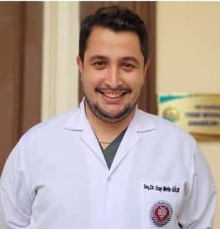 Eray Metin Guler合作论文数Dept Med Biochem, Bezmialem Vakif Univ3Mahmud Esad Pence合作论文数2Ebru Kale合作论文数2Sibel Kuras合作论文数2
 Gulfidan Coskun合作论文数2Halime Hanım Pençe合作论文数Department of Biochemistry, University of Health Sciences Turkey2Gulfem Basol合作论文数2Yilmaz Muserref Banu合作论文数2Sait Polat合作论文数2Halime Tuba Canbaz合作论文数1Gulam HEKİMOĞLU合作论文数1Fatima Efendic合作论文数1
 Dilek Saker合作论文数1Fatma Sena Dumlu合作论文数1Ceyda Hayretdag Ors合作论文数1
 Ayşe Köse合作论文数1
 Duygu Misirli合作论文数1
 Beyza Ozkan合作论文数1
 Hakan Beyaztas合作论文数1
 Nur Elagul Tombul合作论文数1
 Kubra Bozali合作论文数Fac Life Sci & Environm Technol, Avans Univ Appl Sci1
 Kursad Nuri Baydili合作论文数Dept Biostat, Univ Hlth Sci1
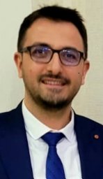 Serhat Aras合作论文数Haydarpasa Numune Training and Research Hospital, University of Health Sciences Turkey1
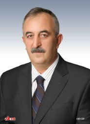 Mahfuz Elmastas合作论文数Faculty of Science and Arts, Gaziosmanpasa University1Alev Kural合作论文数1




