Super-resolution structured illumination microscopy (SR-SIM) enables fluorescence microscopy beyond the diffraction limit at high frame rates. Compared to other super-resolution microscopy techniques, the low photon fluence used in SR-SIM makes it readily compatible with live-cell imaging. Here, we combine SR-SIM with electro-optic fluorescence lifetime imaging (EOFLIM), demonstrating super-resolved multiplexed imaging of spectrally overlapping fluorophores, enhanced contrast due to optical sectioning, and environmental sensing at a spatial resolution of 156 nm. The high photon detection throughput of EOFLIM enables a combination of lifetime precision, size of the field of view, spatial resolution, and speed, which, taken together, is unprecedented, enabling the super-resolved imaging of cellular dynamics. (c) 2026 Optica Publishing Group under the terms of the Optica Open Access Publishing Agreement
Currently, super-resolution optical microscopy is mostly conducted with fluorophores that are excited and that emit in the visible part of the electromagnetic spectrum. Extending this range to the near-infrared (NIR) and even the shortwave-infrared (SWIR) part of the spectrum is highly desirable, because light in this wavelength range is less hazardous for live imaging and can penetrate much deeper into tissue. Here, we detail our development of a novel super-resolution structured illumination microscope that is purpose-built for imaging in the NIR wavelength range. Structured illumination via laser beam interference is accomplished by splitting the excitation light along two arms of a Michelson interferometer. Arbitrary illumination angles and a wide range of pattern spacings can be obtained by controlling a two-axis galvanometric mirror. Phase shifts of the pattern are accomplished by changing the angle of an anti-reflection-coated planoparallel glass plate inserted in one arm of the interferometer. Fluorescence is detected using a NIR-enhanced 26 Megapixel scientific Complementary Metal-Oxide-Semiconductor (sCMOS) camera. We characterize this system by imaging fluorescent beads with a diameter below the diffraction limit, and demonstrate its suitability for biological imaging by resolving cellular nanopores in the plasma membrane of liver sinusoidal endothelial cells.
Advances in cell biology create the demand for developing methods capable of resolving the structure and dynamics of subcellular organelles in living cells, which are beyond the reach of classical microscopy. Live-cell super-resolution fluorescence imaging provides this capability; however, in practice, its application is limited by phototoxicity, which perturbs cellular features and interferes with natural mechanisms of biological processes, providing a biased interpretation. Liver Sinusoidal Endothelial Cells (LSECs), with their nanoscale fenestrations that are physiologically critical and highly dynamic structures in the native state, represent a particularly demanding system for fluorescence-based microscopy. Here, we identify that photoactivation-generated reactive oxygen species (ROS) are the principal cause of fenestration arrest in fluorescence microscopy. By implementing three-dimensional super-resolution structured illumination microscopy (3D SR-SIM), we systematically evaluate a range of fluorophores and ROS scavengers to optimize imaging conditions. By combining BioTracker staining, carbon dioxide-independent medium supplemented with N-acetylcysteine (NAC), we preserved fenestration dynamics without altering the number/size of fenestrations. Complementary atomic force microscopy (AFM) validated that the combination of light and dye exposure impairs fenestration dynamics through ROS, in the absence of antioxidant supplementation. Additionally, AFM provides insights into the cells' nanomechanical changes upon illumination. Our findings confirm the mechanism underlying imaging-induced artifacts in LSECs observed in the literature and provide a broadly applicable framework for extending live-cell super-resolution microscopy of living cells.
The actin cytoskeleton in activated T cells undergoes rapid structural changes during the formation of an immunological synapse. Superresolution fluorescence microscopy provides excellent means to visualize such antibody-triggered changes. Here, we use single-molecule localization microscopy (SMLM) enabled by transparent polymer waveguide chips to resolve the filamentous-actin (F-actin) cytoskeleton in activated Jurkat T cells in comparison to nonactivated T cells across a large field of view. Transparent polymer waveguides enable a wide array of imaging modalities. In combination, these modalities reveal the structural differences between lamellipodial and ramified actin networks within the immunological synapse of activated T cells. SMLM images recorded by using narrow-width waveguide total internal reflection illumination resolve the double-stranded helical structure of actin filaments in activated Jurkat T cells. The average crossover length of the filaments is measured to be ~40 nanometers, which corroborates similar observations of isolated actin filaments by electron microscopy.
Amyloid-β (Aβ1-42) prefibrillar aggregates are considered the most neurotoxic amyloid species, yet their transient and heterogeneous nature makes selective detection challenging. Many fluorescent probes also fail to discriminate Aβ from homologous peptides such as IAPP, leading to poor specificity. We report a peptide-guided late-stage diversification strategy to generate BODIPY-based probes highly selective for prefibrillar Aβ1-42. A rationally engineered cyclic peptide derived from the C-terminal region of Aβ1-42 provides conformational rigidity and precise molecular recognition. Conjugation to BODIPY fluorophores afforded peptide-dye hybrids systematically evaluated for selectivity and photophysical response. A controlled aggregation protocol enabling reproducible generation of prefibrillar Aβ species was established to validate probe performance. A Sonogashira-derived conjugate (probe 8) showed strong fluorescence turn-on and selective affinity for prefibrillar Aβ1-42, with no response to IAPP aggregates. In neuronal cells, probe 8 outperformed conventional antibodies, supporting its potential for mechanistic studies and early Alzheimer's disease diagnostics.
The ultrastructure of endothelial cells (ECs) in situ is of great interest due to their involvement in many physiological processes. In some organs, these cells form transcellular pores or fenestrae, allowing for the rapid exchange of molecules between blood and interstitium. Despite their importance, no optical images of these dynamic morphological structures have yet been acquired in situ. Major obstacles to their in situ imaging are the lack of specifical labels for fenestrae and their size well below the optical diffraction limit. Here, we report how we have overcome these challenges and managed to visualize the EC ultrastructure in situ in 25 μm thick liver sections. To enable this, a lipophilic, fluorescent membrane dye was infused into the portal vein of murine livers to stain the sinusoidal ECs before the organ was harvested. Tissue sections were subsequently imaged using a novel, super-resolution optical sectioning structured illumination microscope (OS-SIM), providing approx. 170 nm spatial resolution with significantly faster image acquisition compared to confocal microscopy.
Background: Reactive oxygen species (ROS) are prevalent in the liver during intoxication, infection, inflammation, and aging. Changes in liver sinusoidal endothelial cells (LSEC) are associated with various liver diseases. Methods: Isolated rat LSEC were studied under oxidative stress induced by H2O2 at different concentrations (0.5–1000 µM) and exposure times (10–120 min). LSEC functions were tested in a dose-dependent and time-dependent manner. Results: (1) Cell viability, reducing potential, and scavenging function decreased as H2O2 concentration and exposure time increased; (2) intracellular ROS levels rose with higher H2O2 concentrations; (3) fenestrations exhibited a dynamic response, initially closing but partially reopening at H2O2 concentrations above 100 µM after about 1 hour; (4) scavenging function was affected after just 10 minutes of exposure, with the impact being irreversible and primarily affecting degradation rather than receptor-mediated uptake; (5) the tubulin network was disrupted in high H2O2 concentration while the actin cytoskeleton appears to remain largely intact. Finally, we found that reducing agents and thiol donors such as n-acetyl cysteine and glutathione (GSH) could protect cells from ROS-induced damage but could not reverse existing damage as pretreatment with n-acetyl cysteine, but not GSH, reduced the negative effects of ROS exposure. Conclusions: The results suggest that LSEC does not store an excess amount of GSH but rather can readily produce it in the occurrence of oxidative stress conditions. Moreover, the observed thresholds in dose-dependent and time-dependent changes, as well as the treatments with n-acetyl cysteine/GSH, confirm the existence of a ROS-depleting system in LSEC.
Liver sinusoidal endothelial cells (LSECs) play a crucial role in hepatic homeostasis, clearance, and microcirculatory regulation. Their fenestrations—patent transcellular pores—are essential for proper liver function, yet disappear in pathological conditions such as liver fibrosis and inflammation through a process known as defenestration. Defenestrated sinusoids are often linked to the liver stiffening that occurs through mechanotransduction-regulated processes. We performed a detailed characterization of polyacrylamide (PAA) hydrogels using atomic force microscopy (AFM), rheometry, scanning electron microscopy, and fluorescence microscopy to assess their potential as biomimetic substrates for LSECs. We additionally implemented AFM; quantitative fluorescence microscopy, including high-resolution structured illumination microscopy (HR-SIM); and an endocytosis assay to characterize the morphology and function of LSECs. Our results revealed significant local variations in hydrogel stiffness and differences in pore sizes. The primary LSECs cultured on these substrates had a range of stiffnesses and were analyzed with regard to their number of fenestrations, cytoskeletal organization, and endocytic function. To explore mechanotransduction in inflammatory liver disease, we investigated LSECs from a genetic model of systemic inflammation triggered by the deletion of Mcpip1 in myeloid leukocytes and examined their ability to restore their fenestrations on soft substrates. Our study demonstrates the beneficial effect of soft hydrogels on LSECs. Control cells exhibited a similar fenestrated morphology and function compared to cells cultured on plastic substrates. However, the pathological LSECs from the genetic model of systemic inflammation regained their fenestrations when cultured on soft hydrogels. This observation supports previous findings on the beneficial effects of soft substrates on LSEC fenestration status.
In living organisms, the natural motion caused by the heartbeat, breathing, or muscle movements leads to the deformation of tissue caused by translation and stretching of the tissue structure. This effect results in the displacement or deformation of the plane of observation for intravital microscopy and causes motion-induced aberrations of the resulting image data. This, in turn, places severe limitations on the time during which specific events can be observed in intravital imaging experiments. These limitations can be overcome if the tissue motion can be compensated such that the plane of observation remains steady. We have developed a mathematical shape space model that can predict the periodic motion of a cylindrical tissue phantom resembling blood vessels. This model is then used to rapidly calculate the future position of the plane of observation of a confocal multiphoton fluorescence microscope. The focal plane is continuously adjusted to the calculated position with a piezo-actuated objective lens holder. We demonstrate active motion compensation for non-harmonic axial displacements of the vessel phantom with a field of view up to 400 $\mu$m $\times$ 400 $\mu$m, vertical amplitudes of more than 100 $\mu$m, and at a rate of 0.5 Hz.
IntroductionDystroglycan (DG) is an adhesion complex comprising two subunits, α-DG and β-DG, which interact non-covalently at the plasma membrane. As a component of the dystrophin-glycoprotein complex DGC, DG plays a crucial role in linking the cytoskeleton to the surrounding basement membranes. Rare primary point mutations in the DAG1 gene have been identified in patients with various forms of neuromuscular dystrophy, ranging in phenotype from mild to severe.MethodsTo gain a deeper understanding of the molecular mechanisms underlying these pathologies, we have designed a series of chimeric GFP-tagged full-length α/β-DG constructs and expressed them in three different cell lines (U-2OS, HEK-293T and C2C12). Wild-type DG constructs were compared to their counterparts carrying pathologic missense mutations previously described in patients, namely, L84F, T190M and C667F and with the mutant I591D, i.e., the topological equivalent of V567D identified in zebrafish.ResultsLive super-resolution fluorescence microscopy showed that the C667F mutant is retained within the ER/Golgi while the T190M and wild-type proteins are correctly localized to the plasma membrane in all 3 cell lines. The L84F mutant exhibits a delay in trafficking to the plasma membrane in two of the cell lines, while localizing strongly at the plasma membrane in the high-expression HEK-293T cells. Similarly, the I591D mutant accumulated at the plasma membrane in the HEK-293T cells, in contrast to the clear retention in the endoplasmic reticulum/Golgi apparatus observed in U-2OS and C2C12 cells.DiscussionOur data demonstrate the importance of using a range of different cell lines for a comprehensive study of DG mutants or variants by live cell optical super-resolution microscopy.
Super-resolution Structured Illumination Microscopy (SR-SIM) enables fluorescence microscopy beyond the diffraction limit at high frame rates. Compared to other super-resolution microscopy techniques, the low photon fluence used in SR-SIM makes it readily compatible with live-cell imaging. Here, we combine SR-SIM with electro-optic fluorescence lifetime imaging (EOFLIM), adding the capability of monitoring physicochemical parameters with 156 nm spatial resolution at high frame rate for live-cell imaging. We demonstrate that our new SIMFLIM technique enables super-resolved multiplexed imaging of spectrally overlapping fluorophores, environmental sensing, and live-cell imaging.
Peroxisomes are ubiquitous cellular organelles with potentially vital roles in lipid and reactive oxygen metabolism. The metabolic demands of the heart are substantial; however, the contribution of peroxisomes to cardiac development, health, and their role in heart failure (HF) remain largely unexplored. We developed and examined a mouse and an engineered human myocardium (EHM) model with a deficiency in cardiac peroxisome biogenesis to investigate the role of peroxisomes in cardiac function and pathology. In the EHM, loss of peroxisome protein import and subsequent peroxisomal metabolic impairment trigger mitochondrial damage and compromise cellular respiration and energy production. Peroxisome dysfunction results in incoherent electrical conduction, defective Ca2+-handling, and ultimately presentation of a HF phenotype with pathological force generation. These phenotypes are mirrored in an orthogonal murine model system with defective cardiac peroxisome biogenesis. Preload-dependent deficits in force generation due to insufficient energy supply are eventually fatal. Thus, peroxisomes play an important role in sustaining normal heart operations. Vice versa, peroxisome maintenance is compromised in pressure overload-induced HF, establishing peroxisomes as potential modulators of pathology and targets of therapy. ### Competing Interest Statement WHZ is founder and equity holder of Myriamed GmbH with an interest in drug development in human iPSC-based cell and tissue models. MT is advisor of Myriamed GmbH. NL is a member of the scientific advisory board of Trace Neuroscience. Deutsche Forschungsgemeinschaft, https://ror.org/018mejw64 Fondation Leducq, https://ror.org/01czwga19 Eva-Luise and Horst Köhler Foundation German Centre for Cardiovascular Research (DZHK) German-Israeli Foundation for Scientific Research and Development (GIF) The F. Thyssen Foundation Marlies und Herbert Repkow Stiftung German Research Foundation Excellence Strategy Interdisziplinäres Zentrum für Klinische Forschung
Super-resolution optical microscopy (SRM) permits the visualization of subcellular structures of biological samples beyond the diffraction limit of light. To evaluate and utilize the specific strengths of each SRM technique a combined approach in the form of correlative super-resolution imaging is essential. Here, the correlative SRM imaging of the ultrastructure of rat liver sinusoidal endothelial cells (LSECs) across a large field of view (FOV) with 3D structured illumination microscopy (3D-SIM) and single-molecule localization microscopy (SMLM), facilitated by a transparent polymer photonic waveguide chip, is presented. This waveguide is not only used for chip-based total internal reflection fluorescence (TIRF) excitation across a large FOV, but also enables the excitation and collection of single-molecule fluorescence via the inverted microscope configuration. Furthermore, the structural design of the waveguides allows to identify and correlate sample positions across multiple microscopes. This correlative SIM and multi-modality SMLM imaging provides a high throughput (FOV of approximate to 180 mu m x 120 mu m) method to analyze the structural morphology of LSECs with high spatial resolution (approximate to 50 nm). Furthermore, waveguide chip-based TIRF excitation also yields a significant reduction of background signals.
Advances in cell biology demand methods that resolve the structure and dynamics of subcellular organelles in living cells. Live-cell super-resolution fluorescence microscopy meets this need but is constrained by phototoxicity, which disturbs cellular function and biases interpretation. Liver sinusoidal endothelial cells (LSECs), with their physiologically critical and highly dynamic fenestrations, represent a particularly challenging model system. We show that photoactivation-generated reactive oxygen species (ROS) are the primary cause of fenestration arrest during fluorescence imaging. Using three-dimensional structured illumination microscopy (3D SR-SIM), we systematically evaluated fluorophores and ROS scavengers to optimize imaging conditions. A combination of BioTracker staining and CO₂-independent medium supplemented with N-acetylcysteine (NAC) preserved fenestration dynamics without altering fenestration number or size. Complementary atomic force microscopy (AFM) confirmed ROS-dependent impairment of fenestration dynamics and revealed nanomechanical changes upon illumination. These findings establish the mechanism underlying imaging-induced artefacts in LSECs and provide a broadly applicable strategy to extend live-cell super-resolution microscopy. ### Competing Interest Statement The authors have declared no competing interest. European Innovation Council, https://ror.org/05cx8cy07, 101046928 Deutsche Forschungsgemeinschaft, 540217954 The Research Council of Norway, https://ror.org/00epmv149, 325446
Macrophage polarization in neurotoxic (M1) or neuroprotective (M2) phenotypes is known to play a significant role in neuropathic pain, but its behavioral dynamics and underlying mechanism remain largely unknown. Two-photon excitation microscopy (2PEM) is a promising functional imaging tool for investigating the mechanism of cellular behavior, as using near-infrared excitation wavelengths is less subjected to light scattering. However, the higher-order photobleaching effect in 2PEM can seriously hamper its applications to long-term live-cell studies. Here, we demonstrate a GHz femtosecond (fs) 2PEM that enables hours-long live-cell imaging of macrophage behavior with reduced higher-order photobleaching effect-by leveraging the repetition rate of fs pulses according to the fluorescence lifetime of fluorophores. Using this new functional 2PEM platform, we measure the polarization characteristics of macrophages, especially the long-term cellular behavior in efferocytosis, unveiling the dynamic mechanism of neuroprotective macrophage polarization in neuropathic pain. These efforts can create new opportunities for understanding long-term cellular dynamic behavior in neuropathic pain, as well as other neurobiological problems, and thus dissecting the underlying complex pathogenesis.
High-resolution optical fluorescence microscopies and, in particular, super-resolution fluorescence microscopy, are rapidly adopting highly sensitive cameras as their preferred photodetectors. Camera-based parallel detection facilitates high-speed live cell imaging with the highest spatial resolution. Here, we show that the drive to use ever more sensitive, photon-counting image sensors in cameras can, however, have detrimental effects on the spatial resolution of the resulting images. This is particularly noticeable in applications that demand a high space-bandwidth product, where the image magnification is close to the Nyquist sampling limit of the sensor. Most scientists will often select image sensors based on parameters such as pixel size, quantum efficiency, signal-to-noise performance, dynamic range, and frame rate of the sensor. A parameter that is, however, typically overlooked is the sensor's modulation transfer function (MTF). We have determined the wavelength-specific MTF of front- and back-illuminated image sensors and evaluated how it affects the spatial resolution that can be achieved in high-resolution fluorescence microscopy modalities. We find significant differences in image sensor performance that cause the resulting spatial resolution to vary by up to 28%. This result shows that the choice of image sensor has a significant impact on the imaging performance of all camera-based optical microscopy modalities. Pixel crosstalk can significantly reduce the image resolution in standard and super-resolution light microscopy using sCMOS cameras up to 28%. The effect for different imaging modalities was evaluated for several wavelengths and image sensor types.
Imaging structures deep within biological tissue and organisms with a spatial resolution well below the optical diffraction limit is still underrepresented. This is mostly due to the complexity and high cost of microscopes that can facilitate such challenges. The majority of deep tissue imaging is still accomplished by exciting fluorescence and other nonlinear light-sample interactions with ultrashort pulsed lasers. Here, we demonstrate a cost efficient and easy to implement method to turn most two-photon laser-scanning microscopes into a super-resolution microscope for deep tissue imaging. We realize this by adding a compact and cost-efficient line-scanning module, a field rotator, and a sCMOS camera to these systems. By combining two-photon excitation with patterned line-scanning and subsequent image reconstruction, we achieve imaging with a spatial resolution well below the diffraction limit. Furthermore, by synchronizing the two-photon line illumination with camera-based line confocalization, a significant contrast enhancement deep within biological tissue can be achieved. We demonstrate this by imaging subwavelength structures in extended Pinus radiata, mouse heart muscle and zebrafish samples.
Recurrence after surgical extraction of middle ear cholesteatoma (MEC) is highly dependent on surgical techniques, specifically aggressiveness of the extraction. Clear visualization of residual MEC mass in the temporal bone would facilitate complete removal of the MEC mass. Regrowth from residual MEC mass is also associated with high level of inflammation/infection. Hence, removal of biofilm infected tissue is crucial in MEC surgery. The aim of this research endeavor is the construction of a prototype for a surgical microscope superimposing common microscopic image with newly developed contrasts based on infrared (IR) light. To acquire these contrasts, two approaches are pursued. The first one utilize IR light (700 nm – 1675 nm) to image tissues relevant in MEC surgery (temporal bone, MEC (N=10), muscle, tendon, fat). These experiments were performed on fixed human samples as well as fresh animal tissue. The second scope of the project is the development of fluorescent IR probes, able to stain bacterial biofilms. These probes are based on drugs and dyes already established in clinical routine and tested on various strains (N=9) isolated during MEC surgery. We were able to discriminate MEC from temporal bone, as well as tendon from muscle and fat using combinations of reflected IR light with different wavelength. It was also possible to demonstrate a distinct binding of our probes to the investigated strains. We think, that the label free contrast between tissues based on IR light as well as fluorescent probes based in clinical approved substance will make the translation of our methods feasible in short period of time. This might help ENT surgeon to optimize their performance and prevent recurrence of MEC.
Das Wiederauftreten eines Mittelohr-Cholesteatoms (MOC) nach der Entfernung hängt von den chirurgischen Methoden und der Aggressivität der Extraktion ab. Eine Visualisierung der MOC-Masse im Schläfenbein würde seine vollständige Entfernung erleichtern. Das Nachwachsen der verbliebenen MOC-Masse ist in einem hohen Maß an Entzündung/Infektion gekoppelt. Daher ist die Entfernung des mit einem Biofilm infizierten Gewebes von entscheidender Bedeutung. Ziel dieses Forschungsvorhabens ist der Bau eines Prototyps eines Operationsmikroskops, das herkömmliche mikroskopische Bilder mit neu entwickelten Kontrasten auf der Grundlage von Infrarotlicht (IR) überlagert. Um diese Kontraste zu erhalten, werden zwei Ansätze verfolgt. Der erste nutzt IR-Licht (700 nm – 1675 nm), um Gewebe abzubilden, die für die MEC-Chirurgie relevant sind (Schläfenbein, MEC (N=10), Muskel, Sehne, Fett). Diese Experimente wurden an fixierten menschlichen Proben und frischem Tiergewebe durchgeführt. Der zweite nutzt fluoreszierende IR-Sonden, die bakterielle Biofilme anfärben können. Diese Sonden basieren auf in der klinischen Routine etablierten Medikamenten und Farbstoffen und wurden an verschiedenen Isolaten (N=9) aus der MEC-Operation getestet. Mithilfe von Kombinationen aus verschiedenen Wellenlängen von reflektiertem IR-Licht konnten wir MEC/Schläfenbein sowie Sehnen/Muskeln/Fett unterscheiden. Eine eindeutige Bindung unserer Sonden an die untersuchten Stämme wurde nachgewiesen. Wir denken, dass der markierungsfreie Kontrast zwischen Geweben auf der Basis von IR-Licht sowie von Fluoreszenzsonden auf der Basis von klinisch zugelassenen Substanzen die klinische Umsetzung unserer Methoden ermöglicht. Dies könnte HNO-Chirurgen helfen, ihre Arbeit zu optimieren und das Wiederauftreten von MEC zu verhindern.
 Wolfgang Hubner合作论文数Biomol Photon, Univ Bielefeld36
 James W. Chan合作论文数Department of Pathology and Laboratory Medicine, University of California, Davis32
 Christopher W. Hollars合作论文数Chemistry, Materials, and Life Sciences, Lawrence Livermore National Laboratory32Christian Pilger合作论文数22
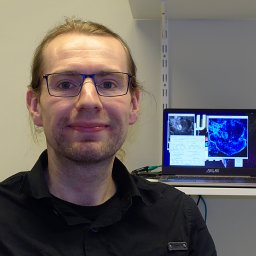 Marcel Muller合作论文数Department of Physics, University of Bielefeld22Chad Talley合作论文数20
 Stephen M Lane合作论文数Lawrence Livermore National Laboratory, University of California20Mark Schüttpelz合作论文数19McNerney Gregory P合作论文数17
 Peter McCourt合作论文数Department of Cell & Systems Biology, University of Toronto17
 Samantha Fore合作论文数17
 Ted A. Laurence合作论文数Lawrence Livermore National Laboratory17Henning Hachmeister合作论文数14
 Benjamin K Chen合作论文数Rockefeller University13
 Balpreet Singh Ahluwalia合作论文数Department of Physics and Technology, The Arctic University of Norway;Chip NanoImaging AS;Karolinska Institutet;Universitetet i Oslo12Gerd Wiebusch合作论文数11Alice Sandmeyer合作论文数11
 Iwan Schie合作论文数University of California Davis Medical Center, University of California11Harry Heinzelmann合作论文数Swiss Center for Electronics and Microtechnology, CSEM SA11S. M. Lane合作论文数10
 Karolina Szafranska合作论文数Department of Medical Biology10
 Tyler James Weeks合作论文数9
 Henning Ortkrass合作论文数9
 Subhash H. Risbud合作论文数Department of Materials Science and Engineering, College of Engineering, University of California, Davis9Chad E. Talley合作论文数9Anders Kokkvoll Engdahl合作论文数8Mario Lachetta合作论文数8
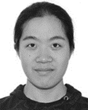 Cihang Kong合作论文数Biomolecular Photonics, Bielefeld University8
 Mönkemöller Viola合作论文数Department of Physics, University of Bielefeld8
 Denise M. Krol合作论文数Department of Materials Science and Engineering, University of California, Davis8
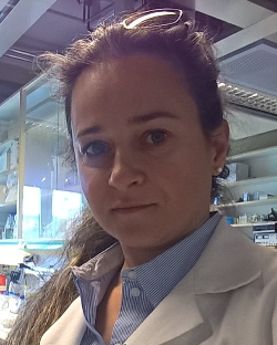 Cristina Ionica Oie合作论文数Department of Medical Biology, The Arctic University of Norway8
 Simon Hennig合作论文数Physics Department, Bielefeld University8
 Barbara Kaltschmidt合作论文数Faculty of Biology, Bielefeld University8
 Xiaoming Wei (韦小明)合作论文数School of Physics and Optoelectronics, South China University of Technology7
 Rod Balhorn合作论文数SHAL Technologies Inc7
 Christian Kaltschmidt合作论文数7
 Sebastian Wachsmann-Hogiu合作论文数Department of Surgery, Cedars-Sinai Medical Center Minimally Invasive Surgical Technologies Institute7
 Adam Schwartzberg合作论文数Lawrence Berkeley National Laboratory, Office of Science, The U.S. Department of Energy7John C. Rutledge合作论文数7
 Stephan Bergmann合作论文数6Robin Diekmann合作论文数Cell Biology and Biophysics Unit6
 John C Voss合作论文数School of Medicine, University of California-Davis6Ly Sonny合作论文数6Schürmann Matthias合作论文数6
 Wolfson Deanna合作论文数NSF Center for Biophotonics Science and Technology, University of California, Davis6
 Jan Schulte am Esch合作论文数6Douglas S. Taylor合作论文数6Thilo D. Lacoste合作论文数6H.-J. Güntherodt合作论文数6




