Introduction: Multivariable analysis of factors associated with ACR showed intensive induction, rATG/rituximab or alemtuzumab, is associated with lowering of ACR risk with rATG/Rituximab associated with less infection risk [1]. Despite induction, rejection remains a major cause of graft and patient loss. It is important to assess role of additional immunosuppression when ACR occurs. Methods: We reviewed ITx cases performed in our center from 2013 to 2020, our most recent Era. Of 126 primary ITx, 113 received induction with rATG/Rituximab +/- Basiliximab/Vedolizumab. Maintenance immunosuppression was tacrolimus/mTOR inhibitor. ACR of any grade was seen in 49(43%) patients with mild rejection most common. 16 episodes of severe ACR in 15(13%) patients were analyzed. Results: Of 15 patients, 5(33%) were MVT and 9(60%) Isolated Intestine. 10 male. Age 1-42yo. Time of rejection post op day 18-698 with the majority of episodes > 6mo due to noncompliance. 4 patients received rATG +/- Simulect/Rituximab/Infliximab and 1 only steroids given co-morbidities. This resulted in 0% graft and 90% patient survival. 2 patients were retransplanted, 2 full PN and 1 death. 3/4 surviving patients had graft removed including partial removal of MVT. 4th patient had granulation of bowel (MVT). Given poor results with rATG, 10 recent cases received Alemtuzumab +/- Vedolizumab. This resulted in 60% graft and 90% patient survival. All responders are on enteral diet with no supplemental PN/IVF. 1 patient had 2nd episode of noncompliance/severe ACR 8mo later; graft was removed. Of non-responders, 2 full PN, 1 retransplanted and 1 death. 2/3 surviving patients had graft removed. 3rd patient had granulation of bowel (MVT). Conclusions: Alemtuzumab results in superior success in the treatment of severe ACR after induction with rATG. Early recognition of treatment failure is key allowing graft removal if tissue does not granulate in order to discontinue or lower immunosuppression. This technique allows improved survival with possibility of PN and/or retransplant in the future.
Introduction: A temporary ostomy to assess the mucosa and easily perform a biopsy remains the diagnostic gold standard in intestinal transplant. However, patients with an ostomy may have high output and decreased renal function. A change in intestinal flora, as stool and mucosa are exposed to air, may trigger rejection. We presented our initial experience with MVT without an ostomy including pediatric patients in 2015 and 5 year outcomes in 2019. We now describe the first series, to the best of our knowledge, of pediatric isolated intestinal transplant (IITx) without an ostomy. Methods: We performed 8 pediatric ITx in 2020. 4 were multivisceral and 4 Isolated Intestinal Transplants (IITx). All 4 IITx were performed without an ostomy. Induction was with ATG/Rituximab/Vedolizumab. Maintenance immunosuppression was with Tacrolimus/mTOR inhibitor. These 4 patients were analyzed further. Results: Of 4 patients, 2 were male. Age 3-12yr. Weight 15.6-43.2kg. All had short bowel syndrome as etiology of intestinal failure (IF). All had negative cross match. Follow up 116-256days. 1(25%) patient had severe acute cellular rejection refractory to medical therapy on POD 21 s/p enterectomy on POD 115. He had a prolonged LOS 167days. He is on full PN planning retransplant. LOS in other patients 26-35days. 1(25%) patient had mild ACR treated with steroids. 2(50%) patients remain rejection free. No patients had PTLD nor GVHD. 3 of 4 patients are on full regular PO diet/enteral with no supplemental PN nor IVF. Initial endoscopy occurred at POD 21-45. Number of scopes performed to date 1-7. Although short follow up, graft and patient survival has been 75% and 100% respectively. Conclusions: Pediatric IITx without an ostomy appears to be a safe and feasible alternative to the current gold standard. Although one patient had severe ACR requiring enterectomy, patient has returned to their pre-transplant status. He is off immunosuppression decreasing risk to baseline as we await a 2nd transplant possibility. More data is needed to accurately compare risk of rejection associated with this technique as well as long term outcomes. Lack of ostomy greatly adds to patient satisfaction and quality of life.
Introduction: Patients with inflammatory bowel disease(IBD) often require operations in their lifetime. Such operations may result in significant resection leading to short bowel syndrome and potentially, intestinal failure(IF). As such, IBD may be an indication for intestinal transplant (Itx). Methods: We reviewed patients transplanted in our center from 2013-2020, our most recent era. Induction was Thymoglobulin/Rituximab/+/- Basilizimab/Vedolizumab. Maintenance immunosuppression was tacrolimus/mTOR inhibitor. Of 126 primary non-renal Itx, 10 were due to IBD. Results: 3 male. 9 adult. Age 4-62yo. All had short bowel syndrome. 8 received an isolated intestine, 2 multivisceral transplant. Positive crossmatch was found in 2 patients. Follow up 373-2756d. Acute rejection occurred in 5(50%) patients. PTLD occurred in 2(20%) patients. Nor Chronic rejection or GVHD was seen in this cohort. 1 graft was lost due to non-compliance and severe rejection and 1 due to thrombosis in perioperative period; both patients are alive on parenteral nutrition(PN). 1 graft and patient were lost due to PTLD. All other patients are on regular diet without supplemental PN/IVF. 1-year patient and graft survival was 100% and 90% respectively. 5-year patient and graft survival was 90% and 70% respectively. Discussion: ITx has become a well-established treatment for those who fail PN and have life-threatening complications. We believe we are entering a new era in ITx with higher patient survival. Although IF as a result of IBD is rare, one should consider ITx in the spectrum of treatment modalities given good survival and quality of life.
Intro: Intestinal failure(IF) continues to be the most common indication for an intestinal transplant (Itx). Given complications of parenteral nutrition (PN) such as liver disease or of the underlying condition such as gastroparesis, en-bloc transplantation including liver/stomach were developed. With evolution of the surgical technique, immunosuppression, graft monitoring and patient management, outcomes of Multivisceral transplantation (MVT) have improved. This allows the possibility to expand indications to other etiologies other than IF. Methods: We reviewed all pediatric Itx performed in our institution from 2013-2020, our most recent era. We performed 51 pediatric Itx. Induction was ATG/Rituximab/+/- Vedolizumab. Maintenance immunosuppression was Tacrolimus/mTOR inhibitor. We identified 4 patients who underwent MVT for hostile abdomen with portal +/-mesenteric thrombosis. Results: 3 female. Age 2-9yo. Weight 8.3-23.4kg. 3 had primary diagnosis of biliary atresia. 2/3 had undergone 2 prior orthotopic liver transplants (OLTs) with graft lost to hepatic artery thrombosis. 1/3 had 1 prior OLT with progressive cholestasis. All 3 patients developed extensive portal +/- mesenteric thrombosis of latest graft progressing to end stage liver disease. Evaluation for retransplant found abdomen to be hostile. 4th patient had sclerosing cholangitis and antithrombin III deficiency leading to portomesenteric thrombosis. Follow up 285-1643d. LOS 46-68d. 3 patients were done without ostomy and 1 with ostomy given size. All patients are on regular/enteral diet. No patients are on PN/IVF supplementation. 2 patients had rejection, 1 mild and 1 moderate. Both were treated successfully with steroids. 1 patient developed PTLD and treated with Rituximab/Cyclophosphamide/Prednisone now in remission for > 3yr. 1 patient developed GVHD of the skin/bone marrow successfully treated with Alemtuzumab. Graft and patient survival are both 100%. 3 of 4 patients are school aged and attending regular school with good quality of life. Conclusions: Despite the high risk of this population, MVT proved to be a successful option in a pediatric population with portomesenteric thrombosis and hostile abdomen. We believe we are entering a new era in MVT with improved patient survival and good quality of life. An expansion of indications for such a procedure may be feasible.
Purpose Combined heart and liver transplantation is a live saving procedure for patients with advanced dual organ failure. Although described 35 years ago, less than 20 cases are performed every year, reflecting the complexity of the procedure. We present the first case series of adult patients receiving En-Bloc Heart-Liver Transplant (En-Bloc HLTx). Methods A present a retrospective analysis of our experience with En-Bloc HLTx in five adult patients, including demographic characteristics, indications, surgical course with operative data, surgica outcomes, and long term follow up with evaluation of rejection by myocardial biopsy results. Results Five patients received En-Block HLTx between June 2016 and December 2017. Mean recipient age was 43 years (26-63), and 3 (60%)patients were male.Indication for dual organ transplantation included hereditary ATTR amyloidosis (n=1), gestations cardiomyopathy with cardiac cirrhosis (n=2), and idiomatic dilated cardiomyopathy with cryptogenic cirrhosis (n=1) or coexistent NASH (n=1).All patients presented NYHA FC IV symptoms, and 4 were admitted to the hospital awaiting transplantation for 63 days (51-89) as status 1A, while 1 patient was on home inotropic support. Sodium MELD score ranged from 8 to 28 (mean = 17.6). Total operative time was 430 min (393-480), and ischemic times (IT) were equal for both organs, with cold IT of 85 min (32-136), and warm IT of 37.5 min (31-47). Patients required CPB support for 107 min (80-138), and aortic cross-clamp of 58 min (52-72). Abdominal fascia remained open in 4 patients, and sternum in 3 patients, with planned definitive wound closure within 48 hours in all patients. Thirty day operative survival was 100%, and 4 (80%) patients were discharged alive from the hospital on POD 36 (17-67). One patient expired on POD 55, due to fungemia and multi system organ failure. Mayor postoperative complications included prolonged ventilation requiring tracheostomy on 3 patients, and renal insufficiency requiring dialysis in 2 patients. Follow up was complete all 4 patients for up to 18 months, All patients remained on low immunosuppression regimen, and there was no evidence of grade 2 or higher rejection on anyone. Conclusion En-Bloc HL Tx is a safe procedure for patients requiring dual organ transplantation. Operative times are significantly decreased, and mid-term outcomes are comparable to single organ transplantation. Combined heart and liver transplantation is a live saving procedure for patients with advanced dual organ failure. Although described 35 years ago, less than 20 cases are performed every year, reflecting the complexity of the procedure. We present the first case series of adult patients receiving En-Bloc Heart-Liver Transplant (En-Bloc HLTx). A present a retrospective analysis of our experience with En-Bloc HLTx in five adult patients, including demographic characteristics, indications, surgical course with operative data, surgica outcomes, and long term follow up with evaluation of rejection by myocardial biopsy results. Five patients received En-Block HLTx between June 2016 and December 2017. Mean recipient age was 43 years (26-63), and 3 (60%)patients were male.Indication for dual organ transplantation included hereditary ATTR amyloidosis (n=1), gestations cardiomyopathy with cardiac cirrhosis (n=2), and idiomatic dilated cardiomyopathy with cryptogenic cirrhosis (n=1) or coexistent NASH (n=1).All patients presented NYHA FC IV symptoms, and 4 were admitted to the hospital awaiting transplantation for 63 days (51-89) as status 1A, while 1 patient was on home inotropic support. Sodium MELD score ranged from 8 to 28 (mean = 17.6). Total operative time was 430 min (393-480), and ischemic times (IT) were equal for both organs, with cold IT of 85 min (32-136), and warm IT of 37.5 min (31-47). Patients required CPB support for 107 min (80-138), and aortic cross-clamp of 58 min (52-72). Abdominal fascia remained open in 4 patients, and sternum in 3 patients, with planned definitive wound closure within 48 hours in all patients. Thirty day operative survival was 100%, and 4 (80%) patients were discharged alive from the hospital on POD 36 (17-67). One patient expired on POD 55, due to fungemia and multi system organ failure. Mayor postoperative complications included prolonged ventilation requiring tracheostomy on 3 patients, and renal insufficiency requiring dialysis in 2 patients. Follow up was complete all 4 patients for up to 18 months, All patients remained on low immunosuppression regimen, and there was no evidence of grade 2 or higher rejection on anyone. En-Bloc HL Tx is a safe procedure for patients requiring dual organ transplantation. Operative times are significantly decreased, and mid-term outcomes are comparable to single organ transplantation.
The management of severe hepatic artery vasospasm soon after liver transplantation (LT) is challenging because it can lead to hepatic artery thrombosis and subsequent graft failure. A 61-year-old man with hepatitis C cirrhosis and portal vein thrombosis received a deceased donor LT. On postoperative day 1, Doppler ultrasonography revealed a high-resistance waveform in the hepatic artery. Angiography showed severe vasospasm of the donor hepatic artery on postoperative day 3. Strong hepatic arterial buffer response (HABR) was considered for this etiology due to high portal vein velocity. Therefore, vasodilators, including nitroglycerin and prostaglandin E1, were initiated. The waveform of the hepatic artery vasospasm gradually improved as portal vein velocity decreased by Doppler ultrasonography within 7 days after LT. In conclusion, hepatic arterial buffer response can induce hepatic artery vasospasm immediately after LT. This vasospasm type may be managed conservatively with a positive outcome.
BACKGROUND Auxiliary partial orthotopic liver transplantation is an alternative technique for the treatment of patients with fulminant hepatic failure and metabolic liver disease. It provides temporary support of liver function until sufficient regeneration of the native liver. Pediatric patients have a long life expectancy and are best candidates to benefit from the interruption of antirejection treatment. DESCRIPTION OF CASE A 4-year-old boy underwent auxiliary partial orthotopic liver transplantation for fulminant hepatic failure using a cadaveric left lateral segment of liver. One year after auxiliary partial orthotopic liver transplantation, the patient's native liver was determined to be completely normal and he was doing well. The patient was then gradually weaned from the immunosuppression over the course of one year. The graft was undetectable on follow-up computerized tomography performed before complete cessation of immunosuppression, leading to the diagnosis of "vanishing graft syndrome". CONCLUSION Graft atrophy commonly occurs after auxiliary partial orthotopic liver transplantation due to cessation of antirejection therapy. But to our knowledge, complete graft disappearance is a rare occurrence reported in the English literature. Timing for withdrawal of the immunosuppression is an important decision to be made in this technique. Hippokratia 2015; 19 (3): 274-277.
BACKGROUND AND PURPOSE:Small infants with biliary atresia and hypoplastic portal veins (PV) are at risk for portal vein thrombosis (PVT) after liver transplantation (LT), which can lead to graft loss and mortality. Extra-anatomical PV reconstruction techniques have been established for adult cases of PVT; however, they have not been widely accepted for infants.METHODS:Here, we report the successful use of an extra-anatomical meso-portal venous jump graft to treat early PVT after LT in a 6-month-old infant with biliary atresia and PV hypoplasia. At the time of LT, despite a reduced-sized left lateral graft, we had to create a temporary abdominal closure with silastic mesh.FINDINGS:On postoperative day 1, PVT was detected by Doppler ultrasound of the liver. Surgical thrombectomy was attempted. We removed the blood clots and reconstructed the PV using an interposition venous graft. As the PV flow was still not sufficient, we performed an extra-anatomical meso-portal venous jump graft procedure from the recipient superior mesenteric vein to the donor PV. This resulted in a significant improvement in PV flow.CONCLUSION:For small infants at high risk for PVT, a detailed pretransplantation surgical plan and treatment options for possible early PVT are mandatory. An extra-anatomical meso-portal venous jump graft is a viable surgical technique for early PVT in infants.
 Gennaro Selvaggi合作论文数61Seigo Nishida合作论文数School of Medicine, University of Miami58P Ruiz合作论文数41David M Levi合作论文数40Andreas G Tzakis合作论文数33Ji Fan合作论文数24
 Andreas Tzakis合作论文数Transplant Center, Department of Surgery, Cleveland Clinic24Panagiotis Tryphonopoulos合作论文数22
 Junseob Moon合作论文数21Deborah Weppler合作论文数20Rodrigo Vianna合作论文数Miami Transplant Institute, University of Miami Miller School of Medicine17Eddie R Island合作论文数17T. Hibi合作论文数15K. Uchida合作论文数13
 Jumilka Garcia合作论文数12Dm Levi合作论文数12
 Venkatasamy Vighnesh Vetrivel合作论文数Graduate, Sri Ramachandra Medical College9
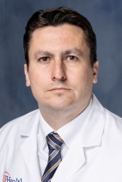 Beduschi Thiago合作论文数8L. Mosna合作论文数6
 Masahiro Ohira合作论文数6
 Lesley Jane Smith合作论文数Department of Pediatrics, University of Miami School of Medicine6
 Tomoaki Kato合作论文数Division of Abdominal Organ Transplantation, Department of Surgery, Columbia University Irving Medical Center;Vagelos College of Physicians and Surgeons, Columbia University6Miyashiro Nunes Dos Santos R合作论文数5
 Joe Tector合作论文数5T. Dohi合作论文数5Matsuura T合作论文数5P. Martin合作论文数4
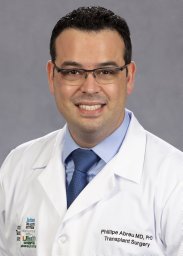 Phillipe Abreu合作论文数HPB Surgical Oncology, University Health Network4Hideki Ohdan合作论文数4
 Camillo Ricordi合作论文数Division of Cellular Transplantation, DeWitt Daughtry Family Department of Surgery, Miller School of Medicine, University of Miami4
 Lynn G. Feun合作论文数Sylvester Comprehensive Cancer Center 4J. Sageshima合作论文数3A. Farag合作论文数3MR Carreno合作论文数3E. R. Island合作论文数3M. Torres合作论文数3
 Linda Chen合作论文数3Ciancio Gaetano合作论文数3Monica Garcia合作论文数3
 George Burke合作论文数Division of Transplantation, and the Departments of Medicine and Pathology, University of Miami School of Medicine3T. Defranc合作论文数3Tadafumi Asaoka合作论文数3I.M. González-Pinto合作论文数3Leopoldo Arosemena合作论文数3W. Kupin合作论文数2C. Twist合作论文数2A. Volsky合作论文数2I. Muraoka合作论文数2




