INTRODUCTION:This study aimed to evaluate whether specific mandibular anatomical features are associated with postoperative neurosensory disturbance (NSD) in patients undergoing sagittal split osteotomy (SSO) and to identify cone-beam computed tomography (CBCT)-based measurements that may improve preoperative risk assessment. METHODS:Preoperative CBCT scans and patient-related variables, including demographic characteristics, type of deformity, side, fixation method, and third molar status, were retrospectively evaluated in patients who underwent SSO between 2016 and 2022. Measurements between the mandibular canal and key anatomical landmarks related to SSO were obtained. Neurosensory disturbance of the inferior alveolar nerve (IAN) was assessed using clinical sensory tests and patient-reported assessments. Outcomes were compared between patients with and without NSD. RESULTS:A total of 52 patients (35 women and 17 men; mean age, 24.2 ± 6.1 years) were included. In univariate analysis, female sex was associated with a higher frequency of NSD. The distance between the lingula and sigmoid notch, total bone thickness inferior to the canal, and cancellous bone thickness buccal to the canal at the mesial aspect of the second molar were significantly shorter in patients with NSD. At the distal aspect of the second molar, total bone thickness buccal and inferior to the mandibular canal, as well as cancellous bone thickness inferior to the canal, were also significantly reduced. However, none of these variables remained significant in multivariate analysis. CONCLUSIONS:Although no independent predictive factor for NSD was identified, female sex and reduced mandibular canal-related bone dimensions may be associated with a higher incidence of postoperative NSD. Preoperative CBCT evaluation of these parameters may support surgical planning and help reduce the risk of inferior alveolar nerve injury.
OBJECTIVE:To compare the clinical, radiographic, esthetic outcomes, and success and survival rates of dental implants placed after bone augmentation techniques. METHODS:This retrospective study included a total of 764 patients receiving 764 dental implants between 2009 and 2019. Four hundred implants were placed without bone augmentation (control), and 364 were placed after bone augmentation. Bone augmentation techniques were guided bone regeneration (GBR), ridge split, and onlay bone grafting. Gingival index (GI), plaque index (PI), probing depth (PD), bleeding on probing (BOP), pink esthetic score (PES) and marginal bone loss (mm) and area (mm2) were measured. The study variables of the implants among augmentation groups were compared statistically. RESULTS:The mean PI and GI scores, and BOP values of the implants in the augmentation and control groups were comparable (p = 0.365, p = 0.230, and p = 0.371 resp.) The mean PD scores of the implants were 2.82 ± 1.22 in the augmentation and 2.54 ± 1.29 in the control groups; the difference was significant (p = 0.002). The mean vertical bone loss of the implants was 0.78 ± 0.70 in augmentation and 0.82 ± 0.82 in the control groups, which was comparable (p = 0.461). The mean PES total values of the implants were 8.30 ± 1.55 in augmentation and 10.04 ± 2.43 in the control groups; the difference was significant (p < 0.001). There were no significant differences between the augmentation and control groups in implant survival (99.18% vs. 98%, p = 0.228) and success (82.97% vs. 85.50%, p = 0.389) rates. Significant differences in some study variables were observed among the augmentation groups. The survival and success rates of the implants in GBR (99.21% and 85.04%), ridge split (99.19% and 79.68%), onlay (99.12% and 84.21%), and control (98.00% and 85.50%) groups were similar (p = 0.630 and p = 0.479, resp.) in the 6-year mean follow-up. CONCLUSION:The implants placed in augmented bone showed similar cumulative success and survival rates compared to implants placed in natural bone with a 6-year mean follow-up. The augmentation group showed lower esthetic scores. There are some differences in clinical parameters among augmentation groups; however, all the augmentation groups showed similar success and survival rates.
Çukurova University Faculty of Dentistry, Department of Oral and Maxillofacial Radiology, Adana, Turkey Çukurova University Faculty of Dentistry, Department of Oral and Maxillofacial Surgery, Adana, Turkey Çukurova University Faculty of Medicine, Physical Medicine and Rehabilitation, Adana, Turkey Kyrenia University Faculty of Dentistry, Department of Oral and Maxillofacial Radiology, Kyrenia, Turkish Republic of Northern Cyprus The efficiency of dental volumetric tomography-derived radiomorphometric indices for diagnosing osteoporosis Osteoporoz Teşhisinde Dental Volümetrik Tomografiden Üretilen Radyomorfometrik İndekslerin Etkinliği
PURPOSE:The effect of botulinum toxin type A (BTX-A) on fracture healing of the long bones is controversial, and no controlled clinical or experimental study has investigated the effect of BTX-A on mandibular fractures. The purpose of this study was to investigate whether BTX-A injection into the masseter muscles affects bone healing by reducing the displacing forces in an unfavorable mandibular fracture model. MATERIALS AND METHODS:Forty-eight male New Zealand white rabbits were used. Ten units of BTX-A was injected into each masseter muscle in the animals in the BTX-A group, whereas saline solution was injected in the animals in the control group. A unilateral osteotomy and fixation with a microplate were performed. Bone healing was evaluated by radiodensitometric, biomechanical, histologic, and histomorphometric methods after 21 days. RESULTS:The mean bone mineral density in the fracture area was significantly higher in the BTX-A group (P = .038). The mean failure load and bending modulus values were significantly higher in the BTX-A group than in the control group (P = .032 and P = .005, respectively). The mean histologic bone healing scores, bone volume-total volume values, and trabecular diameter values were significantly higher in the BTX-A group than in the control group (P = .001, P = .001, and P = .026, respectively). CONCLUSIONS:BTX-A application into the masseter muscles improves bone healing of a unilateral mandibular fracture in rabbits.
Amaç: Bu çalışmanın amacı, transvers maksiller darlığı (TMD) olan yetişkin hastalarda transpalatal distraktör ile yapılan cerrahi destekli maksiller genişletmenin (TPD+CDMG) faringeal havayolu boyutları üzerindeki etkisini araştırmaktır.Gereç ve Yöntem: Bu çalışmaya 5 mm’den fazla TMD’si olan hastalar dahil edildi. Bütün hastalara genel anestezi altında, standart Le-Fort I osteotomisi ve maksillada orta hat osteotomisini takiben maksiller genişletme uygulandı. Dört ay sonrasında apareyler çıkartılarak transpalatal ark uygulandı. Hemen ameliyat öncesinde ve distraktör sökümünden en az 6 ay sonra alınan konik ışınlı bilgisayarlı tomografi görüntüleri üzerinde Dolphin 3D Surgery Görüntüleme yazılımının havayolu modülü kullanılarak, faringeal havayolunun nazofaringeal, retropalatal, retroglossal bölümlerinin hacimleri ve faringeal havayolunun en küçük kesit alanı ölçüldü. Sürekli değişkenler için normal dağılım varsayımı Shapiro-Wilk testi ile kontrol edildi ve dağılım tipine göre testler seçildi. Bağımlı yapıda olan TPD+CDMG öncesi ve sonrası ölçümler arasındaki farklılık test edilirken, normal dağılıma sahip olan ölçümler için eşleştirilmiş t-testi, normal dağılıma sahip olmayan ölçümler için ise Wilcoxon testi kullanıldı. İstatistiksel anlamlılık düzeyi p≤0.05 olarak kabul edildi.Bulgular: Çalışmaya 9’u kadın (%42.9), 12’si erkek (%57.1), toplam 21 hasta dahil edildi. Hastaların yaş ortalaması 22.8±5.7; takip süresi ortalaması 22±11.1 ay olarak bulundu. Retropalatal, retroglossal ve toplam havayolu hacmi ve faringeal havayolunun minimum kesit alanı için TPD+CDMG öncesi ve sonrasında yapılan ölçümler arasında istatistiksel olarak anlamlı fark bulunamadı. Ancak, nazofarinks hacmi için TPD+CDMG öncesi ve sonrası ölçümler arasında anlamlı fark tespit edildi (p≤0.05).Sonuç: TPD+CDMG sonrası nazofarinks hacminde önemli artış olduğu bulunurken, faringeal havayolunun diğer bölgelerindeki hacimsel ve kesitsel değişikliklerin önemli düzeyde olmadığı bulunmuştur.
Objective Our study seeks to investigate the effectiveness of kinesio taping (KT) on postoperative morbidity compared to placebo and control groups after impacted third molar surgery. Methodology Sixty patients with impacted mandibular third molar were included in this prospective, randomized, placebo-controlled clinical study. After surgical extraction of the impacted tooth, patients were allocated into three groups (20 patients each): group 1 received KT (kinesio), group 2 received placebo taping (placebo), and group 3 received no taping (control). The groups were compared regarding facial swelling, pain and trismus. Swelling was evaluated using a tape measuring method. Pain was assessed by a visual analog scale and the number of analgesic tablets taken. Trismus was determined by measuring maximum mouth opening. Results In the KT group, all parameters reduced significantly on 2nd and 4th postoperative days compared to other groups; however, placebo and control groups revealed comparable outcomes. On 7th day, all groups showed comparable results. Conclusions The KT application is an effective method for reducing morbidity after impacted mandibular third molar surgery. However, placebo taping is not as effective as proper taping. Placebo taping shows similar results compared to no taping regarding facial swelling percentage, pain and trismus.
Purpose: Orthognathic surgery not only corrects dentofacial deformities but also affects some vital structures involving voice production. The primary aim of this study was to analyze the effects of bimaxillary orthognathic surgery on voice characteristics of patients with class II and III skeletal deformities; the second aim was to evaluate possible associations among acoustic parameters, pharyngeal airway, and skeletal changes after surgery. Materials and Methods: Using a prospective cohort study design, we enrolled a sample of patients who underwent bimaxillary orthognathic surgery in the university hospital between January 2018 and January 2019. Voice records and cone-beam computed tomography (CBCT) scans of the patients were acquired before surgery and 6 months after surgery. Pharyngeal airway volume, minimum cross-sectional area, hyoid bone position, and skeletal movements of the maxilla and mandible were assessed with Dolphin Imaging software using CBCT data as predictor variables. Acoustic analysis of voice samples (vowel/a/) were performed with Praat software as outcome variables. The within-group and between-group differences in data were analyzed using paired-sample and independent-sample t tests. The degree of relationship between voice and CBCT parameters was assessed using Pearson correlation and multiple regression analysis. Results: The study sample comprised 33 patients; 14 patients had class II skeletal deformity and 19 patients had class III skeletal deformity. Orthognathic surgery in both patients with class II and III skeletal deformities resulted in significant changes in all the voice parameters (All P < .05). Only patients with class II skeletal deformity showed significant changes in airway parameters (increase; all P < .001) and hyoid bone positions (anterosuperior movement; P = .001 and P = .008, respectively). Multiple regression analysis showed that some specific structural changes significantly affected some specific acoustic parameters. Conclusions: Bimaxillary orthognathic surgery significantly changed the acoustic parameters of voice in patients with class II and III skeletal deformities. Some of the structural changes were significantly associated with some of the acoustic parameters. (C) 2020 American Association of Oral and Maxillofacial Surgeons
Dental ve iskeletsel maloklüzyonların ortodontik tedavisinde ankraj kontrolü önemli bir gereksinimdir. Ankraj amaçlı kullanılan intraoral dental elemanların yetersiz olması, ekstraoral apareylerin kullanımının hasta açısından zor olması ve kooperasyon gerekliliği nedeniyle, günümüzde ortodontik tedavide ankraj kontrolünün tam olarak sağlanabilmesi için iskeletsel ankraj üniteleri tercih edilmektedir. Bu amaçla, ortodontik mini vidalar kemik içine yerleştirilen geçici ankraj üniteleri olarak kullanılmaktadır. Mini vidaların boyut olarak küçük olması, kolay uygulama yöntemi, kemiğe mekanik olarak tutunmaları ve tedavi sonunda kolay çıkarılabilmesi klinik uygulamalarda daha popüler hale gelmelerine neden olmaktadır. Bu derlemenin amacı ortodontik ankraj amaçlı kullanılan mini vidaların genel özellikleri, endikasyonları, avantajları ve komplikasyonlarını güncel literatür bilgileri eşliğinde gözden geçirmektir.
Introduction: The primary aim of this in vitro study was to compare the insertion torque (IT) and anchorage force (AF) values of 4 different sizes of orthodontic mini-implants with 2 different angles. The second aim was to evaluate the relationship between IT and AF values under different diameter, length, and insertion angle variables. Methods: A total of 160 mini-implants, including 20 implants in each group, with 4 different sizes (1.6 x 8 mm, 1.6 x 10 mm, 2.0 x 8 mm, and 2.0 x 10 mm) at 2 different angles (70 degrees and 90 degrees), were inserted into bovine iliac bone segments. The IT and AF values leading to 1.5 mm deflection were compared. The correlations between IT and AF values under different variables were also analyzed. Result: The mini-implants with greater diameter and length showed greater IT and AF values (P <0.05). The IT and AF values of mini-implants inserted at 70 degrees angle were significantly greater than those of mini-implants inserted at 90 degrees angle (P <0.001). Significant correlations were found between IT and AF values in all variables. Conclusion: The diameter, length, and insertion angle of orthodontic mini-implants have significant effects on IT and AF values. Insertion angle and diameter of mini-implants are more effective than implant length on skeletal anchorage. Significant correlations are present between IT and AF values of mini-implants regardless of their diameters, lengths, and insertion angles.
Mini-implant anchorage is widely used in our daily clinical practice, however, because of its popularity, basic and clinical research are obviously not enough. This is why we read the article by Talti et al with great interest (Tatli U, Alraawi M, Toroglu MS. Effects of size and insertion angle of orthodontic mini-implants on skeletal anchorage. Am J Orthod Dentofacial Orthop 2019;156:220-8). We learned much from this article about selecting the insertion degree and anchorage size. We would like to ask the following questions. First, in this manuscript, the insertion angle is a fixed angle, which is different from our clinical insertion protocol; hence, it cannot mimic the in-vivo implanting situation very well. Clinically, a mini-implant is first inserted vertical to the cortical bone, then, after the sharp end penetrates the cancellous bone, the insertion angle is changed once more to achieve greater bone contact. Second, the authors chose a 1.6-mm diameter mini-implant rather than a 1.4 mm because of ease in comparing with literature. As the authors mentioned, 1.4 mm is the smallest diameter for commonly used mini-implants, but it is less researched. A discussion of 1.4-mm implants would be valuable. Third, the authors demonstrated that the cortical bone thickness of the bovine iliac bone used in this experiment is similar to that of human jawbones; however, the authors discussed in detail the effect of bone quality on the correlation between insertion torque and anchorage force values by citing the study of McManus et al,1McManus M.M. Qian F. Grosland N.M. Marshall S.D. Southard T.E. Effect of miniscrew placement torque on resistance to miniscrew movement under load.Am J Orthod Dentofacial Orthop. 2011; 140: e93-e98Abstract Full Text Full Text PDF PubMed Scopus (24) Google Scholar which was not confirmed in this study. In other words, if the study could be experimentally verified by taking into account differences in bone quality in different regions of the maxilla and mandible of humans, it might have made a more convincing addition to the orthodontic literature. Effects of size and insertion angle of orthodontic mini-implants on skeletal anchorageAmerican Journal of Orthodontics and Dentofacial OrthopedicsVol. 156Issue 2PreviewThe primary aim of this in vitro study was to compare the insertion torque (IT) and anchorage force (AF) values of 4 different sizes of orthodontic mini-implants with 2 different angles. The second aim was to evaluate the relationship between IT and AF values under different diameter, length, and insertion angle variables. Full-Text PDF Author responseAmerican Journal of Orthodontics and Dentofacial OrthopedicsVol. 156Issue 6PreviewWe thank Dr Liao and Dr Jiang for their interest and comments on our article. They underlined that the fixed-insertion angle we used is a bit different from their clinical protocol. Actually, in our clinical practice, when we insert mini-implants into maxillae, we change the angle as described by the authors. However, in the mandible, we use a fixed-angle protocol to prevent excessive bone compression and implant failure or fracture. Thus, the first reason for choosing a fixed-angle protocol in our study was to provide standardization of exact angles (90° and 70°). Full-Text PDF
Objective: To elucidate the impact of neck pain on cervical alignment in patients with TMDs. Method: Patients diagnosed with TMDs between November 2013 and November 2015 were included. All subjects underwent lateral cervical X-ray evaluation and completed the RDC/TMD Axis II Biobehavioral Questionnaire for TMDs. Patients with neck pain also completed the Neck Pain & Disability Scale (NPDS). Cervical lordosis angle was measured according to C2-C7 Cobb's method. Results: The mean cervical lordosis angle of the whole group (n = 60) revealed hypolordosis (10.9 +/- 12.7 degrees). Cervical lordosis angle did not differ between patients with (n = 28) and without (n = 32) neck pain (12.2 +/- 12.8 degrees versus 9.8 +/- 12.7 degrees, respectively; p = 0.46). TMD-related variables were not correlated with cervical alignment but were moderately correlated with NPDS score, with the exception of TMD-associated disability. Conclusion: Patients with TMDs have hypolordotic cervical malalignment (tendency toward kyphosis) regardless of neck pain.
The aim of this study was to identify the risk factors associated with sinus membrane perforation and the effect of sinus membrane perforation and other risk factors on graft success and postoperative sinusitis. Sinus membrane perforation, graft failure, and postoperative sinusitis were tested for an association with age, sex, operator experience, side of the operation, residual bone height, presence of septa, presence of a mucous retention cyst, and smoking (χ2 test). Logistic regression analysis was used to model the odds ratio (OR) with corresponding risk factors. One hundred and twenty patients were included in this study. A total of 22 (18.3%) perforations occurred. A residual bone height of 3-6mm (OR 6.808, P=0.002) and presence of septa (OR 4.023, P=0.025) were identified as significant risk factors. Twenty-eight (23.3%) sinus grafts were classified as failed. Membrane perforation (OR 16.819, P<0.005) and residual bone height of 3-6mm (OR 5.363, P=0.01) were identified as significant risk factors for graft failure. None of the risk factors investigated in this study was significantly associated with postoperative sinusitis. These results suggest that the presence of septa and a residual bone height of 3-6mm are associated with an increased risk of sinus membrane perforation, and that sinus membrane perforation has a negative effect on graft success.
Objective: To evaluate whether hyperbaric oxygen (HBO) treatment has a favorable effect on implant osseointegration in diabetic rabbits. Material and Methods: An experimental diabetes model was induced in 32 New Zealand rabbits through IV injection of alloxan. After the state of diabetes had been confirmed, one dental implant was placed in the metaphysical region of each animal's tibia. After the implants' placements, the animals were divided into two groups. Half of the animals underwent HBO treatment, while the other group did not receive HBO treatment and served as the control group. The animals were euthanized at the 4th and 8th weeks. The osseointegration of the implants were compared by histomorphometry and resonance frequency analysis (RFA). Results: The Bone Implant Contact (BIC) values were significantly higher in the HBO group than in the control group at the 4th week. There was no difference in the BIC values between the groups at the 8th week. There was no significant difference in the RFA scores between the groups both at the 4th and 8th weeks after the operation. Conclusion: Histomorphometry findings suggest that HBO has positive effect on implant osseointegration in the early healing period in diabetic rabbits. However, implant stability is not affected by HBO treatment.
OBJECTIVESThe aim of this study was to compare autogenous and allograft bone rings in surgically created vertical bone defects.MATERIAL AND METHODSFour male, 1-year-old sheep were used in this study. In each sheep, eight vertical bone defects 7 mm in diameter were created using trephine drill in the iliac wing. Autogenous and allograft bone rings 5 mm in height and 7 mm in diameter were used for vertical augmentation around implants. The study consisted of four groups according to the bone ring type and amount of vertical augmentation, autogenous 2 mm, allograft 2 mm, autogenous 4 mm, and allograft 4 mm. Two of the animals were sacrificed after 4 months, and the remaining two animals were sacrificed after 8 months. Undecalcified sections were prepared from harvested samples. Histological assessment and histomorphometric analysis were performed.RESULTSAutogenous 2 mm group showed higher values than allograft 2 mm group, and autogenous 4 mm group showed higher values than allograft 4 mm group in terms of bone area and bone-to-implant contact (BIC) after 4 months. However, allograft 2 mm group showed higher bone area and BIC values than autogenous 2 mm group after 8 months. Also, autogenous 4 mm and allograft 4 mm groups showed comparable results after 8 months. Allograft 2 mm and allograft 4 mm groups showed higher bone area and BIC values at 8 months compared with 4 months.CONCLUSIONSAllograft bone ring looks promising in augmentation of surgically created vertical bone defects around implants after 8 months of healing.
 Ibrahim Damlar合作论文数21
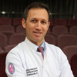 Benlidayi M Emre合作论文数Faculty of Dentistry, Çukurova University17
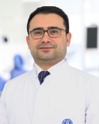 Özgür Erdogan合作论文数Faculty of Dentistry, Istanbul Okan University15
 Mehmet Kurkcu合作论文数13
 Salimov Fariz合作论文数Faculty of Dentistry, Çukurova University13
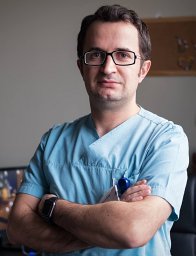 Yakup Ustun合作论文数Faculty of Dentistry, Erciyes University12
 Evlice Burcu合作论文数Faculty of Dentistry, Cukurova University9
 Huseyin Can Tukel合作论文数Department of Oral and Maxillofacial Surgery, Cukurova University8
 Altan Ahmet合作论文数Dentistry Faculty, Mustafa Kemal University6
 halil ibrahim kisa合作论文数5Haluk Oztunc合作论文数5
 Osman Fatih Arpag合作论文数Faculty of Dentistry, Mustafa Kemal University5Akoğlan Mücahide合作论文数4Emin Esen合作论文数Faculty of Dentistry, Çukurova University4
 Ilke Coskun Benlidayi合作论文数Faculty of Medicine, Cukurova University3
 Rengin Guzel合作论文数Faculty of Medicine, Cukurova University3Cem Kurtoglu合作论文数3Mohammed Alraawi合作论文数2
 Halide Namlı合作论文数2
 Orhun Ekren合作论文数Faculty of Dentistry, Cukurova University2
 Sevil Karabag合作论文数2Sürmelioğlu Ozgür合作论文数2
 Halil Murat Aydin合作论文数Bioengineering Division and Center of Bioengineering, Hacettepe University2Kahraman O E合作论文数Faculty of Dentistry, Cukurova University2
 Ahmet Evlice合作论文数2Ilhan Tuncer合作论文数2Kenan Daglioglu合作论文数Experimental Surgery Research Center, Cukurova University2Gulfiliz Gonlusen合作论文数2
 Aslihan Uzel合作论文数2
 Yurdanur Ucar合作论文数Department of Prosthodontics, Cukurova University;College of Dentistry, The Ohio State University;Cukurova University, The Ohio State University2Abdullah Tugrul Coskun合作论文数1Cihan Cem Gürbüz合作论文数1
 Ozlem akinci sozer合作论文数1uygar bakşi合作论文数1Leman Sencar Gundogdu合作论文数1cansu abayli合作论文数1
 Murat Sert合作论文数1
 Ahmet Gunay合作论文数Engn Fac, Balikesir Univ1Sibel Hakverdi合作论文数1
 Metin Sencimen合作论文数Department of Oral and Maxillofacial Surgery, Gulhane Military Medical Academy1Bilgiç Fundagül合作论文数Faculty of Dentistry Department of Orthodontics, Dicle University1
 Vladimir Machon合作论文数1
 Aydin Ozkan合作论文数1
 Meral gunaldı合作论文数1
 Cigdem Usul Afsar合作论文数Faculty of Medicine, Cukurova University Adana1
 Ulas Gormez合作论文数1
 Selen Begum Uzun合作论文数Ministry of Health, General Directorate of Health Research1Iffet yazıcıoglu合作论文数1
 Zeki Arslanoglu合作论文数1




