BACKGROUND:The selection of cardiac phase in coronary computed tomography angiography (CCTA) may affect computational fluid dynamics (CFD)-derived hemodynamic metrics; however, this influence is not well-quantified, particularly in anomalous coronary anatomies. This study aims to evaluate how cardiac phase selection impacts CFD outcomes in normal and anomalous coronary arteries. METHODS:Multiphase CCTA datasets were analyzed from 40 patients: 30 with systolic and diastolic reconstructions (10 inter-arterial anomalous right coronary artery [ARCA], 10 myocardial bridging [MB], 10 normal) and 10 additional normals with mid-diastolic and diastolic reconstructions. Geometric differences among phases were measured. CFD-derived parameters, including time-averaged wall shear stress (TAWSS), oscillatory shear index (OSI), relative residence time (RRT), and CCTA-derived fractional flow reserve (CT-FFR), were calculated. Phase-dependency was assessed by (i) comparing proximal right coronary artery (pRCA) and proximal left anterior descending artery (pLAD) measurements in normal systole vs. diastole and mid-diastole vs. diastole; (ii) comparing pRCA measurements in ARCA vs. normal, and pLAD measurements in MB vs. normal, for systole and diastole. RESULTS:Luminal area differences among phases in pRCA and pLAD segments demonstrated group-distance interactions (p < 0.05). Absolute relative differences in OSI were significantly smaller in pLAD for MB group (median 5.31% [IQR 2.61%-7.46%]) versus normal group (15.99% [6.23%-31.98%]; p = 0.0125). While TAWSS, OSI, and RRT exhibited phase-dependency, this was neither specific to patient cohorts nor coronary artery territories. The variation of relative differences in CT-FFR (maximum 2.4%) was generally lower than that in TAWSS (21.3%), OSI (63.9%), and RRT (28.1%), with all relative differences of the four parameters showing no significant variation from zero. CONCLUSION:The coronary geometry showed clear phase-dependency. The phase dependency should not be ignored when CCTA is used to quantify TAWSS, OSI and RRT. Standardizing cardiac phase selection in CCTA-based CFD analyses is crucial for improving accuracy and clinical consistency.
The objectives were to develop and evaluate a machine learning model based on a combination of biomechanics and image texture analysis, to improve the detection of high-risk carotid plaques. Sixty-five patients, who underwent high-resolution, multi-contrast, magnetic resonance imaging (MRI) of the carotid artery wall within two weeks of a TIA or stroke, were assessed. The MR images were provided by the CARE-II multi-centre carotid imaging trial (ClinicalTrials.gov Identifier: NCT02017756). Following 3D artery construction, parametric maps of structural stress, wall shear stress, and inward remodeling were computed using a one-way fluid structure interaction (FSI) approach. A radiomics pipeline was developed to derive image texture features from mechanics maps and the MR images. Machine learning models were then developed to distinguish non-culprit and culprit carotid plaques, where culprit plaques were deemed responsible for the symptoms associated with the TIA/stroke. The performance of a combined model, developed from the most predictive features of the mechanics map and MR image models, was compared with mechanics and MRI-based models individually. Mechanics [Accuracy = 0.68, AUC = 0.75 ± 0.03] and MRI-based models [Accuracy = 0.67, AUC = 0.71 ± 0.04] showed greater predictive capabilities for culprit lesions than the measurement of vessel stenosis alone [Accuracy = 0.62, AUC = 0.57 ± 0.04] (p < 0.001). The combined mechano-radiomics model [Accuracy = 0.76, AUC= 0.82 ± 0.04] (p=0.037) showed significant improvement in the prediction of culprit plaques compared with MRI and mechanics map features alone, as well as the clinically conventional measurement of vascular stenosis.
Objectives To assess how radiomic features may be combined with plaque morphological and compositional features identified by multi-contrast MRI to improve upon conventional risk assessment models in determining culprit carotid artery lesions.Methods Fifty-five patients (mean age: 62.6; 35 males) with bilateral carotid stenosis who experienced transient ischaemic attack (TIA) or stroke were included from the CARE-II multi-centre carotid imaging trial (ClinicalTrials.gov Identifier: NCT02017756). They underwent MRI within 2 weeks of the event. Classification capability in distinguishing culprit lesions was assessed by machine learning. Repeatability and reproducibility of the results were investigated by assessing the robustness of the radiomic features.Results Radiomics combined with a relatively conventional plaque morphological and compositional metric-based model provided incremental value over a conventional model alone (area under curve [AUC], 0.819 +/- 0.002 vs 0.689 +/- 0.019, respectively, P = .014). The radiomic model alone also provided value over the conventional model (AUC, 0.805 +/- 0.003 vs 0.689 +/- 0.019, respectively, P = .031). T2-weighted imaging-based radiomic features had consistently higher robustness and classification capabilities compared with T1-weighted images. Higher-dimensional radiomic features outperformed first-order features. Grey Level Co-occurrence Matrix, Grey Level Dependence Matrix, and Grey Level Size Zone Matrix sub-types were particularly useful in identifying textures which could detect vulnerable lesions.Conclusions The combination of MRI-based radiomic features and lesion morphological and compositional parameters provided added value to the reference-standard risk assessment for carotid atherosclerosis. This may improve future risk stratification for individuals at risk of major adverse ischaemic cerebrovascular events.Advances in knowledge The clinical relevance of this work is that it addresses the need for a more comprehensive method of risk assessment for patients at risk of ischaemic stroke, beyond conventional stenosis measurement. This paper shows that in the case of carotid stroke, high-dimensional radiomics features can improve classification capabilities compared with stenosis measurement alone.
Background and objectives: Intravascular optical coherence tomography (IVOCT) is capable of delineating periluminal region, including thin fibrous cap, calcium, lipid and thrombus. The segmentation result of these plaques is needed when calculating some useful diagnostic indicators, such as the minimum fiber cap thickness or the maximum Plaque Structural Stress (PSS), to help the diagnosis of vulnerable plaque. Since only some images contain plaques, in order to simplify the network architecture, we designed a three-step framework with single task for each step in this paper to realize the machine learning based pixel-level semantic segmentation of plaque in IVOCT.Methods: A three-step framework is designed: lumen segmentation, image classification and plaque semantic segmentation. Firstly, the lumen of IVOCT is segmented using U-Net. Then the patches are cropped along the lumen boundary, and the plaques in the patches are classified and merged to get whether the original image contains calcium or lipid plaque. In the classification procedure, a self-attention module is introduced into ResNet to form the improved self-attention ResNet. Finally, the selected images with plaque are segmented to get the pixel-level segmentation results of each plaque component, which is realized by an combined network composed by convolutional auto encoder and U-Net.Results and conclusions: In the lumen segmentation step, the Dice coefficient is greater than 95%. In the classification step, the classification precision, sensitivity and specificity of calcium plaque are all 100%; and that of lipid plaque are 97%, 100% and 94% respectively. In the final segmentation step, the Dice coefficient of calcium plaque is 71.8% and that of lipid plaque is 60.5%; the sensitivity of calcium plaque is 78.4% and that of lipid plaque is 80.2%. The experiments show that compared to some commonly used networks the proposed method achieves better performance.
ABSTRACT Objectives To assess how radiomic features may be combined with plaque morphological and compositional features identified by multi-contrast magnetic resonance imaging (MRI) to improve upon conventional risk assessment models in determining culprit lesions. Methods Fifty-five patients (mean age: 62.6; 35 males) with bilateral carotid stenosis who experienced transient ischaemic attack (TIA) or stroke were included from the CARE-II multi-centre carotid imaging trial ( ClinicalTrials.gov Identifier: NCT02017756 ). They underwent MRI within 2 weeks of the event. Classification capability in distinguishing culprit lesions was assessed by machine learning. Repeatability and reproducibility of the results were investigated by assessing the robustness of the radiomic features. Results Radiomics combined with a relatively conventional plaque morphological and compositional metric-based model provided incremental value over a conventional model alone [area under curve (AUC), 0.819 ± 0.002 vs. 0.689 ± 0.019 respectively, p = 0.014]. The radiomic model alone also provided value over the conventional model [AUC, 0.805 ± 0.003 vs. 0.689 ± 0.019 respectively, p = 0.031]. T2-weighted imaging-based radiomic features had consistently higher robustness and classification capabilities compared with T1-weighted images. Higher-dimensional radiomic features outperformed first-order features. Grey Level Co-occurrence Matrix (GLCM), Grey Level Dependence Matrix (GLDM) and Grey Level Size Zone Matrix (GLSZM) sub-types were particularly useful in identifying textures which could detect vulnerable lesions. Conclusions The combination of MRI-based radiomic features and lesion morphological and compositional parameters provided added value to the reference-standard risk assessment for carotid atherosclerosis. This may improve future risk stratification for individuals at risk of major adverse ischemic cerebrovascular events. Clinical Relevance The clinical relevance of this work is that it addresses the need for a more comprehensive method of risk assessment for patients at risk of ischemic stroke, beyond conventional stenosis measurement. Radiomics provides a non-invasive means of assessing plaque vulnerability. Key points T2-weighted imaging-based radiomic features had consistently higher robustness and classification capabilities compared with T1-weighted images. Higher dimensional radiomic features had better performance than first-order features in identifying textures which could detect vulnerable carotid lesions. Radiomic features combined with MRI plaque features may improve atherosclerotic plaque risk stratification.
Introduction: Atheromatous neovessels with high permeability have been implicated in the evolution of intraplaque haemorrhage (IPH). In vivo quantification of vasa vasorum permeability is possible using dynamic-contrast enhanced-magnetic resonance (DCE-MR) imaging. Hypothesis: In vivo relationship of extent of leakiness of neovessels with critical biomechanical forces i.e. stress and strain in atheroma and with IPH remains unreported. Methods: Patients with mild to severe carotid artery stenosis underwent DCE-MR imaging of their bilateral carotid arteries on a 3 Tesla MR system, using TRICKS method. A two-compartment Patlak model was used to generate a parametric map showing partial plasma volume (vp) and K trans. FEA simulations were performed for stress and strain analyses. For histological analysis, carotid plaques were processed and stained. Results: Sixteen patients underwent carotid MR imaging. The two groups were comparable for their demographics and co-morbidities. Peak structural stress (PSS) and peak strain (P1) were significantly higher in symptomatic versus asymptomatic patients (p=0.002 and 0.009 respectively). Carotid atheroma with IPH had higher PSS and P I compared to those without IPH [p=0.036 and 0.009]. Variations in stress during one cardiac cycle was significantly greater among plaques with IPH (p=0.01); variation in strain was greater in atheroma with IPH [p=0.04]. Adventitial K trans was higher in carotid atheroma of symptomatic patients than asymptomatic cohort (p= 0.016). Plaque K trans , adventitial and plaque v p were however comparable for both patient cohorts (p= 0.81, 0.96, 0.36 respectively). Adventitial K trans and plaque v p were higher in atheroma with IPH (p= 0.04, 0.01) but comparable for plaque K trans and adventitial v p (p=0.46, 0.88 respectively). A strong correlation was observed between variations in strain during the cardiac cycle with adventitial K trans for plaques with IPH [Pearson r : 0.84, p=0.03]. Conclusions: A strong correlation exists between adventitial neovessel permeability and variations in strain in atheroma with IPH. Carotid atheroma with IPH have higher biomechanical stresses and strain and variations in these critical biomechanical conditions compared to non-IPH atheroma.
EDITORIAL article Front. Neurosci., 22 November 2022Sec. Brain Imaging Methods Volume 16 - 2022 | https://doi.org/10.3389/fnins.2022.1086022
Advances in medical imaging have enabled patient-specific biomechanical modelling of arterial lesions such as atherosclerosis and aneurysm. Geometry acquired from in-vivo imaging is already pressurized and a zero-pressure computational start shape needs to be identified. The backward displacement algorithm was proposed to solve this inverse problem, utilizing fixed-point iterations to gradually approach the start shape. However, classical fixed-point implementations were reported with suboptimal convergence properties under large deformations. In this paper, a dynamic learning rate guided by the deformation gradient tensor was introduced to control the geometry update. The effectiveness of this new algorithm was demonstrated for both idealized and patient-specific models. The proposed algorithm led to faster convergence by accelerating the initial steps and helped to avoid the non-convergence in large-deformation problems.
Background: Adequate tissue perfusion is an important prognostic and diagnostic factor during the management of lower limb peripheral arterial disease. Convenient and real-time tissue perfusion monitoring remains an elusive challenge. Methods: Tissue perfusion on the dorsal and plantar surfaces of both feet of 20 participants was measured during and after cuff-induced ischemia using a novel 4-channel, laser-based perfusion monitoring device based on diffuse speckle contrast analysis technology (Pedra sensors). Participants were free of significant peripheral arterial disease. Transcutaneous partial pressure of oxygen (TcPO2) measurements were recorded concurrently for comparison. Results: Pedra sensors detected perfusion changes significantly more quickly than TcPO2 sensors. One minute after induced ischemia, the mean percent changes from baseline values (before ischemia) were-22.7 +/- 32.0% and-3.1 +/- 8.8% (P<0.001) for Pedra and TcPO2 sensors, respectively. One minute into induced ischemia, Pedra sensors had reached 50.5% of the 5-minute ischemia reading whereas TcPO2 sensors had reached only 18.6% of the 5-minute reading (P=0.046). Pedra sensors reported hyperemia immediately after cuff release with a mean percent change from baseline of 143.8 +/- 122.3%/173.4 +/- 121.8% on the dorsal/plantar surfaces while TcPO2 measurements were still recording negative changes at that time (-26.7 +/- 19.4%/-18.6 +/- 24.4% dorsal/plantar). Pedra sensors exhibited markedly lower interobserver and intraobserver variability than TcPO2 sensors. Conclusions: A device based on diffuse speckle contrast analysis reported tissue perfusion in real time. Cuff-induced ischemia and hyperemia following cuff release were rapidly and consistently detected on both the dorsal and plantar surfaces of the foot. Diffuse speckle contrast analysis may have value for real-time perfusion monitoring during angiography procedures.
Background and aims Artery is subject to wall shear stress (WSS) and vessel structural stress (VSS) simultaneously. This study is designed to explore the role of VSS in development of atherosclerosis. Methods Silastic collars were deployed on the carotid to create two constrictions on 13 rabbits for a distinct mechanical environment at the constriction. MRI was performed to visualize arteries' configuration. Animals with high fat (n = 9; Model-group) and normal diet (n = 4; Control-group) were sacrificed after 16 weeks. 3D fluid-structure interaction analysis was performed to quantify WSS and VSS simultaneously. Results Twenty plaques were found in Model-group and 3 in Control-group. In Model-group, 8 plaques located proximally to the first constriction (Region-1, close to the heart) and 7 distally to the second (Region-2, close to the head) and 5 plaques were found on the contralateral side of 3 rabbits. Plaques at Region-1 tended to be bigger than those at Region-2 and the macrophage density at these locations was comparable. Minimum time-averaged WSS (TAWSS) in Region-1 was significantly higher than that in Region-2, and both maximum oscillatory shear index (OSI) and particle relative residence time (RRT) were significantly lower. Peak and mean VSS in Region-1 were significantly higher than those in Region-2. Correlation analyses indicated that low TAWSS, high OSI and RRT were only associated with plaque in Region-2, while lesions in Region-1 were only associated with high VSS. Moreover, only VSS was associated with wall thickness of plaque-free regions in both regions. Conclusions VSS might contribute to the initialization and development of atherosclerosis solely or in combination with WSS.
Ferumoxytol is an ultrasmall super paramagnetic particles of iron oxide (USPIO) agent recently used for magnetic resonance (MR) vascular imaging. Other USPIOs have been previously used for assessing inflammation within atheroma. We aim to assess feasibility of ferumoxytol in imaging carotid atheroma (with histological assessment); and the optimum MR imaging time to detect maximum quantitative signal change post-ferumoxytol infusion. Ten patients with carotid artery disease underwent high-resolution MR imaging of their carotid arteries on a 1.5 T MR system. MR imaging was performed before and at 24, 48, 72 and 96 hrs post ferumoxytol infusion. Optimal ferumoxytol uptake time was evaluated by quantitative relaxometry maps indicating the difference in T2* (ΔT2*) and T2 (ΔT2) between baseline and post-Ferumoxytol MR imaging using 3D DANTE MEFGRE qT2*w and iMSDE black-blood qT2w sequences respectively. 20 patients in total (10 symptomatic and 10 with asymptomatic carotid artery disease) had ferumoxytol-enhanced MR imaging at the optimal imaging window. 69 carotid MR imaging studies were completed. Ferumoxytol uptake (determined by a decrease in ΔT2* and ΔT2) was identified in all carotid plaques (symptomatic and asymptomatic). Maximum quantitative decrease in ΔT2* (10.4 [3.5–16.2] ms, p < 0.001) and ΔT2 (13.4 [6.2–18.9] ms; p = 0.001) was found on carotid MR imaging at 48 hrs following the ferumoxytol infusion. Ferumoxytol uptake by carotid plaques was assessed by histopathological analysis of excised atheroma. Ferumoxytol-enhanced MR imaging using quantitative 3D MR pulse sequences allows assessment of inflammation within carotid atheroma in symptomatic and asymptomatic patients. The optimum MR imaging time for carotid atheroma is 48 hrs after its administration.
OBJECTIVES This study determined whether in vivo positron emission tomography (PET) of arterial inflammation (F-18-fluorodeoxyglucose [F-18-FDG]) or microcalcification (F-18-sodium fluoride [F-18-NaF]) could predict restenosis following PTA. BACKGROUND Restenosis following lower limb percutaneous transluminal angioplasty (PTA) is common, unpredictable, and challenging to treat. Currently, it is impossible to predict which patient will suffer from restenosis following angioplasty. METHODS In this prospective observational cohort study, 50 patients with symptomatic peripheral arterial disease underwent F-18-FDG and F-18-NaF PET/computed tomography (CT) imaging of the superficial femoral artery before and 6 weeks after angioplasty. The primary outcome was arterial restenosis at 12 months. RESULTS Forty subjects completed the study protocol with 14 patients (35%) reaching the primary outcome of restenosis. The baseline activities of femoral arterial inflammation (F-18-FDG tissue-to-background ratio [TBR] 2.43 [interquartile range (IQR): 2.29 to 2.61] vs. 1.63 [IQR: 1.52 to 1.78]; p < 0.001) and microcalcification (F-18-NaF TBR 2.61 [IQR: 2.50 to 2.77] vs. 1.69 [IQR: 1.54 to 1.77]; p < 0.001) were higher in patients who developed restenosis. The predictive value of both F-18-FDG (cut-off TBRmax value of 1.98) and F-18-NaF (cut-off TBRmax value of 2.11) uptake demonstrated excellent discrimination in predicting 1-year restenosis (Kaplan Meier estimator, log-rank p < 0.001). CONCLUSIONS Baseline and persistent femoral arterial inflammation and micro-calcification are associated with restenosis following lower limb PTA. For the first time, we describe a method of identifying complex metabolically active plaques and patients at risk of restenosis that has the potential to select patients for intervention and to serve as a biomarker to test novel interventions to prevent restenosis. (C) 2020 The Authors. Published by Elsevier on behalf of the American College of Cardiology Foundation.
The operative caseload of a surgeon has a positive influence on post-operative outcomes. For surgical trainees to progress effectively, maximising operating room exposure is essential, vascular surgery being no exception. Our aim was to ascertain the impact of supervised trainee led vs. expert surgeon led procedures on post-operative outcomes, across three commonly performed vascular operations.A literature search was undertaken using the MEDLINE, Web of Science, and Cochrane databases up to 1 January 2018. Studies reporting outcomes following major lower limb amputation, fistula formation, or carotid endarterectomy (CEA) that involved a direct comparison between supervised trainee and experts were included, with odds ratios (ORs) calculated. Primary outcomes varied depending on the specific procedure: amputations-rate of amputation revision within 30 days; fistula formation-primary patency; CEA-stroke rate at 30 days. Meta-analysis with the Mantel-Haenszel method was performed for each outcome.Sixteen studies were included in the final review. Overall, trainees accounted for a third of all procedures analysed (n = 2 421/7 017; 34.5%). Only one study was identified that described rates of amputation revision, precluding any further analysis. Four studies on fistula formation were included, showing no significant difference in outcomes between trainees and experts in primary patency (OR 1.68, 95% confidence interval [CI] 0.42-6.75). Nine studies were identified reporting post-CEA stroke rates, also demonstrating no difference between trainees and experts (OR 0.89, 95% CI 0.59-1.32).In select cases, with appropriate training and suitable experience, supervised trainees can perform surgical procedures without any detriment to patient care. To ensure high standards for patients of the future, supported training programmes are essential for today's surgical trainees.
OBJECTIVE:The operative caseload of a surgeon has a positive influence on post-operative outcomes. For surgical trainees to progress effectively, maximising operating room exposure is essential, vascular surgery being no exception. Our aim was to ascertain the impact of supervised trainee led vs. expert surgeon led procedures on post-operative outcomes, across three commonly performed vascular operations. METHODS:A literature search was undertaken using the MEDLINE, Web of Science, and Cochrane databases up to 1 January 2018. Studies reporting outcomes following major lower limb amputation, fistula formation, or carotid endarterectomy (CEA) that involved a direct comparison between supervised trainee and experts were included, with odds ratios (ORs) calculated. Primary outcomes varied depending on the specific procedure: amputations-rate of amputation revision within 30 days; fistula formation-primary patency; CEA-stroke rate at 30 days. Meta-analysis with the Mantel-Haenszel method was performed for each outcome. RESULTS:Sixteen studies were included in the final review. Overall, trainees accounted for a third of all procedures analysed (n = 2 421/7 017; 34.5%). Only one study was identified that described rates of amputation revision, precluding any further analysis. Four studies on fistula formation were included, showing no significant difference in outcomes between trainees and experts in primary patency (OR 1.68, 95% confidence interval [CI] 0.42-6.75). Nine studies were identified reporting post-CEA stroke rates, also demonstrating no difference between trainees and experts (OR 0.89, 95% CI 0.59-1.32). CONCLUSION:In select cases, with appropriate training and suitable experience, supervised trainees can perform surgical procedures without any detriment to patient care. To ensure high standards for patients of the future, supported training programmes are essential for today's surgical trainees.
Arterial calcification in different arterial beds has been observed to be an independent predictor of mortality. The association of abdominal visceral artery calcium with all-cause mortality remains unexplored. Patients who had undergone contrast-enhanced computerized tomography (CT) imaging for routine assessment of peripheral arterial disease (PAD) were considered for this study. A novel calcium score (abdominal visceral arteries calcium [AVAC]) for the abdominal visceral arteries (celiac axis, superior mesenteric, and renal arteries) was calculated using a modified Agatston score. Cumulative AVAC was defined as sum total of the calcium score of above individual arteries. The primary outcome was all-cause mortality. The association of AVAC with all-cause mortality was assessed. Of the 134 consecutive patients, 89 were included for analysis. Median follow-up duration was 72 (47-91) months since CT imaging; 35 (39%) patients died during this period. Hypertension and cumulative AVAC score had a significant association with all-cause mortality ( P < .05). Cumulative visceral abdominal artery calcification is associated with all-cause mortality in patients with PAD. Future prospective studies are warranted to investigate this relationship in PAD and other patient cohorts.
Background: Atherosclerosis is a systemic inflammatory disease intertwined with neovascularization. Dynamic contrast-enhanced magnetic resonance imaging (DCE-MRI) enables the assessment of plaque neovascularization. This study aimed to explore the systemic nature of atherosclerosis by assessing difference in severity of neovascularization as quantified by DCE-MRI of vertebral arteries (VAs) between patients with symptomatic and asymptomatic carotid artery disease. Methods: Ten consecutive patients with asymptomatic VA stenosis and concomitant symptomatic carotid artery disease (group 1) and 10 consecutive patients with asymptomatic VA stenosis and concomitant asymptomatic carotid artery disease (group 2) underwent 3-dimensional DCE-MRI of their cervical segment of VAs. A previously validated pharmacokinetic modeling approach was used for DCE-MRI analysis. K-trans was calculated in the adventitia and plaque as a measure of neovessel permeability. Results: Both patient groups were comparable for demographics and comorbidities. Mean luminal stenosis was comparable for both groups (54.4% versus 52.27%, P = .32). Group 1 had higher adventitial K-trans and plaque K-trans (.08 +/- .01 min(-1), .07 +/- .01 min(-1)) compared with Group 2 (.06 +/- .01 mintmin(-1), .06 +/- .01 min(-1)) (P = .004 and .03, respectively). Good correlation was present among the two image analysts (intraclass correlation coefficient = .78). Conclusions: Vertebral Artery atheroma of patients with symptomatic carotid artery disease had increased neovessel permeability compared with the patients with asymptomatic carotid artery disease. These findings are consistent with the hypothesis that atherosclerosis is a systemic inflammatory disease. The VA atherosclerosis is likely to have increased severity of neovascularization if another arterial territory is symptomatic in the same patient cohort.
 Jonathan Gillard合作论文数Department of Radiology, University of Cambridge77
 Martin J Graves合作论文数Department of Radiology, University of Cambridge51
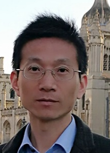 Zhongzhao Teng合作论文数Department of Radiology, School of Clinical Medicine, University of Cambridge42
 K Varty合作论文数38Jonathan R. Boyle合作论文数38
 Stewart R Walsh合作论文数38
 Paul Hayes合作论文数35Tjun Y. Tang合作论文数34Michael E. Gaunt合作论文数20
 Victoria Young合作论文数18
 Andrew J Patterson合作论文数17Usman Ammara合作论文数17
 Zhiyong Li (李志勇)合作论文数School of Biological Science & Medical Engineering, Southeast University14Victoria E Young合作论文数14
 Zhu Chengcheng合作论文数12T.Y. Tang合作论文数12M. E. Gaunt合作论文数10
 Ayesha Noorani合作论文数Addenbrooke’s Hospital, Cambridge University Hospitals10
 Yuan Huang合作论文数Department of Radiology;Department of Radiology, University of Cambridge;Department of Pure Mathematics and Mathematical Statistics, University of Cambridge9David G. Cooper合作论文数Department of Chemical Engineering, McGill University8Jonathan H. Gillard合作论文数7
 Mohammed Chowdhury合作论文数Wolfson College, University of Cambridge;Clinical Medicine School, Cambridge University Hospitals6Nadim Noor合作论文数5A Noorani合作论文数5Bowden David J合作论文数5
 Patrick Coughlin合作论文数5
 Asif Jah合作论文数Pancreatic Cancer Centre, University of Cambridge;Cambridge University Hospital5Simon P.S. Howarth合作论文数5
 Philip W Stather合作论文数Norwich Medical School, University of East Anglia4
 Sheraz Markar合作论文数Surgical Intervention Trials Unit, Nuffield Department of Surgical Sciences, Medical Sciences Division, University of Oxford4Nair Sukumaran K合作论文数4
 Shengyong Chen (陈胜勇)合作论文数School of Computer Science and Engineering, Tianjin University of Technology4
 Valentina Taviani合作论文数Department of Engineering, University of Cambridge4
 James Rudd合作论文数Department of Medicine, University of Cambridge;Addenbrooke’s Hospital4
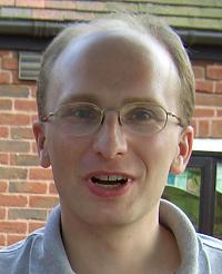 Andrew Priest合作论文数Department of Radiology, University of Cambridge4Claire Cousins合作论文数4Ayoola Awopetu合作论文数3Neethu B.G. Mariam合作论文数3PJ Kirkpatrick合作论文数3J. Naik合作论文数3A. Usman合作论文数3A. Patterson合作论文数3
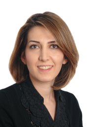 Nasim Sheikh-Bahaei合作论文数Keck Sch Med, Univ Southern Calif3
 Shuo Wang (王烁)合作论文数Digital Medical Research Center, School of Basic Medical Sciences, Fudan University;Shanghai Medical College, Fudan University3Jianmin Yuan合作论文数UIH Grp, Cent Res Inst3Albaghdadi Mazen合作论文数Department of Medicine, Northwestern University Feinberg School of Medicine3
 Elizabeth Warburton合作论文数Neurology Unit, University of Cambridge3Teik Choon See合作论文数3




