More specific targeted drug delivery systems with low immunogenicity and toxicity are expected to increase the efficacy of therapeutic molecules. The extracellular nanovesicles have been introduced as natural delivery systems for therapeutic molecules. More recently, considerable interest has emerged in plant-derived exosomes and their natural commitment to deliver molecules of various origins. Acridine Orange (AO) is an acidophilic dye with a strong tumoricidal action following excitation with a light source at 466 nm, but its clinical use is limited by the potential systemic toxicity. In this study we investigated the ability of exosomes from Citrus Sinensis to be successfully uploaded with AO (Exo-AO). We also studied the ability of the exosomes to be uploaded into target cells, as compared to the free molecules. We found that AO was efficiently uploaded into exosomes through electroporation. In fact, Exo-AO entered into the target cells significantly better than the free molecules, leading to both a marked intracellular AO delivery into target cells and an increase in the cytotoxic effect after excitation under a fluorescence microscope. This study shows a promising new approach for a more effective and less toxic drug delivery through the use of plant-derived extracellular nanovesicles.
Abstract Urinary extracellular vesicles (uEVs) represent a promising source of non-invasive biomarkers; however, their clinical translation is still limited by suboptimal isolation strategies, which often suffer from poor scalability, co-isolation of abundant urinary proteins, and bias toward specific EV subpopulations. Here, we employ a membrane-sensing peptide (MSP)-based affinity approach for uEVs isolation, that exploits the highly lipid membranes curvature of EV as universal target, enabling pan-specific capture independent of surface marker expression. MSP-functionalized beads were applied to minimally processed urine samples and benchmarked against differential ultracentrifugation (dUC) and size-exclusion chromatography (SEC). Comprehensive characterization by nanoparticle tracking analysis, transmission electron microscopy, high-sensitivity flow cytometry, single-molecule array (SiMoA), and fluorescence nanoparticle tracking analysis, demonstrated that MSP-based isolation preserves vesicle integrity and maintains the native distribution of canonical tetraspanins (CD9, CD63, CD81), without evidence of subpopulation bias. Notably, MSP-based isolation significantly reduced co-isolated contaminants, such as uromodulin, resulting in improved sample purity. By combining high recovery, improved purity, and operational simplicity, MSP workflow offers practical advantages, including reduced processing time, scalability, and compatibility with standard laboratory equipment, without the need for extensive pre-processing. These properties characterize MSP-based affinity capture as a robust and versatile alternative to conventional uEVs isolation approaches, with strong potential for translational and clinical applications.
Hypertrophic cardiomyopathy (HCM) is a genetically determined heart disease characterized by marked clinical heterogeneity and limited availability of specific circulating biomarkers that reflect underlying disease mechanisms. This study investigated whether plasma-derived extracellular vesicles (EVs) capture disease-related molecular features in patients with HCM. Plasma EVs were characterized for size, cellular origin, and protein cargo using flow cytometry and targeted and untargeted proteomic analyses. While size and concentration of EVs were comparable between groups, patients with HCM exhibited an enrichment of platelet-derived EVs (CD41a+) and reductions in neutrophil (CD66b+)- and lymphatic-endothelial-cell (CD310+)-derived EVs. Proteomic profiling revealed EV-associated proteins linked to platelet activation and thrombo-inflammation. Multivariable protein signatures derived from EVs discriminated patients from controls with performance comparable to plasma-based models and were not influenced by age. These findings indicate that plasma-derived EVs capture key biological pathways involved in HCM and may complement biomarker discovery strategies.
Extracellular vesicles (EVs) are membrane-bound particles crucial for intercellular communication and serve as promising biomarkers for diseases, including cancer. Isolating and characterizing specific EV subpopulations, particularly those in plasma/serum, enhances biomarker precision and supports targeted therapies. Cancer-derived EVs often express unique surface markers, enabling distinction from other EVs. Accurate sorting of tumor-associated EVs provides insights into cancer progression, metastasis, and treatment response. This study presents a robust method for isolating and sorting CD9 + plasma EVs as a proof-of-concept for broader EV subpopulation analyses. Plasma EVs were isolated via sucrose cushion ultracentrifugation, optimizing purity and yield. Flow cytometry with fluorescence threshold triggering was fine-tuned to detect and sort CD9 + EVs, with instrument calibration and parameter adjustments mitigating swarming and improving sorting accuracy. Size exclusion chromatography further enhanced efficiency by reducing background noise. Sorted CD9 + EVs retained size and marker expression, including Syntenin, Alix, Flotillin-1, and CD9, which were enriched post-sorting. These advancements enable high-purity EV subpopulation isolation, facilitating applications such as identifying cancer biomarkers and developing EV-based targeted therapies.
CD4+CD25hiFoxP3+ regulatory T cells (Treg cells) are key controllers of immune self-tolerance, and their suppressive function is impaired in people with relapsing-remitting multiple sclerosis (pwRR-MS). Because the mechanisms underlying this condition are still ill-defined, we investigated the role of Treg cell-derived extracellular vesicles (Treg-EVs) in Treg cell dysfunction observed in pwRR-MS. We found that Treg-EVs from healthy individuals inhibit CD4+ conventional T (Tconv) cells by shuttling miR-142-3p from the Treg cell to the Tconv cell. There, miR-142-3p down-regulated mRNAs necessary for Tconv cell growth and effector functions, such as the redox controller cystine carrier SLC7A11. However, Treg cells from pwRR-MS released EVs containing reduced amounts of miR-142-3p, resulting in impaired suppressive function. Furthermore, Treg-EV miR-142-3p inversely correlated with the disability score and gadolinium-enhancing lesions in pwRR-MS. Together, our results elucidate a molecular mechanism involving miR-142-3p shuttled by Treg-EVs in the control of immune self-tolerance and unveil its pathogenetic implications in human autoimmunity.
AbstractMicrogravity (µG) experienced during space flights promotes adaptation in several astronauts’ organs and tissues, with skeletal muscles being the most affected. In response to reduced gravitational loading, muscles (especially, lower limb and antigravity muscles) undergo progressive mass loss and alteration in metabolism, myofiber size, and composition. Skeletal muscle precursor cells (MPCs), also known as satellite cells, are responsible for the growth and maintenance of muscle mass in adult life as well as for muscle regeneration following damage and may have a major role in µG-induced muscle wasting. Despite the great relevance for astronaut health, very few data are available about the effects of real µG on human muscles. Based on the MyoGravity project, this study aimed to analyze: (i) the cellular and transcriptional alterations induced by real µG in human MPCs (huMPCs) and (ii) the response of human skeletal muscle to normal gravitational loading after prolonged exposure to µG. We evaluated the transcriptomic changes induced by µG on board the International Space Station (ISS) in differentiating huMPCs isolated from Vastus lateralis muscle biopsies of a pre-flight astronaut and an age- and sex-matched volunteer, in comparison with the same cells cultured on the ground in standard gravity (1×g) conditions. We found that huMPCs differentiated under real µG conditions showed: (i) upregulation of genes related to cell adhesion, plasma membrane components, and ion transport; (ii) strong downregulation of genes related to the muscle contraction machinery and sarcomere organization; and (iii) downregulation of muscle-specific microRNAs (myomiRs). Moreover, we had the unique opportunity to analyze huMPCs and skeletal muscle tissue of the same astronaut before and 30 h after a long-duration space flight on board the ISS. Prolonged exposure to real µG strongly affected the biology and functionality of the astronaut’s satellite cells, which showed a dramatic reduction of responsiveness to activating stimuli and proliferation rate, morphological changes, and almost inability to fuse into myotubes. RNA-Seq analysis of post- vs. pre-flight muscle tissue showed that genes involved in muscle structure and remodeling are promptly activated after landing following a long-duration space mission. Conversely, genes involved in the myelination process or synapse and neuromuscular junction organization appeared downregulated. Although we have investigated only one astronaut, these results point to a prompt readaptation of the skeletal muscle mechanical components to the normal gravitational loading, but the inability to rapidly recover the physiological muscle myelination/innervation pattern after landing from a long-duration space flight. Together with the persistent functional deficit observed in the astronaut’s satellite cells after prolonged exposure to real µG, these results lead us to hypothesize that a condition of inefficient regeneration is likely to occur in the muscles of post-flight astronauts following damage.
Background: Hypertrophic cardiomyopathy (HCM) is diagnosed according to the presence of morphological and functional traits of the heart, often in the presence of genetic mutations. A specific biomarker assessing the aetiopathology of this condition is lacking. Extracellular vesicles (EVs), small particles released by all cells into biological fluids, hold promise as diagnostic and prognostic tools for cardiac diseases. We aim at characterising plasma-derived EVs isolated from 18 consecutive HCM patients and 13 healthy volunteers (CTR). Methods: HCM underwent echocardiographic assessment and genetic testing evaluating 200 genes (NGS). EVs were isolated via ultracentrifugation from platelet-free plasma. Quantitative and qualitative assessments of EVs were performed by nanoparticle tracking analysis and transmission electron microscopy. FACS analysis was used to characterize EV subpopulations. Data are expressed as median and interquartile ranges. Results: Most patients were male (70.8% HCM and 54% CTR) with a median age of 61 (52.5-71) years (HCM) and 47 (44-52) (CTR). Missense mutations in the MYH7 gene were the most found in HCM. The median maximum wall thickness in HCM was 16 mm (15-19) vs 8 mm (7-9) in CTR. No differences were found in EV concentrations between HCM and CTR, respectively, 3.6*109 EV/ml/cell count (2*109-5*109) and 5*10⁹ EV/ml/cell count (4*109-6*109). However, EV concentration was positively associated with the sudden cardiac death risk score in HCM (r= 0.63). Among the EVs positive for CFSE (a specific dye for EVs), those released from platelets, progenitor endothelial cells and neutrophils were increased in HCM patients vs CTR, respectively, by 1.7-, 1.5- and 1.1-fold. A strong negative association (r= -0.74) was found between progenitor endothelial cell-derived EVs and the E/E’ ratio of diastolic function, a strong predictor of first cardiac events. Conclusions: HCM patients present a peculiar phenotypic pattern of EVs that associates which diastolic function and sudden cardiac death.
Extracellular vesicles (EV) have emerged as promising cell-free therapeutics in regenerative medicine. However, translating primary cell line-derived EV to clinical applications requires large-scale manufacturing and several challenges, such as replicative senescence, donor heterogeneity, and genetic instability. To address these limitations, we used a reprogramming approach to generate human induced pluripotent stem cells (hiPSC) from the young source of cord blood mesenchymal stem/stromal cells (CBMSC). Capitalizing on their inexhaustible supply potential, hiPSC offer an attractive EV reservoir. Our approach encompassed an exhaustive characterization of hiPSC-EV, aligning with the rigorous MISEV2023 guidelines. Analyses demonstrated physical features compatible with small EV (sEV) and established their identity and purity. Moreover, the sEV-shuttled non-coding (nc) RNA landscape, focusing on the microRNA and circular RNA cargo, completed the molecular signature. The kinetics of the hiPSC-sEV release and cell internalization assays unveiled robust EV production and consistent uptake by human neurons. Furthermore, hiPSC-sEV demonstrated ex vivo cell tissue-protective properties. Finally, via bioinformatics, the potential involvement of the ncRNA cargo in the hiPSC-sEV biological effects was explored. This study significantly advances the understanding of pluripotent stem cell-derived EV. We propose cord blood MSC-derived hiPSC as a promising source for potentially therapeutic sEV.
Plant-Derived Extracellular Vesicles extracellular vesicles (PDEVs) from organic agriculture (without the use of pesticides and microbicides) contain high levels of antioxidants. Organic PDEVs have shown an increased antioxidant power compared to PDEVs from single plants, suggesting a synergistic effect of the bioactives constitutively expressed in the PDEVs from single fruits. With this study, we wanted to investigate the beneficial effects of a mix of PDEVs on human skin cells. We found detectable levels of citric acid, ascorbic acid, glutathione, catalase, and SOD in a mix of PDEVs deriving from five different fruits (grape, red orange, papaya, pomegranate, and tangerine). We then treated H2O2-conditioned fibroblasts with the mix of PDEVs. The results showed that the PDEVs’ mixture reverted the H2O2-induced redox imbalance, restoring mitochondrial homeostasis, with a strong reduction of mitochondrial anion superoxide and an increase in sirtuin levels. The antioxidant action was consistent with wound repair on a lesion produced in a fibroblast’s monolayer. This result was consistent with an increased level of vimentin and matrix metalloproteinase-9, whose expression is directly related to the efficiency of the reparative processes. These data support a beneficial role of PDEVs in both preventing and treating skin injuries through their potent antioxidant and reparative activities.
BACKGROUND:Acute exacerbations of chronic obstructive pulmonary disease (AE-COPD) are associated with a high rate of cardiovascular events. Thromboinflammation (the interplay between coagulation and inflammation) is probably involved in these events. Extracellular vesicles (EV) increase during AE-COPD, but their role in thromboinflammation in COPD is still unknown. We investigated EV-associated prothrombotic and proinflammatory activity in COPD. METHODS:Patients with AE-COPD, stable COPD (sCOPD) and age- and sex-matched subjects (controls) were enrolled. AE-COPD patients were evaluated at hospital admission and 8 weeks after discharge (recovery; longitudinal arm). In a cross-sectional arm, AE-COPD were compared with sCOPD and controls. EV-mediated prothrombotic activity was tested by measuring the concentration of EV-associated phosphatidylserine, as assessed by a prothrombinase assay, and tissue factor, as assessed by a modified one-stage clotting assay (EV-PS and EV-TF, respectively). Synthesis of interleukin-8 (IL-8) and C-C motif chemokine ligand-2 (CCL-2) by cells of the human bronchial epithelial cell line 16HBE incubated with patients' EV was used to measure EV-mediated proinflammatory activity. RESULTS:Twenty-five AE-COPD (median age [interquartile range] 74.0 [14.0] years), 31 sCOPD (75.0 [9.5] years) and 12 control (67.0 [3.5] years) subjects were enrolled. In the longitudinal arm, EV-PS, EV-TF, IL-8 and CCL-2 levels were all significantly higher at hospital admission than at recovery. Similarly, in the cross-sectional arm, EV-PS, EV-TF and cytokines synthesis were significantly higher in AE-COPD than in sCOPD and controls. CONCLUSIONS:EV exert prothrombotic and proinflammatory activities during AE-COPD and may therefore be effectors of thromboinflammation, thus contributing to the higher cardiovascular risk in AE-COPD.
Human organoids have been proposed to be powerful tools mimicking the physiopathological processes of the organs of origin. Recently, human pancreatic organoids (hPOs) have gained increasing attention due to potential theragnostic and regenerative medicine applications. However, the cellular components of hPOs have not been defined precisely. In this work, we finely characterized these structures, focusing first on morphology and identity-defining molecular features under long-term culture conditions. Next, we focused our attention on hPOs cell type composition using single-cell RNA sequencing founding a complex heterogeneity in ductal components, ranging from progenitor components to terminally differentiated ducts. Furthermore, an extensive comparison of human pancreatic organoids with previously reported transcriptomics signature of human and mouse pancreatic ductal populations, confirmed the functional pancreatic duct subpopulation heterogeneity. Finally, we showed that pancreatic organoid cells follow a precise developmental trajectory and utilize diverse signalling mechanisms, including EGF and SPP1, to facilitate cell-cell communication and maturation. Together our results offer an in-depth description of human pancreatic organoids providing a strong foundation for future in vitro diagnostic and translational studies of pancreatic health and disease. scRNA-seq revealed an intraductal heterogeneity of human pancreatic organoids paving the way to new potential applications of this new technology in developmental biology, regenerative medicine and drug development.
Glioblastoma multiforme (GBM) is the most common primary brain tumor, characterized by a remarkable inner complexity and inter-tumor variability. Moreover, it is very aggressive and resistant to conventional treatments, so that it rapidly relapse. Therefore, there is an immediate need for experimental strategies to enhance our comprehension of GBM, aiming to mitigate its economic and social impact. Here, we described different in vivo and in vitro strategies currently used for the study of GBM. First, we gave a brief and general overview of the classical in vivo models, including xenograft mouse and zebrafish models and canine models, offering a wide range of advantages but also presenting a series of strong limitations. Thus, we described in vitro models, starting from more traditional 2D culture models, comparing different approaches and critically exposing the advantages and disadvantages of using one or the other methods. We also briefly described GBM 2D culture systems that allow recreating multiple cell-cell and cell-extracellular matrix contacts but still do not reflect the complexity of in vivo tumors. We finally described the intricacies of the more novel 3D in vitro models, e.g., spheroids and organoids. These sophisticated models have demonstrated exceptional suitability across a wide spectrum of applications in cancer research, ranging from fundamental scientific inquiries to applications in translational research. Their adaptability and three-dimensional architecture render them invaluable tools, offering new insights and paving the way for advancements in both basic and applied research.
Cancer is a major cause of mortality in humans; often, rather than the primary tumor, it is the presence of metastases that are the cause of death. Extracellular vesicles (EVs) are small structures released by both normal and cancer cells; regarding the latter, they have been demonstrated to modulate almost all cancer-related processes, such as invasion, angiogenesis induction, drug resistance, and immune evasion. In the last years, it has become clear how EVs are widely involved in metastatic dissemination as well as in pre-metastatic niche (PMN) formation. Indeed, in order to achieve a successful metastatic process, i.e., penetration by cancer cells into distant tissues, the shaping of a favorable environment into those distant tissue, i.e., PMN formation, is mandatory. This process consists of an alteration that takes place in a distant organ and paves the way for the engraftment and growth of circulating tumor cells derived from the tumor primary site. This review focuses on the role of EVs in pre-metastatic niche formation and metastatic dissemination, also reporting the last studies suggesting the EVs role as biomarkers of metastatic diseases, possibly in a liquid biopsy approach.
Supplementary Figure 3 from Phosphatidylcholine-Specific Phospholipase C Activation in Epithelial Ovarian Cancer Cells
Supplementary Figure 2 from Phosphatidylcholine-Specific Phospholipase C Activation in Epithelial Ovarian Cancer Cells
Citrus fruits are a natural source of ascorbic acid, and exosome-like nanovesicles obtained from these fruits contain measurable levels of ascorbic acid. We tested the ability of grapefruit-derived extracellular vesicles (EVs) to inhibit the growth of human leukemic cells and leukemic patient-derived bone marrow blasts. Transmission electron microscopy and nanoparticle tracking analysis (NTA) showed that the obtained EVs were homogeneous exosomes, defined as exosome-like plant-derived nanovesicles (ELPDNVs). The analysis of their content has shown measurable amounts of several molecules with potent antioxidant activity. ELPDNVs showed a time-dependent antiproliferative effect in both U937 and K562 leukemic cell lines, comparable with the effect of high-dosage ascorbic acid (2 mM). This result was confirmed by a clear decrease in the number of AML blasts induced by ELPDNVs, which did not affect the number of normal cells. ELPDNVs increased the ROS levels in both AML blast cells and U937 without affecting ROS storage in normal cells, and this effect was comparable to ascorbic acid (2 mM). With our study, we propose ELPDNVs from grapefruits as a combination/supporting therapy for human leukemias with the aim to improve the effectiveness of the current therapies.
Supplementary Figure 1 from Phosphatidylcholine-Specific Phospholipase C Activation in Epithelial Ovarian Cancer Cells
Background and Aims: Extracellular vesicles (EVs), released by almost all cell types, are implicated in cell-to-cell communication. EVs play pro- and anti-atherothrombotic effects depending on their cargo, cell of origin, stimulus triggering the release. Vascular smooth muscle cells (VSMCs) can influence neighboring cells through bioactive molecules packed into EVs. VSMCs express and secrete proprotein convertase subtilisin/kexin type 9 (PCSK9), crucial for VSMCs differentiation, migration and proliferation. This study was aimed at unveiling the influence of intracellular PCSK9 on VSMCs-derived EVs. Methods: EVs were isolated from VSMCs wild-type (VSMCWT-EVs) and VSMCs overexpressing PCSK9 (VSMCPCSK9-EVs). EVs were tested on endothelial cells, monocytes, macrophages and in Danio reriozebrafish embryos. Techniques: flow cytometry, Western blot; nanoparticle tracking analysis; transmission electron microscopy; proteomic analysis; mitochondrial respiration; LDL uptake. Results: VSMCPCSK9-EVs, compared to VSMCWT-EVs, carried a higher amount of PCSK9 and their miRNA content targeted 54 genes associated with atherosclerosis and inflammation. Concerning functional properties, VSMCPCSK9-EVs raised the expression of adhesion molecules in endothelial cells and that of pro-inflammatory cytokines in monocytes and macrophages. Migratory capacity of monocytes was increased with a switch toward a glycolytic phenotype. Secretome of monocytes exposed to VSMCPCSK9-EVs was enriched in pathways involved in immune response, immune effector processes, and cellular response to cytokines. Migratory capacity of macrophages was reduced along with a rise in the expression of CD36 and in the uptake of oxidized LDL. When injected in the hindbrain ventricle of zebrafish embryos, VSMCPCSK9-EVs favored a local recruitment of macrophages. Conclusions: PCSK9 could play an inflammatory role by means of EVs released by VSMCs.
 Ilaria Giusti合作论文数55Sandra D'Ascenzo合作论文数42Antonio Pavan合作论文数38
 Claudio Festuccia合作论文数Dipartimento di Scienze Cliniche Applicate e Biotecnologiche, Università degli Studi dell'Aquila21
 Giovanni Luca Gravina合作论文数Department of Biotechnological and Applied Clinical Sciences, Università di L'Aquila21Danilo Millimaggi合作论文数20
 Mauro Bologna合作论文数19
 Silvana Canevari合作论文数18Angela Ginestra合作论文数15M L Vittorelli合作论文数13Carlo Vicentini合作论文数13Paola Muzi合作论文数12D Millimaggi合作论文数10Marianna Di Francesco合作论文数University of L'Aquila10
 Valentina Bollati合作论文数Laboratorio di Epigenetica Ambientale, Università degli Studi di Milano;Center of Molecular and Genetic Epidemiology, Department of Clinical Sciences and Community Health, University of Milan10
 Egidio Iorio合作论文数9
 Enrico Ricevuto合作论文数9
 Adriano Angelucci合作论文数Dipartimento di Scienze Cliniche Applicate e Biotecnologiche, Università degli Studi dell'Aquila9Silvia Miotti合作论文数9
 Gaspare Carta合作论文数8Francesca Spadaro合作论文数8Carlo Ramoni合作论文数8
 Paola Palumbo合作论文数Department of Aging Medicine and Science, “G. d'Annunzio” University7
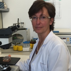 Delia Mezzanzanica合作论文数Fondazione IRCCS Istituto Nazionale dei Tumori7Benedetta Cinque合作论文数Department of Health Sciences, University of l’Aquila7Vincenzo Tombolini合作论文数6Stefania Violini合作论文数6Giuseppina Poppa合作论文数6Lombardi Francesca合作论文数6Anna Rughetti合作论文数6
 Maria Grazia Cifone合作论文数Dipartimento di Medicina Sperimentale, Università di L'Aquila6Giulio Ghersi合作论文数6
 Tina Garofalo合作论文数6M. Letizia Vittorelli合作论文数6Cherubini Alessandro合作论文数5
 Mario Barilani合作论文数Unit of Cell Therapy and Cryobiology, Fondazione IRCCS Ca' Granda Ospedale Maggiore Policlinico5Dell'orso Luigi合作论文数5
 Hideaki Nagase合作论文数The Kennedy Institute of Rheumatology, Nuffield Department of Orthopaedics, Rheumatology, and Musculoskeletal Sciences, Medical Sciences Division, University of Oxford5Simona Taverna合作论文数Dipartimento di Biopatologia e Metodologie Biomediche, Università di Palermo5Giulia Taraboletti合作论文数Mario Negri Institute for Pharmacological Research5
 Serena Cecchetti合作论文数5Pietro Leocata合作论文数Department of Surgery, University of L'Aquila5
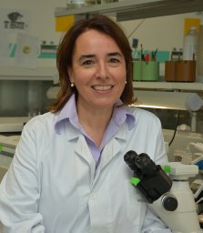 Lorenza Lazzari合作论文数Laboratory of Regenerative Medicine - Cell Factory, Department of Transfusion Medicine and Hematology, Fondazione IRCCS Ca' Granda Ospedale Maggiore Policlinico5Leda Biordi合作论文数5
 Paola Alberti合作论文数Department of Neuroscience and Biomedical Technologies, University of Milan-Bicocca5
 Franca Podo合作论文数Department Cell Biology and Neurosciences, Istituto Superiore di Sanita5M Sorice合作论文数4G Lucania合作论文数4MR Torrisi合作论文数4




