Objectives The aim of this study was to evaluate the rate of timely reperfusion for ST-elevation myocardial infarction (STEMI) with primary percutaneous coronary intervention (PPCI) in regional STEMI Receiving Center (SRC) networks.Background The American College of Cardiology Door-to-Balloon (D2B) Alliance target is a >75% rate of D2B <= 90 min. Independent initiatives nationwide have organized regional SRC networks that coordinate universal access to 9-1-1 with the pre-hospital electrocardiogram (PH-ECG) diagnosis of STEMI and immediate transport to a SRC (designated PPCI-capable hospital).Methods A pooled analysis of 10 independent, prospective, observational registries involving 72 hospitals was performed. Data were collected on all consecutive patients with a PH-ECG diagnosis of STEMI. The D2B and emergency medical services (EMS)-to-balloon (E2B) times were recorded.Results Paramedics transported 2,712 patients with a PH-ECG diagnosis of STEMI directly to the nearest SRC. A PPCI was performed in 2,053 patients (76%) with an 86% rate of D2B <= 90 min (95% confidence interval: 84.4% to 87.4%). Secondary analyses of this cohort demonstrated a 50% rate of D2B <= 60 min (n = 1,031), 25% rate of D2B <= 45 min (n = 517), and an 8% rate of D2B <= 30 min (n = 155). A tertiary analysis restricted to 762 of 2,053 (37%) cases demonstrated a 68% rate of E2B <= 90 min.Conclusions Ten independent regional SRC networks demonstrated a combined 86% rate of D2B <= 90 min, and each region individually surpassed the American College of Cardiology D2B Alliance benchmark. In areas with regional SRC networks, 9-1-1 provides entire communities with timely access to quality STEMI care. (J Am Coll Cardiol Intv 2009;2:339-46) (C) 2009 by the American College of Cardiology Foundation
This study was designed to evaluate the feasibility of applying locally delivered polylactic acid microspheres for drug delivery to the arterial wall. To study drug persistence, rhodamine-loaded microspheres were infused into one carotid artery of 14 rabbits and plain rhodamine solution into the other by using a porous balloon. To study tissue response, plain microspheres and dexamethasone-loaded microspheres were infused into the carotid arteries of another group of rabbits. To study the antiproliferative effects of locally delivered drug, 20 rabbits were subjected to overstretch balloon injury to both carotid arteries and divided into 4 groups: injury alone, plain microspheres, dexamethasone-loaded microspheres, and microspheres containing colchicine and dexamethasone. Fluorescent microspheres persisted in the vessel wall for 4 wk, whereas rhodamine without microspheres disappeared at 72 h. Histopathologic studies in arteries infused with unloaded microspheres showed inflammatory cell infiltrate with polymorphonuclear cells at 1 wk and macrophages and giant cells at 4 wk. Arteries infused with dexamethasone-loaded microspheres did not show any inflammatory cell infiltrate. Local delivery of dexamethasone or dexamethasone plus colchicine did not result in significant change in the intima-to-media ratio or in residual lumen following balloon injury. Polylactic acid microspheres may be used for prolonged delivery of drugs or other bioactive agents locally to the arterial wall. They induce an inflammatory reaction that is suppressable by dexamethasone in the microspheres. Dexamethasone or dexamethasone and colchicine delivered via this system, however, failed to reduce the degree of intimal hyperplasia after overstretch balloon injury to the rabbit carotid arteries.
Our goals were to develop a rabbit model of subacute stent thrombosis and to evaluate the efficacy of a polymer stent coating with covalently bound heparin for its prevention. Bare metal stents (a nitinol wire coil with 3 stainless steel joints) were implanted in a carotid artery of 10 NZW rabbits with a 3.0mm balloon catheter, inflated twice at 6 atm for 1minute. Systemic antiplatelet and antithrombotic therapy was withheld. Animals were sacrificed at 24, 72 and 96 hours. Only the 96 hour group consistently developed occlusive or subocclusive thrombosis. Heparin (0.28μg/cm2) was linked onto the surface of soluble polymer chains of segmented polyurethaneurea (SPUU) or aliphatic Tecoflex polyurethane membrane (50μm thick). using polyethylene oxide (PEO) as a hydrophilic spacer group. Eight stents were coated with this polymer-PEO-Heparin coating. Eight bare metal stents and 8 coated stents were deployed in a carotid artery of 16 NZW rabbits. All animals were sacrificed at 96 hours. At the time of sacrifice, both carotids (stented and contralateral nonstented) were exposed and resting blood flow measured. The amount of thrombus on each stent was assessed by a semiquantitative grading scale (I: no lumen encroachment; II: lumen encroachment; III: subocclusive thrombus; IV: occlusive thrombus) and by the weight of the dry thrombus.
Polymer-coated removable stents were used to deliver 14C-labeled etretinate and 3H-labeled forskolin to the vessel wall in 31 New Zealand White rabbits to study their kinetics. Stents loaded with etretinate (n = 8) and forskolin (n = 14) were implanted in the rabbit carotid arteries, and the animals were euthanized at different time intervals. Drug levels were measured in the media and adventitia of the stented segment, in distant tissues, and in blood. In four rabbits, forskolin-loaded stents were percutaneously retrieved 2 hr after implantation in the carotid artery, and the tissue and blood levels were determined 2 and 24 hr after retrieval. In seven rabbits etretinate-loaded stents were retrieved 72 hr after implantation in abdominal aorta, and drug levels were measured in the tissues and blood immediately after and at 1 and 4 days after retrieval. Levels of etretinate in the vessel wall peaked at 24 hr (250 ng/mg) and remained high up to 72 hr (185 ng/mg) after stent placement. Levels of forskolin peaked within 2 hr of stent placement (135 ng/mg) and rapidly declined to 4.9 ng/mg at 24 hr with the stent in situ. About 50% (1.4 mg) of the original etretinate remained in the stent at 72 hr compared to about 5% (0.08 mg) of forskolin at 24 hr. Ratio of peak drug levels in the vessel wall to those in the blood was 6,000 for etretinate and 780 for forskolin. (ABSTRACT TRUNCATED AT 250 WORDS)
The thrombogenicity of slotted-tube stents made of nitinol or stainless steel (Palmaz Schatz) was assessed in a rabbit carotid artery model. Fourteen nitinol and 8 stainless steel stents (7 mm long single segment stents) were deployed in a carotid artery of 22 NZW rabbits with a 3.0 mm balloon inflated twice at 6 atm for 1 min. No antiplatelet or antithrombotic drugs were given. Eight rabbits with nitinol and 8 with stainless steel stents were euthanized after 4 days. The remaining 6 rabbits with nitinol stents were euthanized after 14 days. At sacrifice, both carotids (stented and control) were exposed and resting blood flow measured. The amount of thrombus on each stent was assessed by a semi-quantitative grading scale (I: no lumen encroachment; II: lumen encroachment; III: subocclusive thrombus; and IV: occlusive thrombus) and by the weight of the dry thrombus.
Permanent intracoronary scents have demonstrated considerable efficacy in the treatment of PTCA‐induced abrupt arterial occlusion, suboptimal dilatation, and restenosis. Despite their benefit, permanent stents have significant limitations including subacute thrombosis, need for anticoagulation with resultant vascular complications, prolonged hospitalization, and high costs. Temporary stem's have the potential for achieving many of the beneficial effects of permanent stenting without some of the iatrogenic complications. The HARTS (Heat Activated Removable Temporary Stent) removable stent employs the shape memory alloy Nitinol, and has the properties of being balloon deployable, but recoverable to a preset “memory” shape by heating it with warmed crystalloid solution. Preclinical experience first with a wire stent design and more recently with a slotted tubular design, has demonstrated the feasibility of implanting and recovering the HARTS device. Speculative applications of this technology include: treatment of acute postangioplasty occlusion and suboptimal angioplasty results; “stentoplasty”—the application of a temporary stent for several hours at the time of a primary angioplasty; and reduction of restenosis rate by inhibition of elastic recoil following short‐term (hours) stenting. Further, the HARTS device may be an important vehicle for local delivery of drugs or other biologically active agents to the arterial wall. All research to date has been preclinical and controlled clinical trials will be necessary to define the safety, efficacy, and potential applications of the HARTS removable stent.
Temporary stenting appears to be an attractive potential therapy that combines some of the benefits of permanent stenting without the need for permanent endovascular prostheses and oral anticoagulation. The devices used for temporary stenting are still evolving. The Flow Support Catheter showed promise in the early clinical trial phase but is not being pursued by the original manufacturer because the device was associated with a high rate of thrombosis and suboptimal overall success rate. The experience with the Instent is limited and the removal of its unattached version relies upon catching the end of the stent in the retrieval device (a wire loop); also, its safety and reliability still need to be proved. The HARTS device is currently in the preclinical phase, but available data from animal studies show it to be a feasible device for coronary use. Its potential applications include use as a permanent stent, a temporary stent, a repositionable stent, a primary lesion-dilating device, or a local drug-delivery vehicle. Clinical trials are likely to commence in the foreseeable future for one or more of these applications.
BACKGROUND:Coronary stenting is associated with two major complications: subacute thrombosis and neointimal proliferation resulting in restenosis. Our hypothesis is that the biocompatibility of metallic stents can be improved by coating with a polymer membrane that delivers agents that favorably modify the local arterial microenvironment. This study evaluates the kinetics, distribution, and bioactivity of the model drug forskolin delivered to the local arterial wall by a polyurethane-coated removable metallic stent.METHODS AND RESULTS:Stents were used in rabbit carotid arteries (n = 20) for as long as 24 hours. The quantity of forskolin bound to the stent decreased exponentially with a half-life of 5.8 hours. Blood concentrations peaked at 140 +/- 39 pg/microL at 4 hours. The adjacent arterial media contained 60 +/- 39 ng/mg, which was 380- and 460-fold greater than the contralateral carotid media and the systemic blood, respectively (P < .0001). Media forskolin concentrations declined exponentially over time with a tissue half-life of 5.0 hours. Drug distributed throughout the vessel wall with decreasing gradients in the radial and axial dimensions consistent with a diffusion process. Removal of the stent was associated with a 100-fold decline in media forskolin concentration within 2 hours. Forskolin release was associated with a sustained 92% increase in carotid blood flow and a 60% decrease in local arterial resistance compared with coated control stents (P < .005). In another set of rabbits (n = 14) using a carotid crush injury, flow-reduction model, forskolin prolonged the time to flow variation and occlusion by 12-fold compared with the use of bare metal stents and 5-fold compared with the use of polyurethane-coated stents (P < .0001).CONCLUSIONS:A polymer-coated metallic stent can deliver forskolin to the local arterial wall in high concentrations relative to the blood or other tissues. High local drug concentrations are dependent on maintaining stent-to-tissue gradients. The delivered drug is biologically active, demonstrating vasodilating and antiplatelet properties.
Cardiac status was assessed noninvasively in a case of thoraco-omphalopagus conjoined twins by two-dimensional and Doppler echocardiography which showed completely separate hearts and major arteries and veins. The twins were successfully separated surgically, without preoperative invasive cardiac investigation. Echocardiography can provide an accurate assessment of cardiac status in conjoined twins and may obviate the need for cardiac catheterization.
 Frank Litvack合作论文数13
 Neal Eigler合作论文数V-Wave10
 James S. Forrester合作论文数Department of Cardiology, Cedars-Sinai Medical Center8
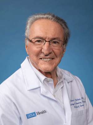 Michael C. Fishbein合作论文数Department of Pathology and Laboratory Medicine, University of California, Los Angeles;Jonsson Comprehensive Cancer Center, University of California, Los Angeles6Yq Tian合作论文数3J FORRESTER合作论文数3S Sheth合作论文数3T Lambert合作论文数3Nl Eigler合作论文数3J KUPFER合作论文数2Aj Hickey合作论文数2Fi Litvack合作论文数2A HICKEY合作论文数2
 Eldad Rechavia合作论文数2Savitri Shrivastava合作论文数2
 Rajendra Makkar合作论文数Smidt Heart Institute, Cedars-Sinai Medical Center2Sushil Sheth合作论文数2Ken Mahrer合作论文数2JM KUPFER合作论文数1Harvey Jacobs合作论文数1Angelo Salvucci合作论文数1MC Fishbein合作论文数1GG LIU合作论文数1
 Bryan McNally合作论文数1M Nakamura合作论文数1H ZENG合作论文数1H Zeng合作论文数1Timothy McPherson合作论文数1Kim Seong Who合作论文数Asan Med Ctr, Univ Ulsan1Eigler N L合作论文数1William Koenig合作论文数1
 Sanjay Kaul合作论文数Cedars-Sinai Heart Institute, Cedars-Sinai Medical Center1Rokos Ivan C合作论文数1
 Spencer B. King合作论文数Emory St. Joseph's Hospital;Southern Regional Medical Center1
 Stephen W. Smith合作论文数Department of Emergency Medicine, Medical School, University of Minnesota;Hennepin County Medical Center1
 Juan J Badimon合作论文数Atherothrombosis Research Unit, Cardiovascular Institute, The Mount Sinai School of Medicine1Ehtisham Mahmud合作论文数1
 Ki Dong Park合作论文数Graduate School of Engineering, Ajou University1Brian Strunk合作论文数1Huai Luo合作论文数1Mridula Rohatgi合作论文数1Paul S. Rostykus合作论文数1Beverly Nighswonger合作论文数1Akinyele O. Aluko合作论文数1Brian W. Gross合作论文数1Joel Kupfer合作论文数1Jackie Jewell合作论文数1William J. French合作论文数1Harvey A. Jacobs合作论文数1




