Rationale: The mechanisms regulating inflammation in sarcoidosis remain poorly understood.Programmed Death-1 (PD-1)+CD8+ T cells exhibit a proinflammatory phenotype that contributes significantly to sarcoidosis progression and reduced lung function.Objective: To better understand the inflammatory signaling processes that precedes Interferon gamma (IFN-γ) production in PD-1+CD8+ T cells.Methods: Publicly available sarcoidosis microarray datasets were downloaded from the Gene Expression Omnibus repository.Data was obtained from lung biopsies of 8 patients with active but self-limiting pulmonary sarcoidosis and 7 patients with active, progressive fibrotic pulmonary sarcoidosis.Flow cytometry and western blot was used to quantify protein levels in CD8+ T cells from sarcoidosis and healthy control subjects.Co-immunuprecipitation (co-IP) analysis was performed to examine direct S-phase kinase-associated protein 2 ( Skp2) and T-box transcription factor TBX21 (T-bet) protein interactions in sarcoidosis CD8+ T cells.Measurements and Main Results: Microarray analysis demonstrated differential expression of the Skp2 gene (P<0.05),suggesting a role for Skp2 in sarcoidosis progression.Flow cytometric analysis showed reduced percent and mean fluorescent intensity of Skp2 protein in sarcoidosis PD-1+CD8+ T cells compared to control cells, with concomitant increases in IFN-γ.Co-IP analysis on sarcoidosis CD8+ T cells revealed direct protein interactions between Skp2 and T-bet.Increased ubiquitination of T-bet in the presence of Skp2 was also noted.Skp2 loss of function decreased T-bet protein and IFN-γ expression, whereas Skp2 overexpression increased T-bet and IFN-γ levels.Conclusion: This work identifies a previously unrecognized role for Skp2 and T-bet ubiqutination in sarcoidosis progression, supporting the use of readily available therapeutics in the treatment of progressive sarcoidosis.
Pulmonary fibrosis is characterized by striking distinctions in mortality according to sex, demonstrating a female predominance and a longer time to death, compared to males. Independent laboratories reported the crucial role of the Programmed Death 1 (PD-1) pathway in pulmonary fibrosis. PD-1+IL-6+CD4+ T cells induce both profibrotic cytokines, IL-17A and TGFβ1, using distinct signaling pathways. Methods: In order to more clearly understand the role of PD-1+CD4+ T cells in the observed sex distinctions and mortality, we investigated IL-6 signaling in PD-1+ CD4+ T cells in humans and murine specimens. Using flow cytometry, ELISA and the Sircol assay to investigate distinctions in pulmonary fibrosis according to sex, we assessed for distinctions among humans with Idiopathic Pulmonary Fibrosis (IPF) , Sarcoidosis and Scleroderma, as well as the bleomycin murine model of pulmonary fibrosis. Results: Despite the male predominance, IPF males possessed higher serum estradiol levels and PD-1+CD4+ T cells, compared to age-matched male controls. We noted significantly higher percentages of PD1+CD4+ T cells with higher IL-6 production in female sarcoidosis subjects, compared to males. While sarcoidosis pulmonary progression was noted in both sexes, males possessed greater percentages of CD4+IL-17A cells compared to females; whereas the female CD4+ T cells expressed significantly higher free TGF-β1. Administration of intranasal bleomycin to male and female mice possessing genetic ablation of the alpha subunit of the Estrogen Receptor (ESRα KO) revealed significant declines in IL-6 but, notably, significant increases in pSTAT3 expression. Surgical removal of ovaries confirmed the observations of significant declines in IL-6 expression from CD4+ T cells and significant increased pSTAT3 and CD4+IL-17A expression. Strikingly, exogenous replacement of estradiol and progesterone in these strains resulted in increased IL-6 expression and reduction of pSTAT3, further confirming the capacity of female hormones to inhibit pSTAT3 expression. PD-1 pathway blockade resulted in reduced collagen production in females compared to males. Conclusion: This work identifies a crucial, previously unrecognized role of female hormones on pSTAT3 signaling pathways relevant to pulmonary fibrosis, supporting a personalized approach to therapeutic intervention according to sex. This abstract is funded by: Foundation of Sarcoidosis Research (FSR) Grant to Ozioma Chioma
 Chioma O合作论文数School of Medicine, Loma Linda University14
 Kevin F. Gibson合作论文数Division of Pulmonary, Allergy, Critical Care, and Sleep Medicine, Department of Medicine, School of Medicine, University of Pittsburgh10
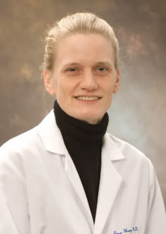 Erica Herzog合作论文数Pathology Faculty, Yale School of Medicine, Yale University10
 Moller D R合作论文数10Laura L. Koth合作论文数San Francisco School of Medicine, University of California Department of Medicine10
 Andrew M Morris合作论文数Division of Infectious Diseases, Department of Medicine, Temerty Faculty of Medicine, University of Toronto;University Health Network;Sinai Health System9
 Naftali Kaminski合作论文数Yale School of Medicine;Boehringer Ingelheim Pharmaceuticals, Inc.9
 Harry S Hochheiser合作论文数Institute for Cyber Law, Policy, and Security, University of Pittsburgh9
 Joe G Garcia合作论文数College of Medicine, University of Arizona;Scripps Research Insitute, University of Florida;The Herbert Wertheim UF Scripps Institute for Biomedical Innovation & Technology., University of Florida9
 Lisa A Maier合作论文数Colorado School of Public Health, University of Colorado Denver;Department of Medicine, National Jewish Health;Division of Environmental and Occupational Health Sciences, National Jewish Health9Edward S. Chen合作论文数The Wentworth Foundation9
 Michael J. Becich合作论文数Department of Biomedical Informatics, School of Medicine, University of Pittsburgh;Hillman Cancer Center, University of Pittsburgh Medical Center;American College of Medical Informatics8
 Stephen R. Wisniewski合作论文数Department of Epidemiology, Graduate School of Public Health, University of Pittsburgh7M. Gulati合作论文数6B. Hu合作论文数6Taylor Adams合作论文数Pulmonary, Critical Care and Sleep Medicine, Yale School of Medicine6M. Vukmirovic合作论文数Sch Med, Yale Univ6
 Xiting Yan合作论文数Section of Pulmonary, Critical Care and Sleep Medicine, Yale School of Medicine, Yale University;Data Analysis and Bioinformatics Hub, The Center for Precision Pulmonary Medicine, Yale University6
 Takis Benos合作论文数Department of Epidemiology, College of Public Health and Health Professions & College of Medicine, University of Florida6Joseph K. Leader合作论文数6RM SENIOR合作论文数5S. O'Neal合作论文数5K. Abel合作论文数5T. Woolard合作论文数4N. Aurelien合作论文数4L. J. Celada合作论文数3M Langford合作论文数3G. Deluliis合作论文数3G. DeIuliis合作论文数3C. B. Strange合作论文数3
 Ronald G. Collman合作论文数Penn Center for AIDS Research, Perelman School of Medicine, University of Pennsylvania3
 Elodie Ghedin合作论文数Laboratory of Parasitic Diseases, National Institute of Allergy and Infectious Diseases;Center for Genomics and Systems Biology, New York University3
 Barbara Methe合作论文数3Milton D. Rossman合作论文数3A. Mihaljinec合作论文数2L. Celada合作论文数2A. Fitch合作论文数2Andrew T Abad合作论文数2W. Luo合作论文数2
 Gordon R Bernard合作论文数Department of Medicine, Vanderbilt University Medical Center;Division of Allergy, Pulmonary and Critical Care Medicine, Vanderbilt University Medical Center2
 Karen Ann Patterson合作论文数2Xiaomei Li (李晓梅)合作论文数The Second Hospital of Shandong University2
 Robert P. Baughman合作论文数College of Medicine, University of Cincinnati;Department of Internal Medicine, University of Cincinnati2Kelvin Li合作论文数2Daniel A. Culver合作论文数2Prescott G. Woodruff合作论文数2Dawn C Newcomb合作论文数2R. A. Sandhaus合作论文数2K. C. Patterson合作论文数1




