Introduction and importance:We report the first case of perioperative zoledronic acid administration in an adolescent with neurofibromatosis type 1 (NF1) complicated by severe scoliosis. This case provides a hypothesis-generating experience for bone turnover screening and bisphosphonate intervention in the management of NF1-related scoliosis. Presentation of case:First case report: adolescent NF1 with severe scoliosis treated by perioperative zoledronic acid plus halo-pelvic traction and spinal fusion. The regimen included preoperative daily traction and 4 mg zoledronic acid, followed by postoperative spinal fusion and 1 mg zoledronic acid. Traction improved spinal alignment and pulmonary function. Bone turnover markers decreased after zoledronic acid administration. Clinical discussion:For the first time, zoledronic acid was administered perioperatively during primary spinal fusion in an adolescent patient with NF1. Bone turnover markers were reduced after zoledronic acid administration, and a temporal decrease in bone turnover was observed during traction and after surgery. The combination of traction and zoledronic acid was associated with favorable bone marker changes in this patient. A preliminary, hypothesis-generating bone management algorithm is proposed based on this single case: screen for bone markers and spinal CT in patients with severe NF1-associated scoliosis; if high bone turnover or low CT values are detected, consider zoledronic acid administration prior to traction. Conclusion:In this adolescent patient with NF1-associated severe scoliosis, perioperative zoledronic acid combined with traction and spinal fusion was associated with reduced bone turnover markers and stable implant fixation at a 12-month follow-up. This strategy may provide a hypothesis-generating option for perioperative bone health management. Further large, controlled studies are needed to confirm its efficacy and safety.
AIM:This study aims to clarify hematological parameters, transfusion requirements, and adverse events of preoperative intravenous (IVIS) versus oral iron supplementation (OIS) in elective surgery patients. METHODS:We conducted a comprehensive literature search across multiple databases up to 10 December 2023. Twelve RCTs involving 930 participants met our eligibility criteria. Our analysis focused on post-treatment hemoglobin levels, changes in hemoglobin from baseline, ferritin levels, hemoglobin attainment rates, transfusion requirements, and adverse events. We employed the random-effects model for data synthesis, calculating pooled standard mean differences (SMD) or mean differences (MD) or risk ratios (RR) with 95% confidence intervals (CI). Methodological quality was assessed using the Cochrane ROB 2 tool. The GRADE approach evaluated the confidence in effect estimates. FINDINGS:IVIS significantly improved post-treatment hemoglobin levels (MD = 0.77 g/dL, 95% CI [0.30 to 1.23]), hemoglobin increments (MD = 0.69 g/dL, 95% CI [0.01 to 1.37]), and ferritin levels (MD = 260.03 ng/mL, 95% CI [119.65 to 400.42]) compared to OIS. IVIS also led to a higher hemoglobin attainment rate (RR = 1.88, 95% CI [1.24 to 2.86]). No significant differences were noted in transfusion rates or volumes. IVIS was associated with fewer digestive (RR = 0.10, 95% CI [0.05 to 0.22]; I2 = 0%) but more pain-related adverse events (RR = 7.79, 95% CI [1.78 to 34.07]; I2 = 0%). Hospital stay durations and mortality rates were similar between the two groups. INTERPRETATION:IVIS offers a superior improvement in hematological parameters for elective surgery patients but not reducing transfusion needs compared to OIS. While IVIS has fewer digestive adverse events, it increases pain-related complications. These findings highlight the importance of personalized approaches in selecting iron supplementation methods, carefully balancing time, efficacy, and adverse event profiles. REGISTRATION:PROSPERO CRD42023483284.
Distal clavicle fractures, accounting for a significant portion of clavicle fractures, present a treatment challenge due to their high non-union rate and the controversy surrounding operative versus non-operative management strategies. This review synthesizes recent studies and clinical evidence to compare the effectiveness of operative and non-operative approaches for distal clavicle fractures. It discusses the anatomical and classification aspects of these fractures, the indications for surgery, and the rationale behind different treatment options. Surgical fixation, predominantly through locking plates or hook plates, generally provides high union rates and stable outcomes; however, complications such as hardware irritation, subacromial impingement, and implant removal rates are significant concerns. Recent surgical advancements, including ligament repair, distal augmentation, and arthroscopic techniques, have shown promise in improving outcomes. Conversely, conservative treatment is recommended for certain fracture types, with emerging evidence suggesting it provides comparable functional outcomes to those of surgical methods. Despite a high non-union rate, non-operative treatment can be effective in restoring functions. However, challenges such as symptomatic nonunion and osteoarthritis are noted. The choice between surgical and conservative treatments depends on multiple factors, including fracture type, patient age, activity level, and overall health status. This review highlights the need for a personalized approach in managing distal clavicle fractures, considering the evolving evidence and advancements in treatment strategies.
Anesthesia for full endoscopic lumbar discectomy (FELD), typically administered locally or regionally, often fails to provide adequate analgesia, necessitating additional analgesics or even sedation. Our previous work has shown that intrathecal morphine (ITM) administration is considered an effective adjuvant approach for managing peri-operative pain for FELD, but the adverse events (AEs) for ITM remain concerns. This study aimed to determine the effective dose of intrathecal morphine required to eliminate intraoperative pain in 90
Study Design: Prospective Cohort Study. Objective: To compare the effectiveness and safety of perioperative denosumab, bisphosphonates, and a control group in promoting lumbar fusion after midline lumbar fusion (MIDLF) surgery in patients with osteopenia or osteoporosis. Methods: This prospective cohort study enrolled 54 patients with osteopenia or osteoporosis undergoing MIDLF surgery. Patients were divided into 3 groups: denosumab (60 mg subcutaneous injection), bisphosphonates (5 mg zoledronic acid intravenous infusion), and control (no anti-osteoporotic drugs, only calcium + vitamin D). Outcomes included fusion rates, clinical scores, bone metabolism markers, and adverse events. Results: 54 patients were included in the study. Among them, 50 patients completed the 6-month postoperative follow-up. Due to the impact of the COVID-19 pandemic, only 29 patients completed the final follow-up. At 6 months, fusion rates were significantly higher in the denosumab (25% complete, 68.6% partial) and bisphosphonate (25% complete, 75% partial) groups compared to the control group (10.5% complete, 57.9% partial, P < 0.001). No significant difference was found between the 2 drug groups. Postoperative fever was more common in the bisphosphonate group (71.4%) than in the denosumab group (11.8%, P =0.004) and control (23.5%, P = 0.011) groups. Conclusions: In the short-term follow-up, both denosumab and bisphosphonates significantly improved the early bone fusion rate after MIDLF surgery, with no significant difference observed between the two. Denosumab was associated with a lower incidence of postoperative fever, making it a recommended choice for perioperative anti-osteoporosis therapy.
BACKGROUD CONTEXT: Conventional pedicle screw (CPS) fixation in osteoporotic spines presents significant challenges. Cortical bone trajectory (CBT) screws can enhance screw holding power by increasing contact with cortical bone. However, the standard CBT (S-CBT) screws may encounter a series of problems such as stress concentration and diminished fatigue resistance. PURPOSE: The S-CBT screw technique has been modified to accommodate longer screws, and the biomechanical behaviors of this modified CBT (M-CBT) screw technique were investigated. STUDY DESIGN: A finite element analysis and biomechanical cadaveric study. METHODS: A validated nonlinearly finite element model spanning L1-S1 was employed in this study. Three L4-5 fusion models, namely CPS, M-CBT, and S-CBT, were generated using inter-body fusion cages and different screw fixations. Next, the models were subjected to loading protocols to simulate flexion, extension, lateral bending, and rotation motion. The range of motion (ROM) and peak von Mises stress of the Cage, rods, screws, and intervertebral discs were analyzed. Besides, 3 types of cadaveric lumbar fusion modes were constructed using diverse screw trajectories. These models were cycled 10,000 times to measure the vertebral body displacement. Afterward, the individual screws were subjected to axial pull-out tests, and the maximum pulling-out force was documented. Finally, the data from the 3 fusion models were compared. RESULTS: Regarding 6 & ring; of freedom movements, the 3 fixation models significantly increased the ROM of the adjacent segments (L3-4 and L5-S1) (p<.01). However, the differences in ROM increments among the 3 models were not statistically significant (p=.815). The peak von Mises stress of the cage for the M-CBT model was lower by 1.06%, 37.75%, 10.28%, and 17.55% compared with the S-CBT model during flexion, extension, right bending, and left rotation directions, respectively. Similarly, the peak von Mises stress of L5 screws for the M-CBT model was lower by 50.57%, 59.98%, 47.29%, 64.07%, 63.24%, and 50.45% compared with S-CBT during flexion, extension, left bending, right bending, left rotation, and right rotation, respectively. In the biomechanical test, the fatigue displacement results revealed that the displacement of M-CBT model was intermediate between the S-CBT and CPS models under both maximum and minimum forces, with statistically significant differences (p<.05). Additionally, the results of the antipullout test following fatigue loads demonstrated that the M-CBT group exhibited the highest maximum pull-out force (Fmax) (381.80 [119.00, 852.20]), followed by the CPS group (329.10 [117.00, 507.80]) and the S-CBT group (321.50 [196.60, 887.20]), but the differences were not statistically significant (p=.665) in the upper vertebral subgroup. Conversely, the Fmax of M-CBT group (384.20 [314.00, 851.20]) was significantly higher than that of S-CBT group (264.70 [118.80, 477.40]) and CPS group (282.20 [50.80, 595.20]) in the lower vertebral subgroup, with a significant difference between M-CBT and S-CBT (p=.037). CONCLUSION: M-CBT could enhance the control force of the anterior column of the vertebral body by increasing the inserted screw length, minimizing the stress on the cages and screws, and optimizing the antifatigue performance of the internal fixation system compared to S-CBT. CLINICAL SIGNIFICANCES: M-CBT screw technique shows better biomechanical properties compared to both S-CBT and CPS techniques, providing a more stable and effective internal fixation option for internal fixation in osteoporotic vertebrae. (c) 2025 Elsevier Inc. All rights are reserved, including those for text and data mining, AI training, and similar technologies.
BACKGROUND:A retrospective cohort study to evaluate the efficacy and safety of midline lumbar fusion (MIDLF) for lumbosacral fusion compared to posterior lumbar interbody fusion (PLIF). METHODS:Patients who had undergone posterior lumbosacral fusion surgery were divided into a MIDLF group (n = 37) and a PLIF group (n = 42). The follow-up time was at least 12 months. The operation data, recovery condition, complications, clinical outcomes, and status of implants and fusion were compared between the 2 groups. RESULTS:The MIDLF group experienced significantly less blood loss, lower postoperative creatine kinase levels and total drainage volume, earlier time to ambulation, and less hospital stay times after surgery compared to the PLIF group (P < 0.05). The mean postoperative back pain visual analog scale scores in the MIDLF group were significantly lower than the PLIF group (P < 0.05). The improvement in Oswestry Disability Index scores during 3-month follow-up displayed a significant difference between the 2 groups (P < 0.05). The fusion rate tended to be higher in the MIDLF group; however, the difference was not significant (P > 0.05). There was no significant difference in respect to screw loosening and cage subsidence rate. There were 2 cases of complications both occurring in the PLIF group. CONCLUSIONS:MIDLF is safe and effective for lumbosacral fusion and in line with the concept of enhanced recovery after surgery.
目的 探讨不同种类阴道微生态异常与人乳头瘤病毒(HPV)感染的相关性,为临床预防和治疗HPV感染提供思路,从而减少宫颈癌的发生.方法 选取2021年1月1日至2021年12月31日要求进行常规妇科检查的患者共2400例,对其同时进行阴道微生态和HPV检查,按照阴道微生态结果分为微生态正常组864例、乳酸杆菌减少组824例、细菌性阴道病(BV)组392例、念珠菌性外阴阴道炎(VVC)组217例、滴虫性阴道炎(TV)组17例、混合性阴道炎组86例,分别计算每组HPV感染率,并将乳酸杆菌减少组(12.01%)、BV 组(24.74%)、VVC 组(13.36%)、TV 组(11.76%)、混合型阴道炎组(20.93%)的 HPV 感染率分别与微生态正常组HPV感染率(11.81%)比较,计算其差异是否具有统计学意义,从而判断上述各种类型阴道微生态异常是否影响HPV感染率.结果 BV组患者HPV感染率24.74%、混合型阴道炎组患者HPV感染率20.93%,与微生态正常组HPV感染率11.81%比较差异有统计学意义(P<0.05);而乳酸杆菌减少组、VVC组、TV组HPV感染率与微生态正常组HPV感染率比较差异无统计学意义(P>0.05).结论 BV及混合型阴道炎与HPV感染有相关性,BV及混合型阴道炎患者中HPV感染率明显升高,而乳酸杆菌减少、VVC和TV患者中HPV感染率未见明显升高.积极治疗BV及混合型阴道炎有助于预防HPV感染及清除HPV,从而减少宫颈癌的发生.
(1)一般情况:患者女性,年龄77岁,身高155 cm,体质量55 kg,因"腹胀10余天"入院。患者10余天前无明显诱因出现腹胀不适,为阵发性隐痛,伴气促不适、食欲减退、恶心呕吐,多次就诊于外院,予以抗感染、补充白蛋白及灌肠等治疗(具体不详)。外院全腹部增强CT提示盆腹腔巨大占位,考虑畸胎瘤(偏恶性)。为进一步诊治入院。拟行开腹盆腔病灶切除术。
目的:探讨妊娠前、后发现脑动静脉畸形(cAVM)孕产妇的临床特征和妊娠结局。方法:对2013年6月至2022年8月在首都医科大学宣武医院诊治的33例cAVM孕产妇的临床资料进行回顾性分析,并对妊娠前、后发现cAVM的孕产妇的临床特征及妊娠结局进行比较。结果:(1)妊娠前发现cAVM孕妇23例次,其中,妊娠前发生过脑出血者10例次,其中积极治疗9例次;妊娠前未发生脑出血13例次,其中积极治疗6例次。妊娠期发生脑出血2例次(9%,2/23),其中1例次行血管内栓塞,1例次血管内栓塞后行手术治疗。妊娠结局:引流产3例次,剖宫产术分娩19例次(足月16例次,早产3例次),自然分娩1例次(早产);围产儿20例,其中死亡1例,余均体健;产妇随访至少半年,健康21例次,有后遗症2例次,无死亡。(2)妊娠后发现cAVM孕产妇10例次(孕早期1例次,孕中期5例次,孕晚期2例次,产后2例次),均表现为脑出血,妊娠期行紧急外科手术3例次,血管内栓塞6例次,血管内栓塞后外科手术1例次。妊娠结局:引流产5例次,剖宫产术分娩4例次(足月2例次,早产2例次),阴道分娩1例次(死产);新生儿5例,均体健;产妇随访健康4例次,有后遗症5例次,死亡1例次。(3)与妊娠前发现cAVM的孕妇相比,妊娠后发现cAVM的孕产妇脑出血的发生率较高、引流产的发生率较高、终止妊娠孕周较早、新生儿出生体重较轻、产妇健康率较低,分别比较,差异均有统计学意义( P均<0.05)。 结论:妊娠合并cAVM孕妇妊娠前治疗可有效预防妊娠后cAVM破裂出血;对于妊娠期及产褥期发生脑出血者,推荐积极的手术治疗;终止妊娠时机及方式需根据孕妇情况个体化处理,由产科、神经外科、麻醉科及胎儿医学科组成的多学科团队协作讨论后决定。
Objective:To investigate the application value of WeChat-based problem-based learning teaching method combined with micro-classroom in the teaching of contrast-enhanced gastrointestinal ultrasound imaging for residents in standardized residency training.Methods:A total of 40 trainees who underwent the standardized residency training in the Department of Ultrasound, The First Affiliated Hospital of Chongqing Medical University from January 2019 to June 2021 were selected as the research objects. They were randomly divided into the experimental group (WeChat-based PBL teaching method combined with micro-classroom) and the control group (traditional teaching method). The teaching content was the diagnosis of gastrointestinal ultrasound imaging. After the theoretical learning and practice, assessments of contrast-enhanced gastrointestinal ultrasound imaging including theory and operating practice were performed to all students. SPSS 22.0 was used for t test and rank sum test.Results:After training, the theoretical test scores of the experimental group were higher than those of the control group [(93.09±2.31) vs. (90.63±2.26)], and the difference was statistically significant ( P<0.05). However, there was no significant difference between the two groups in the number distribution of students at all levels of operational practice ( P>0.05). Conclusion:The WeChat-based PBL teaching method combined with micro-classroom is beneficial to improve students' knowledge of contrast-enhanced gastrointestinal ultrasound imaging, and is worth being popularized and applied in clinical teaching.
子宫内膜癌是妇科常见恶性肿瘤.手术治疗是子宫内膜癌患者重要的治疗方式.为保证手术治疗效果,改善患者预后,需制定严格的手术质量控制标准.子宫内膜癌手术质量涉及术前诊断与评估、手术方式选择及手术范围确定、无瘤手术原则、术后病理诊断及分子分型、手术医生培训与资质等诸多环节.文章根据国内外研究,明确各关键环节要点,总结出明确且可行的质量控制方案.
目的 分析某医院妇科住院诊治的6例骶前肿物患者的病案资料,探讨其临床特点.方法 收集2016年8月1日至2020年8月31日,在某院妇科住院治疗的6例骶前肿物患者临床资料.分析其临床特点、手术情况,以及手术疗效.结果 所有患者通过直肠指诊,盆腔核磁及超声等检查进行了术前肿物与周围组织关系的评估.6例患者均通过腹腔镜下骶前肿物切除,无中转开腹及联用经骶骨治疗等手术方式.骶前肿物大小差异大,最小者3*3cm,最大者25*20cm.手术时间及出血量差异大.出血量最少20ml,最多400ml.手术后住院时间平均为4.333天.术后病理类型多样.所有患者随访至今,无复发.结论 骶前肿物发病比较少见,临床表现及病理学类型多样,易被漏诊.直肠指诊、盆腔核磁是该病术前评估的重要手段.经腹腔镜下骶前肿物切除术,具有创伤小、术后恢复快、并发症低等特点,是一种可靠、有效的治疗手段.
Objective:To investigate the value of periodically rotated overlapping parallel lines with enhanced reconstruction (PROPELLER) in improving ability of demonstrating ocular masses on 3.0 T MR scanner.Methods:This study was a multi-center prospective study involving 6 centers. From July 2018 to January 2020, totally 413 patients with ocular masses from 6 centers were prospectively enrolled, and all of them underwent T 1WI and T 2WI, PROPELLER T 1 FLAIR and T 2WI, and contrast-enhanced scans. The signal intensity of eyelid, vitreous body, lacrimal gland, intraorbital segment of optic nerve, and orbital masses of eyelid, intraocular, lacrimal gland and retrobulbar were measured by two radiologists, and the signal to noise ratio (SNR) and contrast noise ratio (CNR) were calculated. The 5-point scoring method was used to evaluate the motion artefacts, tumor edges and the relationship between the tumor and adjacent structures, and the overall score of image quality was calculated. Paired t-test or Wilcoxon signed rank test was used to compare the image quality between PROPELLER and non-PROPELLER images. Results:The SNR and CNR of PROPELLER T 2WI were higher than those of non-PROPELLER T 2WI (all P<0.001). The SNR and CNR of PROPELLER T 1 FLAIR were lower than those of non-PROPELLER T 1WI (all P<0.05). The scores of artefacts and overall image quality in PROPELLER images were higher than those in non-PROPELLER images (all P<0.001). The tumor edge and the relationship between the tumor and adjacent structures scores of eyelid, intraocular, and lacrimal gland masses in PROPELLER images were higher than those in non-PROPELLER images (all P<0.001),while compared to non-PROPELLER images, retro-global masses in PROPELLER images showed no significant differences (all P>0.05). Conclusion:PROPELLER can reduce ocular motion artefacts, effectively improve image quality and ability of demonstrating anterior (eyelid, intraocular, and lacrimal gland) masses.
目的 探讨不同剂量雌激素联合孕激素对围绝经期综合征患者性激素、骨密度的影响及不良反应的发生情况.方法 回顾性分析2018年8月-2020年8月医院收治的82例围绝经期综合征患者,按照补充雌激素的用量分为低剂量组和高剂量组,每组41例.低剂量组采用1mg雌激素+10mg孕激素治疗,高剂量组采用2mg雌激素+10mg孕激素治疗,2组患者均治疗12个周期(每个周期28天).对比2组患者女性绝经期自测表(KI)评分,临床疗效,性激素水平,骨密度以及不良反应.统计学数据用SPSS22.0软件进行分析,计量资料以t检验,计数资料以x2检验,P<).05为差异有统计学意义.结果 与治疗前相比,治疗后2组患者KI评分均降低(P<0.05),2组间KI评分比较,差异无统计学意义(P>0.05);治疗后2组总有效率比较,差异无统计学意义(P>0.05);与治疗前相比,治疗后2组血清卵泡刺激素(FSH)、黄体生成素(LH)水平均降低(P<0.05),2组间血清FSH、LH水平比较,差异无统计学意义(P>0.05),与治疗前相比,治疗后2组血清雌二醇(E2)水平均升高(P<0.05),且低剂量组血清E2水平低于高剂量组(P<0.05);与治疗前相比,治疗后2组腰椎2~4、双侧髋关节密度均升高(P<0.05),2组间腰椎2~4、双侧髋关节骨密度比较,差异无统计学意义(P>0.05);低剂量组不良反应发生率低于高剂量组(P<0.05).结论 低剂量与高剂量雌激素联合孕激素用于围绝经期综合征患者均可改善临床症状,调节性激素水平,防止骨丢失,但应用低剂量雌激素安全性更好,不良反应发生率更低.
Cervical cancer is a common gynecologic malignant tumor. Surgical treatment is an important treatment for early stage cervical cancer. Whether having lymph node metastasis is of great significance to the assessment of the prognosis of cervical cancer and the choice of treatment options. Preoperative assessment of lymph node metastasis based on identified related risk factors and imaging findings is helpful to the formulation of surgical strategies. In recent years, sentinel lymph node biopsy has been recommended for surgical treatment of patients with early stage cervical cancer due to its safety and feasibility. Patients with cervical cancer can selectively undergo sentinel lymph node biopsy, systematic lymph node resection, or para-aortic lymph node resection based on their preoperative lymph node assessment. According to intraoperative diagnosis of the degree of lymph node involvement, the extent of surgical resection can be determined. So the assessment of lymphatic metastasis, especially the formulation of surgical strategieswere of great significance in the treatment of cervical cancer.
Abstract Background Finite element analyses and biomechanical tests have shown that PEEK rods promote fusion and prevent adjacent segment degeneration. The purpose of this study was to evaluate the effects and complications of hybrid surgery with PEEK rods in lumbar degenerative diseases. Methods From January 2015-December 2017, 28 patients who underwent lumbar posterior hybrid surgery with PEEK rods were included in the study. The patients were diagnosed with lumbar disc herniation, lumbar spinal stenosis, or degenerative grade I spondylolisthesis. Before the operation and at the last follow-up, the patients completed lumbar anteroposterior and lateral X-ray, dynamic X-ray, MRI examinations. In addition, at the last follow-up the patients also completed lumbar CT examinations. The radiographic parameters, clinical visual analog scale (VAS) score and Oswestry disability index (ODI) score were compared. Results The average age of the patients was 44.8 ± 12.6 years, and the average follow-up duration was 26.4 ± 3.6 months. The VAS score improved from 6.3 ± 1.6 to 1.0 ± 0.9, and the ODI score decreased from 38.4 ± 10.8 to 6.8 ± 4.6. The fusion rate of the fused segment was 100%. There were no significant changes in the modified Pfirrmann classifications or disc height index for the nonfused segments and the upper adjacent segments from pre- to postoperatively. No cases of screw loosening, broken screws, broken rods or other mechanical complications were found. Conclusion Hybrid surgery with PEEK rods for lumbar degenerative diseases can yield good clinical results and effectively reduce the incidence of complications such as adjacent segment diseases.
Objective:To assess the clinical and imaging features of NUT gene-related sinonasal carcinomas (NUT midline carcinome).Methods:The clinical data and pretreatment imaging findings of 5 cases with pathologically proven NUT sinonasal carcinomas were analyzed retrospectively in Beijing Tongren Hospital, Capital Medical University from January 2016 to December 2020. Of 5 cases, the tumors affected 4 females and 1 male with an age range of 15 to 48 years (median 19 years). Clinical data of all cases were available before surgery with both CT and MR examination. Tumor location, CT density, boney change, calcification, tumor size, T 1WI, T 2WI and diffusion weighted imaging (DWI) signal intensity, appearance diffusion coefficient (ADC), type of time intensity curve (TIC) of dynamic contrast-enhanced (DCE)-MRI were evaluated. Results:All five cases belonged to T4 stage of the clinic TNM system. The locations were nasal cavity ethmoid, sphenoid and maxillary sinus ( n=1), nasal and maxillary sinus ( n=1), nasal cavity and ethmoid sinus ( n=3). Iso-attenuated in 3 cases, heterogeneous with local necrosis in 2 cases, and heterogeneous with calcification in 3 cases on CT imaging. Bone erosion was found in 4 cases, and bone erosion with destruction in 1 case. The tumor sizes ranged from 4.2 to 4.9 cm (median 4.5 cm) on MR axial imaging. On T 1WI, 5 cases showed isointense compared with adjacent temporal muscles, with focal hypointense in 2 cases. On T 2WI, the tumor was graded as isointense in 3 cases, and hyperintense in 2 cases. Heterogeneous enhancement in all cases with mild in 3 cases, and moderate in 2 cases on postcontrast MR imaging. On DCE-MRI of 5 cases, there were 3 cases of type Ⅲ (washout-shaped curves), and 2 cases of type Ⅱ of the TIC (plateau-shaped curves). The range of ADC values was from 0.63×10 -3 to 1.17×10 -3 mm 2/s, and median ADC value was 0.84×10 -3 mm 2/s, of 5 cases with varying degrees of high signal on DWI. The Ki-67 index ranged from 30% to 80% of the tumor. An immunohistochemical study showed that the tumor cells of 5 cases were all positive for both NUT and INI-1 genes. One case was performed with biopsy and followed by chemotherapy, four cases were performed with surgery, combined with the following chemotherapy, and one also was implemented with radiation therapy. The follow-up time was 7-16 months. Five cases were all alive during the follow-up. Conclusions:The NUT midline sinonasal tract carcinoma is a rare, gene-related solid malignant tumor. The tumor is more commonly seen in young patients, mostly centered in the nasal and ethmoid region with invasive growth, more calcification on CT, and heterogeneous enhancement on MRI. These findings are some characteristics of the tumor.
BACKGROUND:Improving the sagittal lumbar-pelvic parameters after fusion surgery is important for improving clinical outcomes. The impact of midline lumbar fusion (MIDLF) on sagittal lumbar-pelvic alignment for the management of degenerative lumbar diseases is still unknown. AIM:To analyze the effects of short-segment MIDLF and minimally invasive transforaminal lumbar interbody fusion (MIS-TLIF) on sagittal lumbar-pelvic parameters. METHODS:We retrospectively analyzed 63 patients with degenerative lumbar diseases who underwent single-segment MIDLF or MIS-TLIF. The imaging data of patients were collected before surgery and at the final follow-up. The radiological sagittal parameters included the lumbar lordosis (LL), lower LL, L4 slope (L4S), L5 slope (L5S), L5 incidence (L5I), L1 axis and S1 distance (LASD), pelvic incidence (PI), pelvic tilt (PT), sacral slope (SS), and PI-LL mismatch (PI-LL). Additionally, the clinical outcomes, including lower back and leg pain visual analog scale (VAS) and Oswestry disability index (ODI) scores, were also analyzed. RESULTS:In both groups, LL and Lower LL significantly increased, while L5I and LASD significantly decreased at the final follow-up compared to that recorded prior to operation (P < 0.05). In the MIDLF group, L4S significantly decreased compared to that recorded prior to operation (P < 0.05), while the mean SS significantly increased and the PT significantly decreased compared to that recorded prior to operation (P < 0.05). In the MIS-TLIF group, SS slightly increased and the mean PT value decreased compared to that recorded prior to operation, but without a statistically significant difference (P > 0.05). However, the PI-LL in both groups was significantly reduced compared to that recorded prior to operation (P < 0.05). There was no significant difference in the sagittal lumbar-pelvic parameters between the two groups prior to operation and at the final follow-up (P > 0.05). In addition, the change in sagittal lumbar-pelvic parameters did not differ significantly, except for ΔLASD within the two groups (P > 0.05). The mean lower back and leg pain VAS and ODI scores in both groups were significantly improved three months after surgery and at the final follow-up. Though the mean ODI score in the MIDLF group three months after surgery was slightly higher than that in the MIS-TLIF group, there was no significant difference between the two groups at the final follow-up. CONCLUSION:Short-segment MIDLF and MIS-TLIF can equally improve sagittal lumbar parameters such as LL, Lower LL, L5I, and LASD in the treatment of lumbar degenerative diseases. However, MIDLF had a larger impact on pelvic parameters than MIS-TLIF.
Objective:To analyze the relationship between pre-pregnancy body mass index (BMI), gestational random fasting glucose maximum, weight gain during pregnancy, and the occurrence of macrosomia in pregnant women diagnosed with gestational diabetes after 28 weeks gestation.Methods:The clinical data of 310 pregnant women with gestational diabetes after 28 weeks of diagnosis in Xuanwu Hospital of Capital Medical University in 2014 were retrospectively analyzed. They were divided into observation group (96 cases) with macrosomia and control group (214 cases) with normal birth weight according to the weight of newborn. The differences of BMI before pregnancy, the highest value of fasting blood glucose during pregnancy and weight gain during pregnancy between the two groups were analyzed.Results:The pre-pregnancy BMI , the highest value of random fasting blood glucose and weight gain during pregnancy in macrosomia group were significantly higher than those in non macrosomia group ( P<0.05); And the best cut-off point for predicting the delivery of macrosomia in pregnant women with gestational diabetes after 28 weeks of pregnancy was 22.077 kg/m 2, 4.965 mmol/L and 17.400 kg, respectively. The area under the curve (AUC) was 0.646, 0.595 and 0.699 respectively. After correction of confounding factors, the BMI ( OR=1.238, 95% CI: 1.132, 1.354, P<0.001) and weight gain during pregnancy ( OR=1.189, 95% CI: 1.120, 1.262, P<0.001) were risk factors for macrosomia in gestational diabetes mellitus after 28 weeks of gestation ( P<0.05). Conclusions:Pre-pregnancy BMI>22.077 kg/m 2, gestational maximum fasting blood glucose >4.965 mmol/L and gestational weight gain >17.400 kg were all high risk factors for gestational diabetes mellitus pregnant women after 28 weeks. For pregnant women with gestational diabetes, active prenatal intervention and health management are of great significance in reducing the risk of macrosomia.
 Lihui Wei (魏丽惠)合作论文数Peking University People′s Hospital27
 Jianliu Wang (王建六)合作论文数Peking University People's Hospital26
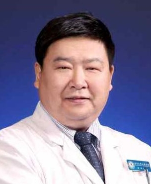 Heng Cui (崔恒)合作论文数Peking University People's Hospital14
 Xiaoping Li (李小平)合作论文数Peking University People's Hospital13
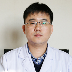 Haolin Sun (孙浩林)合作论文数Peking University First Hospital10
 Qingquan Zhang (张清泉)合作论文数8LianE Zhou (周莲娥)合作论文数8Xian Zhang (张娴)合作论文数6
 Yue Lei合作论文数West China School of Pharmacy, Sichuan University6Shen Xiaguang (沈霞光)合作论文数5
 Chunde Li (李淳德)合作论文数Peking University First Hospital5
 Yi Li (李艺)合作论文数Peking University People's Hospital5
 Danhua Shen (沈丹华)合作论文数Peking University People's Hospital;Peking University Health Science Center5
 Junfang Xian (鲜军舫)合作论文数Beijing Tongren Hospital, Capital Medical University;Graduate School of Capital Medical University4
 Xudong Liang (梁旭东)合作论文数Department of Gynecology, People's Hospital,Peking University4
 Xiuli Sun (孙秀丽)合作论文数Peking University People's Hospital4Longtao Qi合作论文数3Lihong Zheng (郑丽鸿)合作论文数3Chuqi Yang (杨楚琪)合作论文数3Yuetian Wang合作论文数3AnXia Wan (万安霞)合作论文数3Yao Zhao (赵耀)合作论文数3XiaoHong Zhang (张晓红)合作论文数3
 Zhengrong Yu (于峥嵘)合作论文数3
 Yantao Niu (牛延涛)合作论文数Beijing Friendship Hospital, Capital Medical University;Graduate School of Capital Medical University3
 Honglan Zhu (祝洪澜)合作论文数Peking University People's Hospital3Qi-ying Xu合作论文数2Y. Zhao合作论文数2Beiyu Xu合作论文数2Hao-zhe SONG (宋灏哲)合作论文数2Ling-hui HUANG (黄玲惠)合作论文数2Xiaoran Wang (王肖然)合作论文数2JIN Ling (金玲)合作论文数2Dan LI (李丹)合作论文数2Xin-hong ZHANG (张心红)合作论文数2Xuan WANG (王轩)合作论文数2BaoHua Kang (康保华)合作论文数2TianLiang Kang (康天良)合作论文数2YongXian Zhang (张永县)合作论文数2SenLin Guo (郭森林)合作论文数2Yan Wang (王雁)合作论文数2Hong Jiang (姜虹)合作论文数2Wu Jian-Xing合作论文数2Yan Zhao (赵彦)合作论文数2
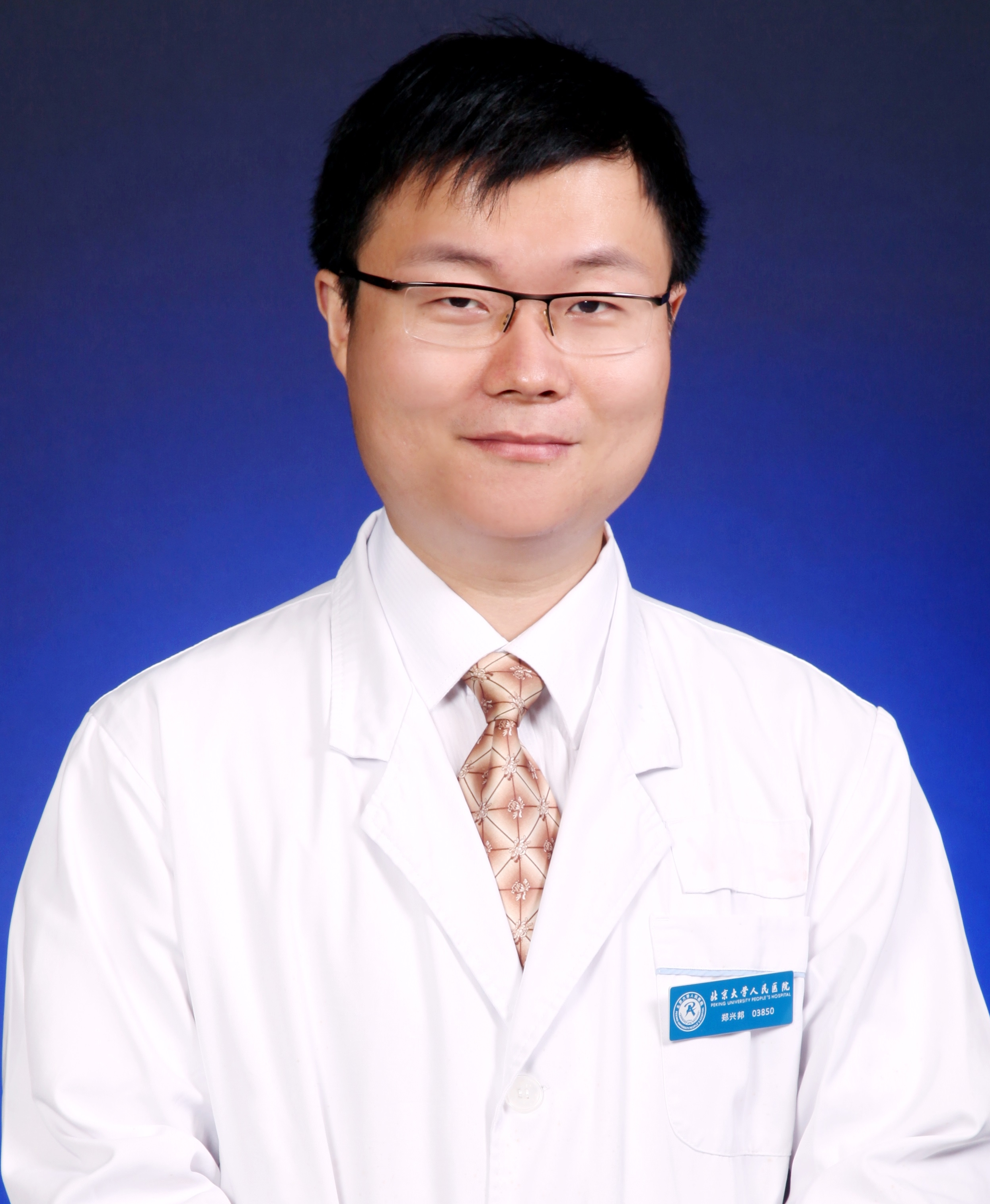 Xingbang Zheng (郑兴邦)合作论文数Peking University People's Hospital2Li Dong (董丽)合作论文数2
 Yue Wang (王悦)合作论文数Peking University Third Hospital2




