Background The incidence of brain metastases from gastric origin is less than 1% in those with primary gastric cancer. Given this exceedingly rare presentation, there is limited literature describing the outcomes of their neurosurgical treatment. We wish to identify the role of surgical intervention for brain lesions in metastatic gastric cancer via institutional case series and systematic review. Methods This study was divided into two sections: (1) a retrospective, single-center patient series assessing outcomes of neurosurgical treatment modalities in patients with malignancy arising from the stomach with brain metastases and (2) a systematic review abiding by the Preferred Reporting Items for Systematic Reviews and Meta-Analyses (PRISMA) guidelines between the years of 1980 and 2021 assessing outcomes of patients with primary stomach cancer with metastasis to the brain treated with surgery. Results Four patients with gastric brain metastases were treated at our institution, and 16 patients were identified in literature from a total of 9 studies and case reports. The mean age at the time of stomach cancer diagnosis was 57.3 years, with a mean time to brain metastases of 14.8 months. The primary gastric cancer was most commonly adenocarcinoma (70%). Patients most presented with single lesions (58%) and were treated with multimodal neurosurgical intervention (65%). Mean overall survival following neurosurgery was 12.45 months. Conclusion Brain metastases from gastric origin are extremely rare. Surgical resection of metastatic brain lesions should be considered as a treatment modality in surgical candidates. Future attention should be given to the effect of adjuvant therapies and surgical techniques on survival and quality of life.
INTRODUCTION: Controllable factors associated with surgical site infections (SSIs) have focused on reducing contamination of the surgical field with potential pathogens. The aim of this prospective study is to determine the incidence of glove contamination in a series of elective neurosurgical operations and determine the relationship of such glove contamination to subsequent SSI. We hypothesize that contamination of surgical gloves is associated with subsequent SSI.METHODS: In this prospective quality improvement study, gloves of the surgical team were swabbed for standard culture just prior to wound closure of elective neurosurgical operations. Patient characteristics, surgical details, and occurrence of subsequent SSIs were collected retrospectively from the electronic medical records. Data were analyzed with c2 with Fisher's exact test and Student's t test.RESULTS: Surgical glove contamination occurred in 10 of 96 elective neurosurgical cases (10.4%). SSIs occurred in 6 cases (6.2%), but no SSI occurred in a case in which surgical glove contamination occurred (P = 1). SSI was associated with younger patient age (P = 0.0448), and surgical glove contamination was associated with less resident experience (P = 0.0354).CONCLUSIONS: Surgical glove contamination identified at the time of wound closure does not correlate with the development of subsequent SSI in elective neurosurgical operations.
Background and purpose The goal of this study was to systematically review the metabolic profile of meningiomas using magnetic resonance spectroscopy in comparison to gliomas, as measured by mean metabolite ratios. Methods Following the PRISMA guidelines, a systematic literature review was performed using the PubMed, Ovid Embase, Web of Science, and the Cochrane databases from inception to May 2021. Studies were selected based on predetermined inclusion and exclusion criteria. Results Eight studies were ultimately selected with 207 patients included. Fifty-nine patients were diagnosed with meningioma (age = 48.4, 66.7% female) and 148 patients diagnosed with glioma (age = 56.4, 49.2% female). Three studies reported elevated Cho/Cr in meningiomas compared to gliomas (5.71 vs. 1.46, p < 0.05, 7.02 vs. 2.62, p < 0.05, and 4.64 vs. 2.52, p = 0.001). One study reported Ala/Cr to be significantly elevated in meningiomas compared to gliomas (1.30 vs. undetectable, p < 0.001). One study reported myo-Inositol/Cr to be significantly elevated in meningiomas in comparison to gliomas (1.44 vs. 1.08, p < 0.05). One study reported Glu/Cr to be significantly elevated in meningiomas in comparison to gliomas (3.47 vs. 0.89, p = 0.002). Two studies reported Cho/NAA to be significantly elevated in meningiomas in comparison to gliomas (4.46 vs. 2.6, p = 0.004, and 5.8 vs. 2.55, p < 0.05). Two studies reported NAA/Cr was significantly elevated in gliomas compared to meningiomas (undetectable vs. 1.54, p < 0.001 and undetectable vs. 0.58, p < 0.05). Conclusions Significant differences in metabolite ratios between tumor types were reported in Cho/Cr, Ala/Cr, Glu/Cr, Cho/NAA, myoI/Cr and NAA/Cr between meningiomas and gliomas.
Study Design Bibliometric literature review. Objective Total disc arthroplasty (TDA) is an evolving technique used by surgeons to preserve neural elements and range of motion in patients with degenerative disc disease, spondylosis and more. In comparison to discectomy, disc arthroplasty has been shown to decrease morbidity, reduce adjacent-level disease, and preserve pre-operative range of motion. The objective of this manuscript is to provide a bibliometric analysis of the use of disc arthroplasty in scientific literature and highlight the main contributing authors and their publication characteristics. Methods The Scopus database was used to perform a title-specific, keyword-based search for all publications until June 2022. The keyword "total disc arthroplasty" was used. The most-cited 100 articles were selected for analysis. Parameters included the following: title, citation count, citations per year, authors, specialty first author, institution, country of origin, publishing journal, Source Normalized, Impact Per Paper (SNIP), and Hirsch index. Results Our search on disc arthroplasty yielded 580 articles that were published, with the first publication on the topic found in 1966. The most cited 100 articles received a total of 8694 citations, with an average of 86.94 citations per article. After assessing countries with the greatest contributions, United States, China, and Germany were top 3 with 54, 14, and 7 articles, respectively. Conclusion The bibliometric analysis provides an overview of how medical research is analyzed in academic medicine. In the present study, we evaluated the global trends in disc arthroplasty for the treatment of degenerative disc disease.
-BACKGROUND: Lumbosacral plexus tumors are un-common, and because of their deep location and proximity to critical nerves subserving lower extremity function, -nderstanding surgical approaches and short-term out-comes is important.-METHODS: In a retrospective case series of lumbosa-cral plexus tumor surgeries performed from May 2000 to July 2021 by a single neurosurgeon, demographic infor-mation, clinical presentation, imaging studies, and opera-tive outcomes were analyzed. -RESULTS: A total of 42 patients with mean age of 48.3 years (range, 16-84 years) underwent surgery for a lumbosacral plexus tumor. Patients presented with leg pain (n = 25; 59.5%), followed by back/flank pain (n = 5; 11.9%), abdominal/pelvic pain (n = 5; 11.9%), leg weak-ness (n = 5; 11.9%), and leg numbness (n = 3; 7.1%). The most common tumor pathology was schwannoma (n = 20; 50.0%) followed by neurofibroma (n = 9; 22.5%). A retro-peritoneal approach was used in all cases. Gross total resection was achieved in 23 (54.8%) patients, and only 1 (2.4%) patient exhibited symptomatic tumor recurrence af-ter subtotal resection of a malignant tumor. Mean follow-up was 33.1 months (range, 1-96 months). Postoperatively, patient neurological status remained unchanged or improved (n = 37; 88.1%). Complications were infrequent, with 4 (9.5%) patients experiencing new sensory symptoms and 1 patient (2.4%) experiencing new anticipated motor weakness after en bloc resection of a malignant tumor. -CONCLUSIONS: Indications for surgery include pain and/or neurological symptoms attributable to the lesion or large size if asymptomatic. Careful study of preoperative imaging is necessary to determine the best approach. Intraoperative nerve stimulation is essential to preserve function and guide extent of resection in benign tumors.
The nervus conarii provides sympathetic nerve innervation to the pineal gland, which is thought to be the primary type of stimulus to this gland. This underreported nerve has been mostly studied in animals. One function of the nervus conarii may be to activate pinealocytes to produce melatonin. Others have also found substance P and calcitonin gene-related peptide from the nervus conarii ending in the pineal gland. The following paper reviews the extant medical literature on the nervus conarii including its anatomy and potential function.
Variations of the cervical plexus are not uncommon. Therefore, a detailed knowledge of such variants can prevent patient complications.
BACKGROUND: Bone density has been associated with a successful fusion rate in spine surgery. Hounsfield units (HUs) have more recently been evaluated as an indirect representation of bone density. Low preoperative HUs may be an early indicator of global disease and chronic process and, therefore, indicative of the need for future reoperation. OBJECTIVE: To assess preoperative HUs and their association with future adjacent segment disease requiring surgical intervention through retrospective study. METHODS: Patients who underwent lumbar interbody fusion at a single institution between 2007 and 2016 were retrospectively reviewed. Hounsfield unit values were measured from preoperative computed tomography (CT) using sagittal images, encircling cancellous portion of the vertebral body. Patient charts were reviewed for follow-up data and adjacent-level disease development. RESULTS: A total of 793 patients (age: 56.1 ± 13.7 years, 54.4% female) were included in this study. Twenty-two patients required surgical intervention for adjacent segment disease. Patients who underwent lumbar interbody fusion and did not subsequently require surgical intervention for adjacent-level disease were found to have a higher mean preoperative HU than patients who did require reoperation (180.7 ± 70.0 vs 148.4 ± 8.1, P = .032). Preoperative CT HU was a significant independent predictor for the requirement of adjacent-level surgery after spinal arthrodesis (odds ratio = 0.891 [0.883-0.899], P = .029). CONCLUSION: Patients who underwent lumbar interbody fusion that did not require reoperation for adjacent-level degeneration were found to have a higher mean preoperative HU than patients who did require surgical intervention. Lower preoperative CT HU was a significant independent predictor for the requirement of adjacent-level surgery after spinal arthrodesis.
The sciatic nerve typically follows its course through the greater sciatic foramen, below the piriformis muscle, and down the posterior aspect of the thigh, but many anatomical variations exist. Herein, we report an unusual relationship between the sciatic nerve and piriformis muscle in which the split common fibular nerve went through the piriformis and had a variant communication with the tibial nerve. To our knowledge, this anatomical variation has not been previously reported. Such variants are important to fully understand pathologies involving the sciatic nerve.
The possibility of these treatments being noninvasive can eliminate surgical costs and operative complications, while the potential to restore the microenvironment of the IVD can lead to unprecedented levels of functional improvement in patients.
The connections between the cervical plexus and sympathetic trunk are important in understanding the details of the cervical plexus. Such knowledge can aid in preventing patient complications.
-BACKGROUND: Computed tomography Hounsfield unit (HU) is an alternative tool to the dual energy x-ray absorptiometry scan T-score to quantify the bone quality. DEXA scan can overestimate the bone mineral density. We studied the correlation of the vertebral mid-axial and midsagittal HU to the T-score. - METHODS: We retrospectively reviewed the electronic medical records of patients who underwent dual energy xray absorptiometry and computed tomography scans within 1 year. Age, sex, mid-axial and mid-sagittal HU, and T-score were collected and statistically analyzed. - RESULTS: We identified 100 patients with computed tomography of the L1-L4. There were 80 female and 20 male patients. The average age was 60.25 years (+/- 12.54 years). Nineteen patients were diagnosed with degenerative spine disease. There were 56 patients with a normal T-score, 33 with osteopenia, and 11 with osteoporosis. The average HU for the mid-sagittal view was 163 (+/- 59) for L1, 159 (+/- 62) for L2, 151 (+/- 59) for L3, and 150 (+/- 62) for L4. The mid-axial and mid-sagittal HU were positively correlated to the DEXA T-score (P < 0.001). The HU was 177 (95% CI: 160-194) for normal T-scores, 132 (120-143) for T-scores indicating osteopenia, and 106 (88-124) for T-scores indicating osteoporosis (P < 0.001). Post hoc analysis showed no difference between osteopenia and osteoporosis (P [ 0.46). - CONCLUSIONS: Mid-sagittal HU is positively correlated to the T-score, similar to the mid-axial HU. Therefore, it can be used to acquire a general overview of the bone quality before spinal instrumentation.
The nerve to obturator internus lacks detailed anatomical descriptions in reports in the literature. This chapter discusses this nerve and its related anatomy.
Caudal regression syndrome (CRS) represents a spectrum of clinical phenotypes with varying degrees of malformation of the lower body with involvement of structures deriving from all 3 layers of the trilaminar embryo. We review areas of active investigation in the diagnosis, etiology, epidemiology, and treatment of the disease with a focus on underlying genetics. CRS pathobiology is complex and multifactorial with a significant contribution from environmental factors as evidenced in twin studies. Contemporary genomic and genetic investigations in both human primary tissue and murine in vitro and in vivo models implicate various genes associated with caudal differentiation and neural cell migration in embryogenesis. A large number of identified targets center around the metabolic regulation of retinoic acid and its derivatives. Dysregulation of retinoic acid homeostasis has been associated with abnormal embryonic cell migration, differentiation, and organogenesis with resulting malformations and agenesis in both a laboratory and a clinical setting. There appears to be a significant overlap in potential genetic targets with CRS and other developmental syndromes with similar presentations, such as VACTERL (vertebral defects, anal atresia, cardiac defects, tracheo-esophageal fistula, renal anomalies, and limb abnormalities) association. CRS represents a spectrum of caudal developmental abnormalities with treatment options limited to mild and moderate expressions of disease. Continued research is necessary to further clarify mechanisms of disease pathobiology and complex polygenetic and environmental interaction. Despite this, progress has been made in identifying genetic targets and downstream effectors contributing to preclinical and clinical progression.
The carotid sinus nerve (CSN) originates from the glossopharyngeal nerve, and consists of afferent fibers responsible for mediating the baroreceptor reflex. This case study provides the first report of a neuronal loop linking the glossopharyngeal nerve to the vagus nerve, and supplying dual innervation to the CSN. Although few anatomical studies exist on the nerve, anatomical variations of the CSN have been documented, and may be clinically relevant in procedures with close proximity to this nerve.
BACKGROUND: The proximal course of the obturator nerve as related to the psoas major has been described differently among various authors. Because this relationship is important for better understanding of clinical presentations and during surgical approaches, this study aimed to elucidate this anatomy via cadaveric dissection. METHODS: Twenty obturator nerves from 10 white cadaveric specimens underwent dissection. Observations were made of the relationship between the nerve and psoas major muscle. RESULTS: On all sides, the obturator nerve descended posterior to the psoas major and never through it. CONCLUSIONS: These results might be important to clinicians who interpret radiology of this region, to clinicians who treat patients with presumed obturator compression syndromes, or to surgeons who operate near the intracavitary part of the obturator nerve.
The purpose of this paper is to provide a comprehensive review encompassing the syndromes associated with the lower cranial nerves (LCNs). We will discuss the anatomy of some of these syndromes and the historical contributors after whom they were named. The LCNs can be affected individually or in combination, since the cranial nerves at this level share their courses through the jugular foramen and hypoglossal canal and the extracranial spaces. Numerous alterations affecting them have been described in the literature, but much remains to be discovered on this topic. This paper will highlight some of the subtle differences among these syndromes. Symptoms and signs that have localization value for LCN lesions include impaired speech, deglutition, sensory functions, alterations in taste, autonomic dysfunction, neuralgic pain, dysphagia, head or neck pain, cardiac or gastrointestinal compromise, and weakness of the tongue, trapezius, or sternocleidomastoid muscles. To assess the manifestations of LCN lesions correctly, precise knowledge of the anatomy and physiology of the area is required. Treatments currently used for these conditions will also be addressed here. Effective treatments are available in several such cases, but a precondition for complete recovery is a correct and swift diagnosis.
•Arachnoid villi are outpouchings of the subarachnoid space into the dura mater.•The arachnoid villi greater than 2 mm transform into arachnoid granulations.•The hypertrophied arachnoid villi 3 cm from midline are termed “ectopic”.•Arachnoid villi can potentially develop throughout one's adult life.•Patients are usually asymptomatic but can represent non-specific symptoms.
 Tubbs R Shane合作论文数Seattle Science Foundation10
 Joe Iwanaga合作论文数Dept Neurosurg, Sch Med, Tulane Univ8
 Aaron S. Dumont合作论文数Department of Neurosurgery, School of Medicine, Tulane University5
 El Arjani Turki合作论文数3
 Marios Loukas合作论文数Department of Anatomical Sciences St. George’s University3
 Taylor Reardon合作论文数2Frank De Stefano合作论文数2
 Erin McCormack合作论文数Dept Neurosurg, Tulane Univ2
 Sauson Soldozy合作论文数University of Miami2Ryan Jarrah合作论文数2
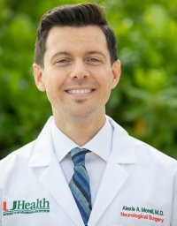 Alexis A Morell合作论文数Miller Sch Med, Univ Miami2
 Brian Fiani合作论文数2
 Timur Urakov合作论文数Dept Neurol Surg, Univ Miami2
 Mansour Mathkour合作论文数Correspondence to: Department of Neurosurgery, Ochsner Health System2
 Tyler Scullen合作论文数2
 Cuong J. Bui合作论文数2
 Santiago Gutierrez Gomez合作论文数2
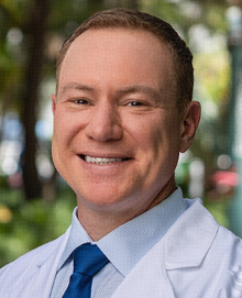 Ricardo Jorge Komotar合作论文数Department of Neurological Surgery, Miller School of Medicine, University of Miami2Karishma Mehta合作论文数1Timothy Mayo合作论文数1Ahdarsh Vallabhaneni合作论文数1Dominique Higgins合作论文数1Juan J Cardona合作论文数1Joseph A Yunge Tigre合作论文数1
 Bianca Marquez合作论文数1
 Ashley K. Yearwood合作论文数1Chase Schlesselman合作论文数1Adam S Levy合作论文数1
 Tiffany Eatz合作论文数Miller School of Medicine, University of Miami1Arada Chaiyamoon合作论文数Khon Kaen University1
 Martín A Merenzon合作论文数1
 Cassidy Werner合作论文数Tulane Center for Clinical Neurosciences, Tulane University School of Medicine1
 Sumedh Shah合作论文数Department of Neurosurgery, University of Miami Miller School of Medicine1Joseph Herbert合作论文数Division of Neurological Surgery, University of Missouri School of Medicine1
 Gregory W Basil合作论文数Miller School of Medicine, University of Miami1Daniel G Eichberg合作论文数Department of Neurosurgery, University of Miami Miller School of Medicine1
 Evan Luther合作论文数Department of Neurosurgery, University of Miami Miller School of Medicine1
 David J. Cohen合作论文数University of Missouri-Kansas City1
 Ivan Michael E合作论文数Dept Neurol Surg, Univ Miami1
 Burks S Shelby合作论文数Department of Neurological Surgery and the Miami Project to Cure Paralysis, University of Miami Miller School of Medicine1
 N. Scott Litofsky合作论文数1Aysun Uz合作论文数1Basem Ishak合作论文数1
 Grace Li Smith合作论文数Division of Radiation Oncology, Department of GI Radiation Oncology, MD Anderson Cancer Center, The University of Texas1Allan D. Levi合作论文数1




