BACKGROUND:Surgical resection is the standard treatment for stage I non-small cell lung cancer (NSCLC). Radiofrequency ablation (RFA) is an option in high-risk patients who cannot undergo surgical resection of stage I NSCLC, but prognostic factors and long-term oncologic results have not been fully evaluated. We evaluated outcomes after image-guided RFA and factors associated with survival in high-risk patients with stage I NSCLC. METHODS:We evaluated the outcomes of image-guided-RFA performed by thoracic surgeons for biopsy-proven stage I NSCLC in high-risk patients over a 17-year period. The primary endpoint evaluated was overall survival (OS), studied using Kaplan-Meier analysis. Covariates associated with OS were analyzed with univariate proportional hazards regression and multivariate Cox regression. RESULTS:One hundred and eleven patients (median age, 74 years) underwent image-guided RFA. After a median follow-up of 30 months, estimated OS was 86% at 1 year (95% confidence interval [CI] 80%-93%) and 54% at 3 years (95% CI, 46%-64%). During follow-up, local progression in the treated lesion, as per imaging criteria, occurred in 44 patients (40%) among whom 14 had histologically proven recurrence (13%). Covariates associated with improved OS in multivariate analysis included size <2 cm (P = .043) and adenocarcinoma histology (P = .013). CONCLUSIONS:Although surgical resection remains the standard, image-guided RFA is effective for high-risk patients with stage I NSCLC who are not surgical candidates. Analysis of covariates associated with survival identified lesion size and histology as important prognostic factors. Prospective studies of RFA are needed to further define patient selection in this high-risk group.
e18054 Background: HNSCC is increasingly being diagnosed in a younger population, with a variable prognosis. While studies have evaluated outcomes in young compared to older patients, evaluation of prognostic factors specifically in younger patients, that may help guide treatment decisions, is needed. Therefore, we conducted a retrospective study to investigate the association of patient and tumor characteristics with disease specific survival (DSS) in HNSCC patients ≤45 years old (yo). Methods: Patients treated at the University of Pittsburgh Medical Center from January 1, 2007 to June 30, 2020, that were ≤45 yo at time of diagnosis with HNSCC without distant metastatic disease, were included. Those who received treatments at other institutions and who had no follow-up after index treatments were excluded. Patient demographics, social history, tumor pathology and treatment course were collected. The primary endpoint of DSS was defined as the interval between the time of pathologic diagnosis and the time of cancer-related death. Proportional hazards regression was applied separately for surgically and non-surgically treated patients. A multivariate analysis was conducted for surgically treated patients only. Results: 230 patients were included. Median age was 42, 73% male, 45% oral cavity primary, 35% oropharynx (89% Human papillomavirus positive), 14% larynx, 69% were smokers, and 73% received combined modality therapy. 35% of patients had a recurrence with the majority locoregional only (61%). Median DSS was not reached. The 5 year DSS was 73% (95% CI 67% to 80%). There was no difference in DSS between surgically treated (n = 169) and non-surgically treated (n = 61, p = 0.94) patients. For surgically treated patients, univariate analysis showed significantly worse DSS in those with higher pathologic T and N stages, positive margins, perineural invasion (PNI), extranodal extension (ENE), and negative HPV status. For non-surgically treated patients, only negative HPV status was associated with significantly worse DSS (p = 0.034). In the multivariate analysis for surgical treated patients, which included all significant characteristics from univariate analysis, pathologic T and N stage, positive margins, (HR 4.92, 95% CI 1.98-12.25, p = 0.006) and PNI (HR 2.66, 95% CI 1.17-6.08, p = 0.02) were significantly associated with worse DSS. Conclusions: Pathologic T and N stage, positive margins, and PNI are independently associated with significantly worse DSS in surgically treated HNSCC patients aged ≤45. Compared with classic prognostic factors seen in the overall population with HNSCC, PNI may be a uniquely strong prognostic factor in younger-aged HNSCC patients.
Purpose/Objective(s) There is a great need for improvement in outcomes in pts with R/M HNSCC that have progressed on anti-PD-1 mAb (IO) and the development of a more personalized treatment (Tx) approach. Materials/Methods Our phase II prospective study evaluated a drug selection strategy based on immune gene expression. Included pts had progressed on prior IO and received ≤3 lines of Tx. All pts underwent a biopsy during screening that was analyzed via the OmniSeq immune report card (IRC). The IRC reports a panel of 397 genes as a relative rank score (RRS) where expression of each gene is compared to a reference population, normalized to a value between 1 and 100. If the CTLA4 vs LAG3 RRS difference was ≥ 15.2, pts were “selected” to receive Nivolumab (N) plus Ipilimumab (I) (if CTLA4 higher) or N + Relatlimab (R) (if LAG3 higher). If the CTLA4 vs. LAG3 RRS difference was < 15.2, pts were randomized 1:1 to N+I or N+R. The primary endpoint was the ORR by RECIST 1.1 of Tx in those “selected” for Tx. DCR (CR/PR/SD) was estimated with Wilson Score 95% confidence intervals. For RNA Seq analysis, proportion analysis of highly expressed genes (≥75 RRS) across groups were compared to <75 RRS. Fisher exact test was performed on each gene for the comparisons, genes with p<0.05 were filtered out to examine whether the proportion is higher or lower in a particular category. Results 20 pts were enrolled, and 18 were evaluable for ORR. Primary site was larynx/hypopharynx (5), oral cavity (2), oropharynx [11 (10 HPV+)]. Most recent Tx was IO monotherapy (11), chemo plus IO (5), or chemo (2). 9/18 pts were also platinum failure. Best response was SD in 5 patients. The DCR was similar in all 18 pts (28%) and by regimen N+R (n=11 27%), N+I (n=7, 28%). 6 pts were “selected” for Tx, all receiving N+R with a DCR of 33% (95%CI: 9.7, 70), median duration of SD of 133.5 days. 12 patients were randomized, with DCR of 25% (95%CI:8.9-53.2), median duration of SD of 55 days. We analyzed the highly expressed genes (≥75 RRS) from 26 pts [enrolled(20) + screened(6)]. Genes highly expressed in >90% of pts included: KRT5, TRIM29, and >50%: EGFR, GITR, KREMEN1, S100A8, TGFB1, CCNB2, LEXM. PRDM1 was expressed in a higher proportion of progressing pts (62%) vs. SD (0%) (p=.04). Comparing the most recent prior Tx, IO alone (n=16) vs. chemo (alone or with IO)(n=10), a higher proportion of chemo pts expressed: GNLY, OAS1, IFIT3, IDO2, IL12B (p<0.05) while TGFB1 was higher after IO alone (p=0.04). Conclusion Our unique study prospectively evaluated a Tx selection strategy based on immune gene expression. Although efficacy was low with both N+I and N+R, prospective selection was feasible and selected pts had a numerically higher DCR, with a longer duration of SD. This represents a first step toward a more personalized approach to treatment. Further gene expression analysis of this cohort is ongoing.
PDF file - 122K, STAT3 decoy treatment does not inhibit cell viability or STAT3 target gene expression in A4 STAT3 null cells
6018 Background: PD-1 inhibition using nivolumab (nivo) monotherapy is modestly effective in metastatic HNSCC. Neoadjuvant trials using nivo may permit development of more effective combinations in surgically resectable HNSCC. We evaluated combinations with nivo plus additional immune checkpoints, CTLA-4 (ipilimumab, ipi) and LAG-3 (relatlimab, rela). Methods: Phase II randomized trial of neoadjuvant nivo alone (240 mg q2 weeks), or with ipi (1 mg/kg q3 weeks) or rela (160 mg q4 weeks) for 4 weeks prior to surgery. Response was scored using RECIST and standard pathologic response criteria. Patients were stratified by p16, PD-L1, and LAG-3, with staining assessed by immunohistochemistry. Freshly digested tumors were subjected to single cell RNA sequencing (scRNAseq) for T cell receptors and gene pathways to identify biomarkers of response or progression. The Cochran-Armitage trend test was used to explore associations with increasing pathological response efficacy. Results: 41 patients (pts) have been enrolled, with 33 evaluable for this analysis. Of these 33, median age is 63 (32-81), primary site oral cavity (n=25), oropharynx (n=5, 3 HPV), larynx (n=3), clinical T2 (n = 5), T3 (n = 12), T4 (n = 13), and cN0/1 (n = 22), cN2 (n = 9), and PD-L1 CPS > 1 (n=25). There were no serious study drug-related AEs or unexpected surgical delays/complications. Pathologic response (Table) was more frequent with nivo/rela (11/13) vs. nivo/ipi (6/10) or nivo (4/10). Partial (>50%) or major (>90%) pathologic responses were more frequent and deeper in the combination arms. Minor (<50%) pathologic responses were more frequent with nivo/rela, and similar between nivo/ipi and nivo. There was no association between RECIST response, PD-L1, or LAG3 and pathologic response. Combined PD-L1 and LAG3 expression was not associated with pathologic response in the nivo/rela arm, however more patients with combined positivity had a >50% response (4 vs. 0). Expansion of CD8 + T cells, as well as expanded proportion of CD8 + CXCL13 + T cells in responder tumors were identified in post-treatment specimens using scRNAseq. Conclusions: Neoadjuvant nivo/ipi or nivo/rela combinations were safe and associated with promising pathologic responses compared to nivo monotherapy. Anti-tumor CD8 + T cell populations and targetable pathways are emerging in responder patients. The trial continues to enroll and further evaluation of this strategy is warranted. Clinical trial information: NCT04080804 . [Table: see text]
PDF file - 61 KB, Percentages of ANX-binding cells within the peripheral memory (TPM) and terminally differentiating (TTD)subsets in the circulation of HNSCC patients and normal controls
Background: Proinflammatory chemokines/cytokines support development and maturation of tertiary lymphoid structures (TLS) within the tumor microenvironment (TME). In the current study, we sought to investigate the prognostic value of TLS- associated chemokines/ cytokines (TLS-kines) expression levels in melanoma patients by performing serum protein and tissue transcriptomic analyses, and to then correlate these data with patients clinicopathological and TME characteristics. Methods: Levels of TLS-kines in patients' sera were quantitated using a custom Luminex Multiplex Assay. The Cancer Genomic Atlas melanoma cohort (TCGASKCM) and a Moffitt Melanoma cohort were used for tissue transcriptomic analyses. Associations between target analytes and survival outcomes, clinicopathological variables, and correlations between TLS-kines were statistically analyzed. Results: Serum of 95 patients with melanoma were evaluated; 48 (50%) female, median age of 63, IQR 51-70 years. Serum levels of APRIL/TNFSF13 were positively correlated with levels of both CXCL10 and CXCL13. In multivariate analyses, high levels of serum APRIL/TNFSF13 were associated with improved event-free survival after adjusting for age and stage (HR = 0.64, 95% CI 0.430.95; p = 0.03). High expression of APRIL/TNFSF13 tumor transcripts was significantly associated with improved OS in TCGA-SKCM (HR = 0.69, 95% CI 0.52-0.93; p = 0.01) and in Moffitt Melanoma patients (HR = 0.51, 95% CI: 0.320.82; p = 0.006). Further incorporation of CXCL13 and CXCL10 tumor transcript levels in a 3-gene index revealed that high APRIL/CXCL10/CXCL13 expression was associated with improved OS in the TCGA SKCM cohort (HR = 0.42, 95% CI 0.19- 0.94; p = 0.035). Melanoma differentially expressed genes positively associated with high APRIL/CXCL10/CXCL13 tumor expression were linked to tumor infiltration by a diverse array of proinflammatory immune cell types. Conclusion: Serum protein and tumor transcript levels of APRIL/TNFSF13 are associated with improved survival outcomes. Patients exhibiting high coordinate expression of APRIL/CXCL10/CXCL13 transcripts in their tumors displayed superior OS. Further investigation of TLS-kine expression profiles related to clinical outcomes in larger cohort studies is warranted.
PDF file - 1.2MB, Modified STAT3 decoys bind to pSTAT3 protein with similar affinity as the parental STAT3 decoy
PDF file - 1978 KB, Decreased percentages of circulating apoptosis-resistant CD8+CCR7+ T cells in HNSCC patients and NC
PDF file - 57 KB, Results of the experimental verification of the two predictive models
PDF file - 2.2MB, Modified STAT3 decoys demonstrate enhanced resistance to thermal denaturation
PDF file - 45K, In-vitro expansion of EGFR-specific CTL in the presence of NK cells.
PDF file - 441K, Importance of IFN-γ released by cetuximab activated NK cells in the enhancement of DC maturation.
PDF file, 216K, Decreased STAT3 target gene expression in erlotinib sensitive (or resistant) HNSCC cells treated with the STAT3 decoy.
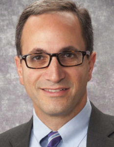 Robert L. Ferris合作论文数School of Medicine, UNC114
 Jennifer Grandis合作论文数School of Medicine, University of California, San Francisco83
 Theresa L. Whiteside合作论文数Department of Immunology, University of Pittsburgh58
 Jonas T. Johnson合作论文数Department of Otolaryngology, School of Medicine, University of Pittsburgh55
 James D. Luketich合作论文数Department of Cardiothoracic Surgery, School of Medicine, University of Pittsburgh;McGowan Institute for Regenerative Medicine, University of Pittsburgh46
 Athanassios Argiris合作论文数Department of Medical Oncology, Sidney Kimmel Medical College, Thomas Jefferson University;1st Department of Medical Oncology, Hygeia Hospital42
 Arjun Pennathur合作论文数40
 Raja R. Seethala合作论文数Department of Pathology, Division of Anatomic Pathology, School of Medicine, University of Pittsburgh38Kim Seungwon合作论文数37
 Sufi M. Thomas合作论文数Department of Otolaryngology, University of Pittsburgh37Duvvuri Umamaheswar合作论文数34
 Tony E. Godfrey合作论文数Chobanian & Avedisian School of Medicine, Boston University;Department of Surgery, Boston University School of Medicine34Chiosea Simion I合作论文数Dept Pathol, Univ Pittsburgh32
 Julie Elaine Bauman合作论文数Division of Hematology and Oncology, Department of Medicine, The University of Arizona College of Medicine - Tucson;The University of Arizona Cancer Center30
 Malabika Sen合作论文数27Daniel E. Johnson合作论文数25Maria L. Freilino合作论文数25
 John M. Kirkwood合作论文数Division of Malignant Hematology and Medical Oncology, Department of Medicine, School of Medicine, University of Pittsburgh;Department of Pathology, School of Medicine, University of Pittsburgh24Wang Lin合作论文数22
 Ahmad Tarhini合作论文数Moffitt Cancer Center;University of South Florida21
 Dwight E. Heron合作论文数American College of Radiation Oncology;NRG Oncology;Bon Secours Mercy Health;REDJuvenate, LLC20
 Jill M. Siegfried合作论文数Department of Pharmacology, Medical School, University of Minnesota19Joyce Sonali合作论文数17Matthew J. Schuchert合作论文数17Neil A. Christie合作论文数17L Wang合作论文数16Changyou Li合作论文数16
 Steven J Hughes合作论文数16Ohr James合作论文数15
 Liqiang Xi合作论文数14Albert B. Deleo合作论文数14Siva Raja合作论文数13
 Jessica Lee合作论文数San Jose/ and NASA Ames Research Center, San Jose State University13
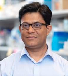 Raghvendra M. Srivastava合作论文数Department of Otolaryngology, University of Pittsburgh12
 Omar Awais合作论文数12Jonathan P Etter合作论文数11
 Naftali Kaminski合作论文数Yale School of Medicine;Boehringer Ingelheim Pharmaceuticals, Inc.11
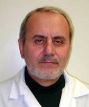 Soldano Ferrone合作论文数Departments of Surgery and of Orthopedic Surgery, Massachusetts General Hospital, Harvard Medical School11
 Barton Branstetter合作论文数Department of Radiology, University of Pittsburgh11
 Danith Ly合作论文数Department of Chemistry, Mellon College of Science, Carnegie Mellon University11Rapireddy Srinivas合作论文数11Pandit Kusum V合作论文数Div Pulm Allergy & Crit Care Med, Univ Pittsburgh11Sahu Nivedita合作论文数11
 Rodney Landreneau合作论文数University of Pittsburgh Medical Center;The Lung Center, Penn Highlands Healthcare11
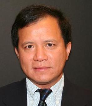 Pui Kai (Tom) Li合作论文数Division of Medicinal Chemistry and Pharmacognosy, College of Pharmacy, The Ohio State University11
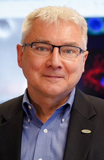 Pawel Kalinski合作论文数Department of Immunology, Roswell Park Comprehensive Cancer Center11
 Walter J. Storkus合作论文数Department of Dermatology, School of Medicine, University of Pittsburgh;Department of Immunology, School of Medicine, University of Pittsburgh11Xiaomin Chen合作论文数11




