Background While gut microbiota dysbiosis is a hallmark of inflammatory bowel disease (IBD), the causal microbial drivers and their host-mediated mechanisms remain elusive. This study leverages an integrated multi-omics approach, combining Mendelian randomization (MR) and transcriptome analysis, to bridge the gap from microbial causality to host molecular pathways. Methods We performed a two-sample MR analysis using large-scale genome-wide association study (GWAS) data to identify specific gut microbiota taxa with a causal effect on IBD risk. Subsequently, we conducted a multi-level bioinformatic analysis of IBD patient transcriptomes to elucidate the downstream host genes, regulatory networks, and immune cell interactions modulated by these causal microbes. Results Our MR analysis established a robust causal protective effect of the family Bifidobacteriaceae against IBD. Integrating this finding with transcriptomic data, we identified three key host genes as potential mediators acting through distinct mechanisms: LCT, whose regulation may foster a protective prebiotic niche; MCM6, which appears to function as a hub driving the proliferation of pathogenic immune infiltrates; and UBXN4, a critical regulator of cellular proteostasis, the failure of which can precipitate inflammatory stress. Conclusions This study moves beyond association to delineate a causal protective role for Bifidobacteriaceae in IBD and pinpoints specific host genes (LCT, MCM6, UBXN4) through which this effect is likely orchestrated. These findings provide a novel mechanistic framework for host-microbiota interactions and highlight new pathways for therapeutic intervention in IBD.
BACKGROUND Hepatic ischemia-reperfusion injury (IRI) is a critical pathological process associated with trauma, sepsis, and liver surgeries, including transplantation. During reperfusion, oxidative stress in sinusoidal endothelial cells triggers cell death, however, the underlying regulatory mechanisms remain poorly defined. Previous studies have implicated leukocyte-derived chemotaxin-2 (LECT2) in various diseases and identified it as a ligand for the orphan receptor Tie1, yet its role in endothelial cell injury during liver IRI remains unclear. AIM To investigate whether LECT2 exacerbates liver IRI by regulating oxidative stress in endothelial cells through the Tie1/Src signaling pathway and to evaluate the therapeutic potential of targeting this axis. METHODS In vitro hypoxia-reoxygenation injury was modeled in EA.hy926 endothelial cells, followed by LECT2 knockdown or recombinant LECT2 treatment, Tie1 silencing, and Tie1-Ig3 segment protein treatment to block LECT2/Tie1 binding. Src kinase activity was inhibited using dasatinib. Cell viability, oxidative stress, cytotoxicity, and signaling pathway activation were assessed. In vivo , LECT2 knockout mice underwent hepatic ischemia-reperfusion, while injury markers, inflammatory cytokines, and endothelial damage were evaluated. RESULTS LECT2 knockdown reduced oxidative stress and endothelial cell damage following hypoxia-reoxygenation, whereas recombinant LECT2 exacerbated these effects. Disruption of LECT2/Tie1 binding, via either Tie1 knockdown or Tie1-Ig3 treatment mitigated injury. Mechanistically, LECT2 activated Src kinase phosphorylation in a Tie1-dependent manner, and Src inhibition reversed LECT2-induced cell damage. In mice, LECT2 deletion attenuated liver IRI, decreased apoptosis and inflammation, and better preserved sinusoidal endothelial integrity. CONCLUSION The LECT2/Tie1/Src signaling axis plays a critical role in regulating oxidative stress and endothelial cell injury during hepatic ischemia-reperfusion. Targeting the LECT2/Tie1/Src signaling pathway may offer a novel therapeutic strategy for mitigating liver IRI in clinical settings.
Liver fibrosis, marked by excessive ECM deposition, can progress to cirrhosis and hepatocellular carcinoma, yet effective treatments are lacking. Since hepatic stellate cell (HSC) activation is central to fibrosis, inhibiting it is a key therapeutic strategy. Vitamin D receptor (VDR) activation can suppress HSC activation by inhibiting the TGFβ/SMAD3 pathway, making it a promising target. However, steroidal VDR agonists’ clinical use is limited by hypercalcemia caused by upregulation of calcium metabolism genes. To overcome this, we designed novel steroidal VDR modulators by modifying the side chain to selectively impair transactivation of calcium-related genes while preserving antifibrotic signaling. Among 30 synthesized compounds, D13 exhibited strong VDR affinity and potent antifibrotic activity in vitro. In a bile duct ligation mouse model, D13 significantly alleviated liver fibrosis without inducing hypercalcemia, unlike calcipotriol. Mechanistically, D13 inhibited the TGFβ/SMAD3 pathway without excessively upregulating calcium metabolism genes. Thus, D13 represents a promising antifibrotic candidate warranting further investigation.
Background and Objectives Non-alcoholic fatty liver disease (NAFLD) has become a growing global public health concern. Effective therapeutic strategies for NAFLD remain urgently needed. Liver-on-a-chip (LC) technology offers an innovative platform for NAFLD modeling and drug development. This study aimed to develop a biomimetic liver-chip using co-cultured human hepatocyte (HepaRG) with hepatic stellate and endothelial cells to model NAFLD, and evaluate the therapeutic potential of scalable telomerase reverse transcriptase (hTERT)-immortalized umbilical cord mesenchymal stem cell-derived exosomes (TMSC-Exo).Methods HepaRG cells, hepatic stellate cells, and endothelial cells were used to construct a dual-chamber biocompatible LC. The NAFLD model was induced by free fatty acid (FFA) and applied to evaluate the efficacy of resmetirom and TMSC-Exo for the treatment of NAFLD. Moreover, the high-fat (HF) diet-induced mouse model was analyzed to verify the in vitro results. Proteomic analyses were performed to explore the molecular mechanisms involved in the development of NAFLD and the effect of TMSC-Exo in treating NAFLD.Results Cells cultured in LC showed better viability compared to those in the Transwell system. The on-chip NAFLD model mimicked the characteristics of NAFLD in vivo, including intracellular lipid accumulation and impaired hepatocyte functions in albumin synthesis, levels of urea, CYP1A2, and CYP3A4. Both TMSC-Exo and resmetirom displayed a significant effect in reducing the lipid accumulation in the on-chip NAFLD model. The TMSC-Exo showed superior effects in elevating the levels of albumin, urea, CYP1A2, and CYP3A4. The therapeutic effects of TMSC-Exo were also confirmed in the NAFLD mouse models. Proteomic analysis found that the top 15 up- and down-regulated differentially expressed proteins in NAFLD models compared to the control group were mainly associated with lipid metabolism, endoplasmic reticulum stress, and inflammation.Conclusions Our on-chip NAFLD model successfully recapitulated key pathological features of hepatic steatosis and functional impairment. Using this model, we evaluated TMSC-Exo and demonstrated its significant therapeutic efficacy against NAFLD.
Colorectal cancer (CRC) remains largely refractory to immune-checkpoint blockade, with adenomatous polyposis coli (APC) mutations present in 80%–90% of cases. Loss of APC was previously thought to promote tumor progression mainly through deregulated Wnt/β-catenin signaling. Here, we report that APC loss leads to inhibition of CD8+ T cell infiltration and CRC immune evasion through the dephosphorylation of signal transducers and activators of transcription 1 (STAT1) by protein tyrosine phosphatase non-receptor type 13 (PTPN13), independently of β-catenin. Peptides containing the last 11 C-terminal amino acid (aa) residues of APC (APC11) bind directly to PTPN13 to block PTPN13–STAT1 interactions and facilitate STAT1 phosphorylation, interferon regulatory factor-1 (IRF1) expression, major histocompatibility complex (MHC) class I antigen presentation, and T cell intratumoral infiltration, all of which eventually inhibit tumor progression and enhance the effects of programmed cell death 1 (PD1) blockade. Thus, we have identified a previously unknown APC/PTPN13/STAT1-dependent tumor immune-suppressive mechanism. The potent tumor-suppressing effect of combining anti-PD1 antibodies with APC11 peptides provides a compelling target and rationale for future development of anti-tumor drugs for patients with CRC.
Purpose.Develop a multi-scale fusion model (MSFM) based on multi-phase contrast-enhanced computed tomography (CECT) to predict pancreatic cancer (PC) resectability, thereby assisting expert decision-making.Methods.This retrospective study enrolled 280 patients with PC from four institutions, which were randomly divided into a training cohort (202 patients) and an independent test cohort (78 patients). Three-phase CECT images (arterial, venous, and delayed phases) were used for modeling. The MSFM comprises two sub-networks: (1) a multi-phase fusion network for extracting cross-phase shared fusion features, (2) a phase-specific branch network for capturing phase-specific features; and a post-fusion strategy to generate the final predictive score by integrating the shared fusion features and three groups of phase-specific features. Additionally, a human-machine fusion deep learning model (HMfDL) was constructed by fusing the predictive score of the MSFM with expert assessments.Results.In the independent test, the MSFM achieved an AUC (area under the receiver operating characteristic curve) of 0.8385 (95% CI: 0.7521-0.9249), accuracy of 84.62%, sensitivity of 72.00%, and specificity of 90.57%. This performance outperformed single-phase models (AUC range: 0.7638-0.7781), two-phase models (AUC range: 0.7826-0.7864), and ten states-of-the-art classifiers (AUC range: 0.7404-0.7796). The HMfDL further improved the performance, reaching an AUC of 0.8626 (95% CI: 0.7853-0.9400), accuracy of 91.03%, sensitivity of 80.00%, and specificity of 96.23%. Notably, the HMfDL corrected 58.82% of misdiagnosis made by experts.Conclusions. The MSFM effectively fuses multi-phase CECT to enable highly accurate predictions of PC resectability, and provides valuable support for expert decision-making through HMfDL.
Hepatic fibrosis, a pathological consequence of chronic liver injury, is characterized by excessive deposition of extracellular matrix (ECM). Activation of hepatic stellate cells (HSCs) is critical to the pathogenesis. Vitamin D receptor (VDR) agonists have been demonstrated to inhibit transforming growth factor-beta 1 (TGF-β1) induced HSC activation, thereby reducing ECM deposition and attenuating the progression of liver fibrosis. Despite their broad therapeutic potential, the clinical translation of VDR agonists has been hampered by the risk of inducing hypercalcemia. To address this limitation, we designed a series of novel non-steroidal VDR agonists based on a phenylindole scaffold and evaluated their anti-fibrotic properties. Among them, compounds I-7, II-4, II-6, and II-8, exhibited significant inhibition of HSC activation in vitro. Owing to its robust activity, compound II-8 was selected for further investigation in a bile duct ligation (BDL)-induced liver fibrosis model. Histological analysis confirmed that treatment with II-8 significantly inhibited the fibrosis progression. Crucially, the hypercalcemia typically associated with VDR agonist therapy was not observed. Hypercalcemia is a major drawback of currently marketed steroidal VDR agonists, which significantly limits their broad clinical application. The compounds we have designed can effectively avoid this hypercalcemia side effect. These findings underscore the potential of II-8 as a potent therapeutic agent for the treatment of liver fibrosis.
Sepsis-associated acute kidney injury (SA-AKI) is a life-threatening clinical condition with limited therapeutic options, where ferroptosis serves as a key pathogenic driver. In this study, we conducted clinical sample analyses and proteomic profiling, and identified that fatty acid-binding protein 4 (FABP4) was significantly upregulated in the plasma of patients with SA-AKI, as well as in the renal tissues of SA-AKI model mice. To explore the functional role of FABP4, we administered BMS-309403 (BMS), a selective FABP4 inhibitor, in both a lipopolysaccharide (LPS)-induced murine SA-AKI model and an in vitro model of RAS-selective lethal 3 (RSL3)-treated human renal tubular epithelial (HK-2) cells. Our results demonstrated that BMS treatment markedly improved renal function, reduced tubular injury, and alleviated renal inflammation in mice. These protective effects were tightly associated with the inhibition of ferroptosis, as evidenced by reduced lipid peroxidation, decreased iron deposition, improved mitochondrial function, and the reversal of abnormal expression of key ferroptosis-related proteins, glutathione peroxidase 4 (GPX4) and acyl-CoA synthetase long chain family member 4 (ACSL4). Furthermore, in vitro experiments confirmed that BMS effectively mitigated RSL3-induced ferroptosis in HK-2 cells. To our knowledge, this study is the first to demonstrate that FABP4 exacerbates SA-AKI by promoting ferroptosis, suggesting that targeted inhibition of FABP4 may represent a promising novel therapeutic strategy for SA-AKI.
Orthotopic liver transplantation (OLT) remains the gold standard treatment for end-stage liver disease but is severely limited by donor scarcity and immune rejection. Consequently, bioartificial liver (BAL) systems and hepatocyte transplantation have emerged as promising alternatives, yet their clinical efficacy hinges on the unresolved challenge of generating functional hepatocytes at scale. This review systematically examines the molecular landscape governing hepatocyte proliferation, dissecting the key regulatory roles of Wnt/β-catenin, Hippo-YAP, Notch, and TGF-β signaling pathways in balancing proliferation and differentiation. We critically evaluate current in vitro expansion strategies, contrasting viral-mediated genetic modification approaches with emerging non-genome-editing strategies. Particular emphasis is placed on “next-generation” methodologies, including chemically defined media, bioreactor-based dynamic cultures, and multicellular co-culture systems that recapitulate the native hepatic niche. Furthermore, we highlight recent breakthroughs in liver organoid technology—such as the construction of multi-zonal and vascularized architectures—that bridge the gap between in vitro expansion and in vivo physiological relevance. Finally, we summarize the translational progress of these expanded hepatocytes in BAL devices, cell transplantation trials, and high-fidelity pharmacotoxicological models. We further discuss how emerging technologies, including CRISPR-Cas9-mediated gene correction and bioengineering innovations, may help address current limitations, while acknowledging that substantial preclinical validation remains necessary.
Current nanoparticle-based drug delivery systems present significant challenges in treating solid tumors, primarily due to a variety of physiological and pathological barriers that hinder the deep penetration of drugs. Herein, we demonstrate bio-inspired fractal-structured gel-drugs (DOX@HPLA, DHPL) that significantly enhance deep tumor penetration, thereby greatly improving the chemotherapy efficiency for hepatocellular carcinoma (HCC). The fabrication of DHPL is achieved through a crosslinking of 8-arm polyethylene glycol (PEG) and polysaccharide heparin in the presence of DOX, which is subsequently modified with targeting lactobionic acid (LA). During the process of penetration, these fractal-structured gel-drugs could leverage their soft and rough surfaces endowed by their unique fractal structures to increase the interfacial contact sites with the tumor cells through topographic interactions, which optimizes the cell adhesion in the local area. This results in a significant enhancement of interfacial adhesion and internalization, thereby substantially improving receptor-mediated endocytosis associated with subsequent transcytosis. In the process of transcytosis, the fractal-structured geldrugs facilitate the early endosomes/recycling endosomes pathway for exocytosis and concurrently diminish degradation in the early endosomes/late endosomes/lysosome pathway, resulting in deeper tumor penetration and thereby enhancing the chemotherapy efficiency for HCC. It is believed that the bio-inspired fractal-structured gel-drugs enable efficient amplification of interfacial interactions with cells through fractal-structuremediated topographic interactions, providing an avenue for deep penetration treatment of solid tumors.
The role of human umbilical cord mesenchymal stem cell-derived extracellular vesicles (hUCMSC-EVs) in liver regeneration is promising, yet their clinical translation is hampered by insufficient production. Current strategies targeting their secretion are inefficient and lack a clear mechanistic understanding. We isolated and characterized hUCMSC-EVs pretreated with the H89 and other mTORC1 inhibitors. Our findings revealed that H89 effectively enhances the secretion of hUCMSC-EVs across diverse cell types, demonstrating universal efficacy. Importantly, H89 upregulates GABARAPL1 expression, a key negative regulator of the PKA/mTORC1 pathway, to inhibit mTORC1 activity and promote the formation of amphisomes and SNARE-mediated hUCMSC-EVs release. Furthermore, EVs derived from H89-pretreated hUCMSCs (H-EVs) exhibited altered cargo composition, significantly increased proliferative activity, and potentiated liver regeneration via the RELA/miR-29a axis, which regulates the homeostasis of hepatic stellate cells. Our results highlight that H89 enhances hUCMSC-EV secretion through mTORC1 inhibition, with the resulting benefits for liver regeneration mediated by the RELA/miR-29a network. These findings demonstrate the great promise of H89 in EV-based liver regeneration, offering a promising platform for clinical translation.
Background Endoscopic submucosal dissection (ESD) has been widely accepted as the gold standard curative approach for early oesophageal cancer and precancerous lesions. However, post-ESD oesophageal stricture remains a challenging clinical complication. In this study, a novel oesophageal catheter for stricture prevention after oesophageal ESD is introduced, and a preliminary description of the feasibility and safety of this new method are provided. Methods In total, five patients who underwent oesophageal ESD and received the novel oesophageal catheter for stricture prevention or a nasogastric tube between September 2024 and October 2025 at Jiangyin Hospital Affiliated to Nantong University were included. The patients’ clinical characteristics, endoscopic treatment and follow-up data were analysed. The primary outcome was the proportion of patients who developed oesophageal stricture after ESD. Secondary outcomes included the number of endoscopic balloon dilation or endoscopic radial incision (ERI) sessions required for stricture resolution and procedure-related adverse events. Results Four patients who underwent catheter placement immediately after ESD did not develop stricture during follow-up (average follow-up period: 17.25 months). The fifth patient with delayed catheter placement (due to concerns about pharyngeal pain symptoms) developed stricture, which was temporarily resolved by ERI but recurred six months later. Subsequent placement of the same catheter controlled the stricture, and this patient was followed up for 6 months after catheter removal. Conclusion The novel oesophageal catheter for stricture prevention represents potentially safe, low-cost and readily available prophylactic strategy for patients undergoing oesophageal ESD. This simple and accessible approach may offer preliminary insights for clinical practice, suggesting the possibility of reducing post-ESD stricture rates and improving long-term quality of life, although further validation in larger cohorts is needed. Trail registration This study was retrospectively registered at the Chinese Clinical Trial Registry site. [Registration number: ChiCTR2500112721(https://www.chictr.org.cn/); registration date: November 19, 2025].
The specific operational diagrams for the website's predictive classification results.
Hepatic progenitor cells (HPCs) are frequently overactivated, and their differentiation into hepatocytes is impaired in advanced liver diseases. To explore the effects of intestinal epithelial cells and their exosomes on the hepatic differentiation of HPCs, co-culture systems of Caco-2/HepaRG cell lines and intestine/HPC organoids are established in a novel gut-liver-on-a-chip. Exosomes derived from intestinal organoids are administered to mice with carbon tetrachloride (CCL4)-induced liver fibrosis. The results showed that the co-culture of HPCs and intestinal epithelial cells promoted the hepatic differentiation of HPCs, mediated by exosomes derived from intestinal epithelial cells. Treatment with exosomes derived from intestinal organoids ameliorated liver fibrosis in a mouse model of CCL4-induced liver fibrosis. A cluster of miRNAs, miR-371-373, is identified within the exosomes of the intestinal epithelial cells, which target RPS6KA2 to modulate hepatic differentiation. This findings demonstrate that exosomes from intestinal epithelial cells promote the hepatic differentiation of HPCs. Exosomes from intestinal organoids may be a novel therapeutic strategy for the treatment of advanced liver diseases.
Comparison of clinical characteristics for the external validation set 2 patients with different DLRI risk groups.
Clinical characteristics of patients according to the anti-inflammatory TCM treatment in DLRI high risk patients in the internal validation set 2 after propensity score matching.
Deep learning radiomic feature selection and the establishment of DLRI using the LASSO logistic regression model.
Purpose: From 8% to 28% of patients with papillary thyroid carcinoma (PTC) experience recurrence, complicating risk stratification and treatment. We previously identified an inflammatory molecular subtype of PTC associated with poor prognosis. Based on this subtype, we aimed to develop and validate a noninvasive radiomic signature to predict prognosis and treatment response in patients with PTC. Experimental Design: We collected preoperative ultrasound images from two large independent centers (n = 2,506) to develop and validate a deep learning radiomics signature of inflammation (DLRI) for predicting the inflammatory subtype of PTC, including its correlation with prognosis and anti-inflammatory traditional Chinese medicine (TCM) treatment. Training set 1 (n = 64) and internal validation set 2 (n = 1,108) were from Tianjin Medical University Cancer Institute and Hospital. External validation sets 1 (n = 76) and 2 (n = 1,258) were from Fudan University Shanghai Cancer Center. Results: We developed a DLRI to accurately predict PTC's inflammatory subtype (AUC = 0.97 in training set 1 and AUC = 0.82 in external validation set 1). High-risk DLRI was significantly associated with poor disease-free survival in the first cohort [HR = 16.49, 95% confidence interval (CI), 7.92-34.35, P < 0.001] and second cohort (HR = 5.42, 95% CI, 3.67-8.02, P < 0.001). The DLRI independently predicted disease-free survival, irrespective of clinicopathologic variables (P < 0.001 for all). Furthermore, patients with high-risk DLRI were likely to benefit from anti-inflammatory TCM treatment (HR = 0.19, 95% CI, 0.06-0.55, P = 0.002), whereas those with low-risk DLRI did not. Conclusions: DLRI is a reliable noninvasive tool for evaluating prognosis and guiding anti-inflammatory TCM treatment in patients with PTC. Prospective studies are needed to confirm these findings.
 Mingxin Pan (潘明新)合作论文数112Zesheng Jiang合作论文数48Guolin He合作论文数46Qing Peng合作论文数39Chen-jie ZHOU (周陈杰)合作论文数32
 Yuan Cheng (程远)合作论文数30Jizhen YANG (杨继震)合作论文数27
 Yan Wang (汪艳)合作论文数Southern Medical University25
 Erwei Sun (孙尔维)合作论文数24Jun Weng合作论文数22
 Shao Li (李梢)合作论文数Department of Automation, Tsinghua University;Institute of TCM-X, Beijing, Tsinghua University22Lei Feng合作论文数21ZiLin Zhao (赵子粼)合作论文数19Lei Cai合作论文数18Li Yang合作论文数17JinMin Xie (谢金敏)合作论文数17BaoHua Chen (陈保华)合作论文数17Zhi Zhang (张志)合作论文数17
 Yu Wang (王宇)合作论文数Department of Head & Neck Surgery, Cancer Hospital,Fudan University16
 Anqi Jin合作论文数15
 Weituo Zhang (张维拓)合作论文数Shanghai Jiao Tong University School of Medicine15Ning Qu合作论文数Fudan Univ, Shanghai Canc Ctr, Dept Head & Neck Surg, Shanghai 200032, Peoples R China15Ben Ma合作论文数15Dapeng Li合作论文数15Jingzhu Zhao合作论文数15Tian Liao合作论文数15Shuang Wang (汪爽)合作论文数15FanDong Kong (孔凡东)合作论文数15
 Qiang Li (李强)合作论文数第二军医大学第一附属医院呼吸内科15
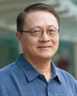 Herbert Yu合作论文数University of Hawaiʻi Cancer Center;School of Public Health, Yale University15
 Ming Gao (高明)合作论文数Cancer Hospital, Tianjin Medical University;Nankai University15
 Biyun Qian (钱碧云)合作论文数Shanghai Hospital Development Center;School of Health Science and Engineering, University of Shanghai for Science and Technology;Shanghai Jiao Tong University School of Medicine15
 Xiangqian Zheng (郑向前)合作论文数Department of Thyroid and Neck Tumor, Tianjin Medical University Cancer Institute and Hospital15Xu Han合作论文数14JianFeng Chen (陈建锋)合作论文数14Jiasheng Qin合作论文数13YuQiang Dan (单毓强)合作论文数12JinLong Yu (俞金龙)合作论文数12Kunhou Yao (姚坤厚)合作论文数河南大学临床医学院12
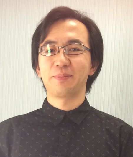 Weijie Zhou (周伟杰)合作论文数Southern Medical University12Kebo Zhong合作论文数11Huan Zhang合作论文数11AiHui Li (李爱辉)合作论文数11Yi Zhang (张翌)合作论文数11QingJun Zhang (张清军)合作论文数11XiaoPing Xu (徐小平)合作论文数11
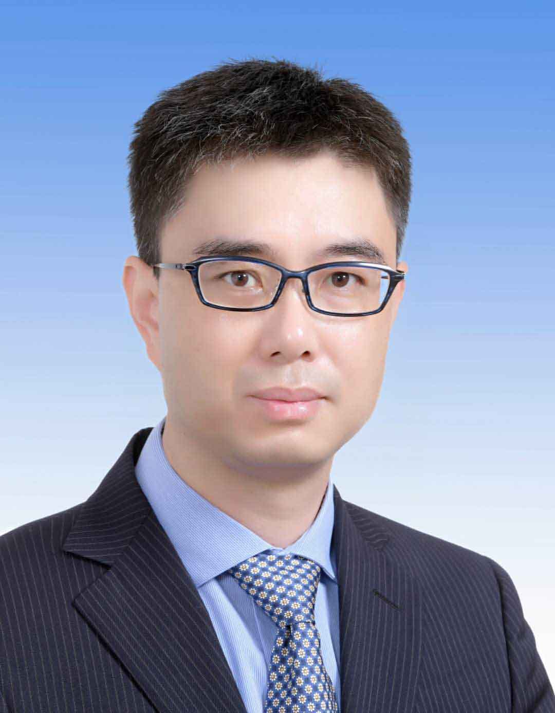 Yu Fu (傅钰)合作论文数State Key Laboratory of Microbial Resources, Institute of Microbiology, Chinese Academy of Sciences;University of Chinese Academy of Sciences11Fanhong Zeng合作论文数10Min Zeng合作论文数10




