As large language models (LLMs) continue to revolutionize AI research, there is a growing interest in building large-scale brain foundation models to advance neuroscience. While most existing brain foundation models are pre-trained on time-series signals or connectome features, we propose a novel graph-based pre-training paradigm for constructing a brain graph foundation model. In this paper, we introduce the Brain Graph Foundation Model, termed BrainGFM, a unified framework that leverages graph contrastive learning and graph masked autoencoders for large-scale fMRI-based pre-training. BrainGFM is pre-trained on a diverse mixture of brain atlases with varying parcellations, significantly expanding the pre-training corpus and enhancing the model’s ability to generalize across heterogeneous fMRI-derived brain representations. To support efficient and versatile downstream transfer, we integrate both graph prompts and language prompts into the model design, enabling BrainGFM to flexibly adapt to a wide range of atlases, neurological and psychiatric disorders, and task settings. Furthermore, we employ meta-learning to optimize the graph prompts, facilitating strong generalization to previously unseen disorders under both few-shot and zero-shot learning conditions via language-guided prompting. BrainGFM is established on 27 neuroimaging datasets spanning 25 common neurological and psychiatric disorders, encompassing 2 types of brain atlases (functional and anatomical) across 8 widely used parcellations, and covering over 25,000 subjects, 60,000 fMRI scans, and a total of 400,000 graph samples aggregated across all atlases and parcellations.
With the growing interest in foundation models for brain signals, graph-based pretraining has emerged as a promising paradigm for learning transferable representations from connectome data. However, existing contrastive and masked autoencoder methods typically rely on naive random dropping or masking for augmentation, which is ill-suited for brain graphs and hypergraphs as it disrupts semantically meaningful connectivity patterns. Moreover, commonly used graph-level readout and reconstruction schemes fail to capture global structural information, limiting the robustness of learned representations. In this work, we propose a unified diffusion-based pretraining framework that addresses both limitations. First, diffusion is designed to guide structure-aware dropping and masking strategies, preserving brain graph semantics while maintaining effective pretraining diversity. Second, diffusion enables topology-aware graph-level readout and node-level global reconstruction by allowing graph embeddings and masked nodes to aggregate information from globally related regions. Extensive experiments across multiple neuroimaging datasets with over 25,000 subjects and 60,000 scans involving various mental disorders and brain atlases demonstrate consistent performance improvements.
Introduction:Cognitive impairment is a common complication of chronic kidney disease (CKD), but its underlying mechanisms are not fully understood. This study aims to investigate the glymphatic system function in CKD patients with and without cognitive impairment (CI) by analyzing the coupling between the global blood oxygen level-dependent (gBOLD) signal and the cerebrospinal fluid (CSF) signal using resting-state functional magnetic resonance imaging (rs-fMRI). Methods:Twenty-nine patients with CKD were enrolled (19 with CI and 10 without), along with 22 healthy controls (HCs). All patients underwent high-resolution structural MRI and rs-fMRI scans. The gBOLD-CSF coupling was quantified by calculating the maximum negative correlation within a predefined time-lag range between the gBOLD signal and the fourth ventricular CSF signal. The gBOLD-CSF coupling was compared between the CKD and HC groups using analysis of covariance (ANCOVA), adjusting for age, sex, education, and mean framewise displacement (FD). The difference between patients with CKD with and without CI was assessed using ANCOVA, after adjusting for age, sex, education, hypertension, diabetes, and mean FD. Partial correlation analysis was performed to explore the associations between gBOLD-CSF coupling and clinical indicators, such as estimated glomerular filtration rate (eGFR), Montreal Cognitive Assessment (MoCA) scores, and other laboratory data. Results:After adjusting for covariates, gBOLD-CSF coupling was significantly lower in the CKD group than in the HC group (β = -0.178, p = 0.003). This finding remained robust in sensitivity analyses adjusting for hypertension and diabetes. Within the CKD group, patients with CI had significantly lower gBOLD-CSF coupling than those without CI (β = -0.135, p = 0.040). Correlation analyses revealed that gBOLD-CSF coupling tended to be positively associated with hemoglobin, MoCA score, and eGFR, and negatively associated with blood urea and creatinine; however, none of these correlations reached statistical significance after false discovery rate correction (all q > 0.05). Conclusion:Patients with CKD exhibit impaired glymphatic system function, manifested as reduced gBOLD-CSF coupling, which is associated with the severity of CI. These findings support the hypothesis that impaired glymphatic clearance may contribute to cognitive decline in CKD via the kidney-brain axis. Larger longitudinal studies are needed to validate its clinical significance.
The direct cognitive benefits of interventions targeting cognitive aging highly vary across individuals due to individual heterogeneity in aging-related comorbidities. This variation necessitates a better characterization of brain representations reflecting how older individuals uniquely benefit from interventions, thereby revealing the true effect of these interventions. Resting-state functional MRI (rsfMRI) has potential to uncover meaningful brain representation; however, traditional methods analyzing summary features derived from rsfMRI data are limited in capturing both within-individual variations across timepoints and between-individual difference, particularly in small and heterogeneous local intervention studies. Recent rsfMRI foundation models pretrained on large-scale observational cohorts, such ask UK Biobank, offer a potential alternative, yet their validity and generalizability in local intervention studies remain unclear. In this study, we systematically evaluated existing rsfMRI foundation models for classifying intervention responses to changes of episodic memory using data from two independent randomized controlled trials targeting cognitive aging among older adults with mild cognitive impairment. RsfMRI foundation models outperformed conventional machine learning and deep learning approaches, while performance differed across foundation models with different pretraining objectives. Clinically informed fine-tuning using an external Alzheimer's disease cohort further improved performance and demonstrated robustness to confounders (e.g., head motion during MRI data acquisition, data collection site, intervention arm). Interpretability analyses confirmed that brain representations learned by the model reflect the brain networks supporting cognitive aging or episodic memory. Together, these findings provide an empirical evaluation of current rsfMRI foundation models and support nuanced interpretation of heterogeneous intervention outcomes by linking individual differences in longitudinal brain dynamics to cognitive change. ### Competing Interest Statement The authors have declared no competing interest. National Institutes of Health, https://ror.org/01cwqze88
Neonatal hyperbilirubinemia is common in early neonatal life, and delayed recognition of high-risk infants may result in bilirubin-induced neurological injury. Prediction models based on routinely available maternal, perinatal, neonatal, and standardized laboratory variables may support early risk stratification as an adjunct to bilirubin measurement and clinical assessment. This single-center retrospective cohort included 986 neonates admitted between January 1, 2016, and March 1, 2026. The hemolytic-disease variable was excluded before preprocessing and model fitting. Admissions from 2016 through 2023 formed the development cohort (n = 776), and those from 2024 through 2026 formed a held-out temporal validation cohort (n = 210). Stability was assessed using repeated stratified 5-fold cross-validation repeated 5 times. The primary outcome was peak total bilirubin of at least 230 µmol/L during hospitalization. Only predictors documented by the predefined early-admission prediction time were used. Tabular prior-data fitted network (TabPFN) was compared with 5 conventional machine learning models. Performance included discrimination, calibration, Brier score, decision-curve analysis, bootstrap confidence intervals (CIs), and grouped permutation importance. The development and temporal validation cohorts included 468 and 54 events, respectively. Across 25 held-out development folds, TabPFN achieved a mean area under the receiver operating characteristic curve of 0.997 (standard deviation [SD], 0.002), mean area under the precision-recall curve of 0.998 (SD, 0.001), and mean Brier score of 0.023 (SD, 0.009). In temporal validation, TabPFN achieved an area under the receiver operating characteristic curve of 0.999 (95% CI, 0.997–1.000), area under the precision-recall curve of 0.997 (95% CI, 0.991–1.000), and Brier score of 0.013. At the development-derived threshold of 0.538, sensitivity was 1.000, specificity 0.981, positive predictive value 0.947, negative predictive value 1.000, and F1 score 0.973. Random forest had marginally higher temporal discrimination, whereas TabPFN had the lowest Brier score. Leading predictors were maternal disease count, abortion or preterm history, postnatal abnormality, and birth-weight category. After exclusion of the hemolytic-disease variable, TabPFN showed stable repeated-cross-validation performance and strong temporal discrimination, calibration, and clinical net benefit. These single-center findings support TabPFN as a candidate decision-support approach requiring independent multicenter validation and prospective benchmarking against standard-of-care bilirubin risk assessment.
Brain modulation interventions (BMIs) targeting cognitive and neuropsychiatric symptoms have shown substantial heterogeneity in response, limiting their clinical utility. Resting-state functional connectivity (rsFC) may capture BMI-induced neuroplasticity and support patient stratification. However, examining these biomarkers within small, heterogeneous intervention samples remains challenging. The objective of this study is to present BRAIN-DISC, an analytic framework that links large-scale cohort-derived rsFC patterns with evaluation in targeted BMI trials. Three demonstrations were conducted. In the CogTE trial (n = 74), the Alzheimer’s-resilient connectome (ARC), derived from cohort contrasts of Superagers and Alzheimer’s disease, was evaluated as a response biomarker for cognitive training in mild cognitive impairment (MCI). In the BEEM trial (n = 26), a brain-derived neuropsychiatric phenotyping (BNP) subtype was used to stratify response to transcranial direct current stimulation combined with training. In a third demonstration, we conducted an end-to-end implementation by discovering rsFC biotypes jointly informed by autonomic nervous system (ANS) and cognitive function (rsFC-AC) in the MIDUS cohort (n = 208), and evaluating it as a predictive biomarker in BREATHE trial (n = 56). In CogTE, greater shifts toward the ARC pattern were associated with improvements in executive function and episodic memory. In BEEM, individuals with the affective dysregulation subtype showed greater improvement in corresponding neuropsychiatric domains. In the third demo, three rsFC-AC biotypes were discovered in MIDUS cohort; the high-ANS subtype showed greater improvement in episodic memory in BREATHE trial. BRAIN-DISC provides a scalable framework for translating cohort-derived rsFC signatures into intervention settings to support both response monitoring and patient stratification.
The benefits of interventions targeting cognitive aging vary substantially across individuals, largely owing to heterogeneity in aging-related comorbidities. It is necessary to robustly identify neural patterns underlying intervention response and test their generalizability across heterogeneous cohorts. Resting-state functional MRI (rsfMRI) offers a potential pathway, but relying on predefined summary features with conventional methods has limited capacity to capture both within-individual longitudinal variation and between-individual differences, particularly in small and heterogeneous studies. Recent rsfMRI foundation models pretrained on large observational cohorts present a promising alternative by learning transferable spatiotemporal representations from time-series signals. Yet their validity and generalizability in local intervention settings remain unclear. Here, we systematically evaluated rsfMRI foundation models using data from two independent randomized controlled trials of older adults with mild cognitive impairment, testing whether these models can robustly extract longitudinal brain representations that predict post-intervention changes in episodic memory across trials. Foundation models outperformed conventional machine learning and deep learning approaches across both trials. Clinically informed adaptation using an external Alzheimer's disease cohort further improved performance and robustness to confounders (i.e., head motion, site, and intervention arm), with accuracy up to 82%. Multivariate decomposition of foundation model embeddings identified latent neural patterns associated with episodic memory change with cross-study consistency at baseline that became more spatially distributed at post-intervention. These findings show that rsfMRI foundation models can enable robust and generalizable identification of latent neural patterns linking longitudinal brain dynamics to individual intervention response, laying the foundation for precision-driven neural target discovery in cognitive aging research.
Event-related potential (ERP), a specialized paradigm of electroencephalographic (EEG), reflects neurological responses to external stimuli or events, generally associated with the brain's processing of specific cognitive tasks. ERP plays a critical role in cognitive analysis, the detection of neurological diseases, and the assessment of psychological states. Recent years have seen substantial advances in deep learning-based methods for spontaneous EEG and other non-time-locked task-related EEG signals. However, their effectiveness on ERP data remains underexplored, and many existing ERP studies still rely heavily on manually extracted features. In this paper, we conduct a comprehensive benchmark study that systematically compares traditional manual features (followed by a linear classifier), deep learning models, and pre-trained EEG foundation models for ERP analysis. We establish a unified data preprocessing and training pipeline and evaluate these approaches on two representative tasks, ERP stimulus classification and ERP-based brain disease detection, across 12 publicly available datasets. Furthermore, we investigate various token-embedding strategies within advanced Transformer architectures to identify embedding designs that better suit ERP data. Our study provides a landmark framework to guide method selection and tailored model design for future ERP analysis.
Transcranial magnetic stimulation (TMS) is a cornerstone tool for causal inference in human brain function and an increasingly used neuromodulation therapy, yet it induces well-recognized discomfort that may systematically bias measured outcomes. Despite its ubiquity, a critical gap remains in understanding how TMS-induced discomfort is represented across the brain and to what extent it contributes to TMS-evoked neural responses. Using concurrent TMS-fMRI across 11 cortical targets, we collected an unprecedented dataset (165 participants; 1,535 runs) spanning healthy participants and those with elevated affective symptoms. Cross-validated multivariate modeling revealed that TMS-induced discomfort engages distributed cortical and subcortical regions across sensorimotor, attentional, default mode, and limbic networks, with both shared and group-specific patterns. Discomfort-related activity accounted for approximately 12% and 25% of TMS-evoked responses in healthy and elevated-symptom groups, respectively, and varied systematically across stimulating sites. These findings identify TMS-induced discomfort as a substantial and previously under-characterized component of TMS-evoked neural responses, underscoring the need to explicitly measure and model it. By providing a whole-brain map of regional contributions associated with TMS-induced discomfort and an analytic framework to dissociate direct neuromodulatory effects from discomfort-related responses, this work improves the interpretability of TMS-evoked signals and supports more rigorous causal inference and therapeutic applications.
We determined brain microstructure alterations in early-stage Parkinson's disease (PD) using diffusion tensor imaging and neurite orientation dispersion and density imaging (NODDI). Additionally, dopamine transporter (DAT) PET in PD was also performed to evaluate whether microstructural changes and dopaminergic losses within the brain contribute independently to PD motor severity. Mean diffusivity was significantly higher in PD in many mid-brain, nigrostriatal, sub-cortical, cortical and white matter regions. However, Viso (NODDI outcome for cerebrospinal-fluid volume fraction) in the motor cortices and parietal lobe were more strongly correlated (r ~ 0.5, p < 0.01) with PD motor severity. Most DAT PET and diffusion MRI measures were uncorrelated and stepwise multiple linear regression analysis determined a combination of DAT availability in the putamen and Viso in the precentral gyrus (motor cortex) as the best predictor of PD motor severity (55% variance explained); inclusion of Viso in precentral gyrus independently accounted for 11% variance in motor severity.
INTRODUCTION:In the context of non-pharmacological interventions (NPIs), cognitive resilience refers to the capacity to enhance cognition from NPIs in the presence of neurodegeneration, and it is closely linked to brain network topological characteristics. This study aims to examine the roles of plasma biomarkers and brain topology in NPI-related cognitive resilience by developing a plasma biomarker-informed brain topology for cognitive resilience (PBIT-CR) index. METHODS:We first identified latent brain topology components from brain topological features by associating them with five plasma biomarkers in a predefined discovery sample and then evaluated and validated the interaction between these latent components and baseline neurodegeneration in two additional samples. RESULTS:One brain topology component informed by plasma biomarkers, which indicates greater neuroprotection, interacted with baseline neurodegeneration to predict cognitive enhancement following two distinct NPIs. DISCUSSION:Baseline brain topological characteristics informed by plasma information could provide insights for identifying individuals who may benefit from NPIs.
BACKGROUND AND PURPOSE: Chronic multisymptom illness (CMI) includes symptoms of fatigue, pain, and sleep difficulties, as well as neurologic, respiratory, and gastrointestinal problems and is particularly common in veterans from the 1990?1991 Gulf War and the Afghanistan and Iraq Wars. Glymphatic system function may play an important role in the etiopathology of CMI but has not been addressed. DTI-derived analysis along the perivascular space provides a promising proxy for glymphatic system function by evaluating the status of perivascular space fluid flow. The objective of this study was to compare this DTI-derived glymphatic index in veterans with CMI and healthy controls, and to reveal possible correlations between this index and the severity of CMI symptoms. MATERIALS AND METHODS: DTI-derived indices were extracted from imaging data of 203 veterans who met clinical diagnostic criteria for CMI, and 224 age-matched healthy control subjects from multiple public research databases. Severity of CMI, sleep difficulty, pain intensity, and the degree of chronic fatigue were based on self-report measures. MRI scanner and site variations were harmonized. Statistical analyses were performed adjusting for demographic confounding factors. RESULTS: Both healthy controls and veterans showed significantly reduced glymphatic indices associated with increased age. Compared with controls, veterans showed bilaterally lower indices (Cohen d = ?0.47; P < .001) after adjusting for age, sex, and education. Across the entire sample of veterans, negative correlations were observed between glymphatic indices and pain intensities (r = ?0.17; P = .01), sleep disturbances (r = ?0.17; P = 0.02), degree of fatigue (r = ?0.20; P = 0.006), severity of CMI (r = ?0.17; P = 0.02), and the indices were positively correlated with medullar volumes (r = ?0.19; P = .007). Note, these results showing significant outcomes for a group of patients do not guarantee the same outcome for individual patients. CONCLUSIONS: This study suggests that impaired glymphatic functions are strongly associated with CMI. These findings improve our understanding of the pathologic mechanism underlying CMI and point to DTI-based metrics as a potential biomarker for disease severity in this condition.
INTRODUCTION:Functional brain network alterations associated with Alzheimer's disease (AD) pathology, including amyloid beta (Aβ) and phosphorylated tau (p-tau), are difficult to interpret due to overlapping aging-associated and non-amyloid biological processes. METHODS:We analyzed resting-state functional magnetic resonance imaging (fMRI) from 289 older adults classified as Aβ-positive (A+, n = 129) or Aβ-negative (A-, n = 160) based on cerebrospinal fluid biomarkers. A contrastive deep learning framework was used to identify A+-specific network dimensions and predict individual Aβ and p-tau levels. RESULTS:A+-specific signatures were localized to the right superior temporal and anterior cingulate cortices and linked to attention and memory functions, with transcriptomic enrichment implicating synaptic dysfunction and glial activity. In contrast, signature dimensions shared between A+ and A- individuals involved language-related regions and aging-associated molecular pathways. CONCLUSION:These findings suggest that contrastive graph learning may help separate amyloid-associated functional network variation from broader background biological variability, providing insight into the heterogeneity of AD-related biomarkers and cognitive dysfunction.
Behavioural variant frontotemporal dementia (bvFTD), marked by profound changes in behaviour and personality, is the most common subtype of frontotemporal dementia, driven by neurodegeneration in frontotemporal regions. This neurodegeneration pattern is partially shaped by white matter abnormalities arising from the spread of protein aggregates along axonal pathways. While prior studies mainly focused on diffusion tensor imaging metrics such as fractional anisotropy and mean diffusivity, the alteration in local white matter geometry remains largely unexplored. Using a novel Director Field Analysis (DFA) method, 51 patients with bvFTD and 51 healthy controls were studied to examine alterations in the local geometry of white matter fibres in bvFTD, and their associations with macrostructural morphology, global network parameters, and clinical manifestations. Unlike the unidirectional decrease in fractional anisotropy and increase in mean diffusivity, we identified significant bidirectional alterations in white matter local geometry, characterized by increased geometric distortion in the forceps minor and dorsal cingulum and decreased distortion in widespread frontotemporal association tracts, including the inferior fronto-occipital fasciculus, superior longitudinal fasciculus, uncinate fasciculus, frontal aslant tract, and arcuate fasciculus. Patients with bvFTD also showed reduced cerebral white and grey matter volumes (both P < 0.0026), enlarged lateral ventricles and choroid plexus (both P < 0.0001), decreased global network efficiency (P = 0.0010), and increased local efficiency (P = 0.0014). Importantly, decreased white matter geometric distortion across affected tracts was strongly associated with greater clinical severity, as reflected by higher Clinical Dementia Rating scores (r = -0.68, P < 0.0001). Mediation analyses further demonstrated that white matter geometric distortion significantly mediated the effects of macrostructural atrophy and reduced global network efficiency on clinical severity. Furthermore, neuroimaging-transcriptional association analysis on the group differences in nodal efficiency of the white matter networks identified several biological processes/pathways critical for the formation and propagation of TAR-DNA-binding protein 43/microtubule-associated protein tau pathologies along axonal pathways, as well as processes related to cellular homeostasis and oligodendrocyte-related pathways that may exacerbate these proteinopathies. Our findings advance understanding of the neural bases of the functional impairments in bvFTD and suggest potential mechanistic pathways for developing novel treatment strategies.
We present a data-driven inverse construction of the dilaton field in a bottom-up AdS/QCD description of heavy vector quarkonia. Instead of adopting an \emph{ad hoc} analytic ansatz, we use a multilayer perceptron to learn \(Φ'(z)\) as a smooth function of the holographic coordinate, with \(Φ(0)=0\) imposed to ensure ultraviolet consistency. The dilaton and its derivatives obtained by automatic differentiation generate the holographic potential \(U(z)\), and the associated Schrödinger-like equation is discretized and diagonalized to extract the low-lying eigenmodes. Masses and decay constants are then evaluated from the eigenvalues and the near-boundary behavior of the bulk-to-boundary modes. Training on PDG data for charmonium and bottomonium yields a non-quadratic dilaton profile that resolves the longstanding difficulty of simultaneously reproducing both the heavy-quarkonium spectrum and the monotonic suppression of leptonic decay constants with radial excitation. The combined fit achieves RMS deviations of \(1.26\%\) (charmonium) and \(3.32\%\) (bottomonium). This work establishes neural-network reconstruction as a flexible tool for holographic modeling and provides a basis for future extensions incorporating additional channels, lattice constraints, or finite-temperature backgrounds.
Accumulating evidence implicates cholinergic dysfunction in freezing of gait (FOG) in Parkinson’s disease (PD). Given the basal forebrain (BF) provides the primary cholinergic input to the cortex, elucidating BF-cortical connectivity and its dynamic reconfiguration during motor challenges in PD-FOG is important. This study included 44 PD-FOG, 45 without FOG (PD-NFOG), and 27 healthy controls (HC). All underwent diffusion and resting-state functional MRI. A subgroup (27 PD-FOG, 20 PD-NFOG, 27 HC) also underwent gait-related task-based fMRI. Multimodal gradients of BF connectivity were compared across the three groups and between task and rest. BF free-water was elevated in both PD groups versus HC. Connectivity gradient analysis further revealed reduced structure-function coupling along the anteromedial-to-posterolateral BF axis, with weakest coupling in posterolateral subregions. Cortically, coupling progressively reduced from unimodal sensory to transmodal association cortex. While this topographic architecture was qualitatively preserved in both PD groups, during turning imagery, PD-FOG patients exhibited impaired adaptive decoupling in the somatomotor network, localized to an insular hub with attenuated task-evoked nodal responsivity. BF free-water elevation and hub dysfunction independently predicted clinical status along the continuum from healthy to PD-NFOG to PD-FOG. Our findings reveal a state-dependent reconfiguration failure in PD-FOG, suggesting a circuit-level, neurodynamic mechanism for FOG.
The development of large-scale artificial intelligence (AI) models is influencing neuroscience research by enabling end-to-end learning from raw brain signals and neural data. In this paper, we review applications of large-scale AI models across four major neuroscience domains: neuroimaging and data processing, brain-computer interfaces and neural decoding, clinical decision support and translational frameworks, and disease-specific applications across neurological and psychiatric disorders. These models show great potential for addressing major computational neuroscience challenges, including multimodal neural data integration, spatiotemporal pattern interpretation, and the development of translational frameworks for clinical research. Moreover, the interaction between neuroscience and AI has become increasingly reciprocal, as biologically informed architectural constraints are now incorporated to develop more interpretable and computationally efficient models. This review highlights both the promise of such technologies and critical implementation considerations, with particular emphasis on rigorous evaluation frameworks, effective integration of domain knowledge, prospective clinical validation, and comprehensive ethical guidelines. Finally, we provide a systematic listing of critical neuroscience datasets used to develop and evaluate large-scale AI models across diverse research applications.
Multimodal neuroimaging provides complementary insights for Alzheimer's disease diagnosis, yet clinical datasets frequently suffer from missing modalities. We propose ACADiff, a framework that synthesizes missing brain imaging modalities through adaptive clinical-aware diffusion. ACADiff learns mappings between incomplete multimodal observations and target modalities by progressively denoising latent representations while attending to available imaging data and clinical metadata. The framework employs adaptive fusion that dynamically reconfigures based on input availability, coupled with semantic clinical guidance via GPT-4o-encoded prompts. Three specialized generators enable bidirectional synthesis among sMRI, FDG-PET, and AV45-PET. Evaluated on ADNI subjects, ACADiff achieves superior generation quality and maintains robust diagnostic performance even under extreme 80% missing scenarios, outperforming all existing baselines. To promote reproducibility, code is available at https://github.com/rongzhou7/ACADiff
The present study introduces a novel rehabilitation wheelchair control system that integrates electroencephalogram (EEG)-augmented electromyogram (EMG) signals with the Wavelet Channel Attention-Hybrid Transform Test-Time Training (WCA-HTT) model. The system has been designed to optimise channel attention mechanisms and test-time training in a synergistic manner, thereby achieving enhanced pattern recognition accuracy for the control of wheelchair movements, including gear shifting, turning, and braking. Within the WCA-HTT framework, wavelet transforms are employed to decompose multi-scale frequency features, while channel attention mechanisms highlight the most salient signal components. The employment of hybrid temporal modelling with test-time training facilitates the capture of intricate dependencies within biosignal sequences. The experimental evaluations demonstrate a classification accuracy of 97.5% across six action categories and a command validation rate exceeding 95.8% for wheelchair operations. These findings provide substantiation for the reliability and safety of the proposed system for use by individuals with disabilities.
Neonatal hyperbilirubinemia is common in early neonatal life, and delayed recognition of high-risk infants may result in bilirubin-induced neurological injury. Prediction models based on routinely available maternal, perinatal, neonatal, and standardized laboratory variables may support early risk stratification as an adjunct to bilirubin measurement and clinical assessment. This single-center retrospective cohort included 986 neonates admitted between January 1, 2016, and March 1, 2026. The hemolytic-disease variable was excluded before preprocessing and model fitting. Admissions from 2016 through 2023 formed the development cohort (n = 776), and those from 2024 through 2026 formed a held-out temporal validation cohort (n = 210). Stability was assessed using repeated stratified 5-fold cross-validation repeated 5 times. The primary outcome was peak total bilirubin of at least 230 µmol/L during hospitalization. Only predictors documented by the predefined early-admission prediction time were used. Tabular prior-data fitted network (TabPFN) was compared with 5 conventional machine learning models. Performance included discrimination, calibration, Brier score, decision-curve analysis, bootstrap confidence intervals (CIs), and grouped permutation importance. The development and temporal validation cohorts included 468 and 54 events, respectively. Across 25 held-out development folds, TabPFN achieved a mean area under the receiver operating characteristic curve of 0.997 (standard deviation [SD], 0.002), mean area under the precision-recall curve of 0.998 (SD, 0.001), and mean Brier score of 0.023 (SD, 0.009). In temporal validation, TabPFN achieved an area under the receiver operating characteristic curve of 0.999 (95% CI, 0.997-1.000), area under the precision-recall curve of 0.997 (95% CI, 0.991-1.000), and Brier score of 0.013. At the development-derived threshold of 0.538, sensitivity was 1.000, specificity 0.981, positive predictive value 0.947, negative predictive value 1.000, and F1 score 0.973. Random forest had marginally higher temporal discrimination, whereas TabPFN had the lowest Brier score. Leading predictors were maternal disease count, abortion or preterm history, postnatal abnormality, and birth-weight category. After exclusion of the hemolytic-disease variable, TabPFN showed stable repeated-cross-validation performance and strong temporal discrimination, calibration, and clinical net benefit. These single-center findings support TabPFN as a candidate decision-support approach requiring independent multicenter validation and prospective benchmarking against standard-of-care bilirubin risk assessment.
 Xingyu Wang (王行愚)合作论文数East China University of Science and Technology65
 Jing Jin (金晶)合作论文数Department of Automation, School of information Science and Engineering, East China University of Science and Technology;Key Laboratory of Smart Manufacturing in Energy Chemical Process, Ministry of Education, School of information Science and Engineering, East China University of Science and Technology65
 Andrzej Cichocki合作论文数Peraton Labs;Systems Research Institute Polish Academy of Sciences33
 Norbert Schuff合作论文数Department of Radiology & Biomedical Imaging, School of Medicine, University of California, San Francisco27
 Guoxu Zhou (周郭许)合作论文数School of Automation, Guangdong University of Technology26
 Michael Weiner合作论文数School of Medicine, University of California, San Francisco24
 Greg Fonzo合作论文数21
 Hua Xie合作论文数Electr. Eng. & Comput. Sci. Dept, Michigan Univ.;c;Electr. Eng. & Comput. Sci. Dept, Michigan Univ.20
 Carlisle Nancy B合作论文数Center for Mind and Brain, University of California, Davis20
 Kanhao Zhao合作论文数19
 Qibin Zhao合作论文数RIKEN Center for Advanced Intelligence Project18
 Lina Yao (姚丽娜)合作论文数School of Computer Science and Engineering, The University of New South Wales;CSIRO’s Data61;Macquarie University17
 Amit Etkin合作论文数Wu Tsai Neurosciences Institute, Stanford University;Alto Neuroscience16
 Lifang He (何丽芳)合作论文数P.C. Rossin College of Engineering and Applied Science, Lehigh University15
 Feng Duan (段峰)合作论文数College of Artificial Intelligence, Nankai University;School of Medicine, Nankai University13
 Jordi Sole-Casals合作论文数Signal Processing Group, University of Vic13
 Bei Wang (王蓓)合作论文数School of Information Science and Engineering, East China University of Science and Technology;Key Laboratory of Smart Manufacturing in Energy Chemical Process, Ministry of Education, East China University of Science and Technology12Corey J. Keller合作论文数11Wang Zhan合作论文数11
 Xiaoyu Tong合作论文数Department of Bioengineering, Lehigh University10
 Zhe Sun (孙哲)合作论文数School of Modern Posts, Nanjing University of Posts and Telecommunications10Desmond J. Oathes合作论文数Department of Psychiatry, Perelman School of Medicine, University of Pennsylvania10
 Ansgar Furst合作论文数Neurology & Neurological Sciences Department, Stanford University10
 Bruce L. Miller合作论文数Weill Institute for Neurosciences, University of California San Francisco;School of Medicine, University of California San Francisco10
 Cesar F. Caiafa合作论文数Instituto Argentino de Radioastronomía, Consejo Nacional de Investigaciones Cientificas y Técnicas10
 Howie Rosen合作论文数Memory and Aging Center, Weill Institute for Neurosciences, University of California San Francisco;School of Medicine, University of California San Francisco9
 Wei Wu (吴微)合作论文数School of Mathematical Sciences, Dalian University of Technology9
 Dinggang Shen (沈定刚)合作论文数School of Biomedical Engineering, ShanghaiTech University;Image Display, Enhancement and Analysis Lab, ShanghaiTech University9
 Hao Jia (贾浩)合作论文数School of Medicine, Nankai University8
 John Wesson Ashford合作论文数Department of Psychiatry and Behavioral Science, School of Medicine, Stanford University8
 Dezhong Yao (尧德中)合作论文数School of Life Science and Technology, University of Electronic Science and Technology of China;Center for Information in Medicine, University of Electronic Science and Technology of China8
 Brian Y. Chen合作论文数Department of of Computer Science and Engineering, P.C. Rossin College of Engineering and Applied Science, Lehigh University8
 Brendan Allison合作论文数Department of Cognitive Science, University of California, San Diego;Allison Consulting, LLC8
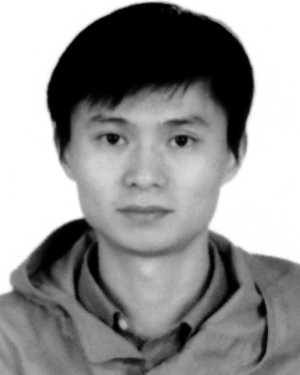 Yangsong Zhang合作论文数School of Computer Science and Technology, Southwest University of Science and Technology8
 Ian Daly合作论文数University of Reading8
 Ehsan Adeli合作论文数Department of Psychiatry and Behavioral Sciences, School of Medicine, Stanford University8
 Xiaobo Chen (陈小波)合作论文数School of Computer Science and Technology, Shandong Technology and Business University;Automotive Engineering Research Institute, School of Automotive and Traffic Engineering, Jiangsu University7
 Andrei A. Vakhtin合作论文数7
 Peng Xu (徐鹏)合作论文数School of Life Science and Technology, University of Electronic Science and Technology of China7
 Joel Kramer合作论文数Memory and Aging Center, Weill Institute for Neurosciences, University of California San Francisco7Maria Luisa Gorno-Tempini合作论文数7
 Li Shen合作论文数Division of Informatics, Department of Biostatistics, Epidemiology and Informatics, Perelman School of Medicine, University of Pennsylvania7
 Susanne G Mueller合作论文数6
 Russell T Toll合作论文数Department of Psychiatry, University of Texas Southwestern Medical Centre at Dallas6Zhou, Sijie合作论文数6
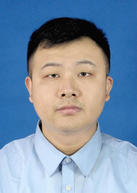 Long Chen (陈龙)合作论文数Medical School of Tianjin University6
 Tao Zhou (周涛)合作论文数School of Computer Science and Engineering, Nanjing University of Science & Technology6
 Marzieh Nezamzadeh合作论文数6
 Baiying Lei (雷柏英)合作论文数School of Biomedical Engineering, Shenzhen University Medical School6




