BACKGROUND:Patients undergoing first-time atrial fibrillation (AF) ablation can benefit from targeting non-pulmonary vein (PV) triggers. Preprocedural identification of high-risk individuals can guide planning of ablation strategy. This study aimed to create a preprocedural screening tool to identify patients at risk of non-PV triggers during first-time AF ablation.METHODS:All patients who underwent first-time AF ablation at the Hospital of the University of Pennsylvania between 2018 and 2022 were identified. Those who underwent non-PV trigger provocative maneuvers or had spontaneous non-PV trigger firing were included. Non-PV triggers were defined as non-PV ectopic beats triggering AF or sustained focal atrial tachycardia that occurred spontaneously, after AF cardioversion, or after standard provocative maneuvers. The provocative maneuvers included incremental isoproterenol infusion (3, 6, 12, and 20-30 mu g/min) and an atrial burst pacing protocol. Risk factors associated with non-PV triggers in a stepwise multivariable logistic regression model with backward elimination were used to create a risk score.RESULTS:A total of 163 (8.0%) of 2038 patients had non-PV triggers during first-time AF ablation. Based on the multivariable model, we created a risk score using female sex (1 point; odds ratio [OR], 1.90 [95% CI, 1.36-2.67]), sinus node dysfunction (1 point; OR, 1.84 [95% CI, 1.04-3.24]), prior cardiac surgery (1 point; OR, 2.26 [95% CI, 1.45-3.53]), moderate to severe left atrial enlargement (2 points; OR, 3.43 [95% CI, 2.46-4.79]), and cardiac sarcoidosis/amyloidosis (4 points; OR, 7.24 [95% CI, 3.03-17.33]). Internal validation using bootstrap resampling showed an optimism-adjusted C statistic of 0.715 (95% CI, 0.678-0.751). Among all first-time AF ablations, 68.1% of procedures were low-risk for non-PV triggers (scores 0-1, 4.3% risk), 17.8% were intermediate-risk (score 2, 10.5% risk), and 14.1% were high-risk (score >= 3, 22.6% risk).CONCLUSIONS:A preprocedural screening tool can classify patients based on their risk of non-PV triggers during first-time AF ablation. This risk score can guide operators to identify patients who would benefit most from adjunctive non-PV trigger testing. However, further validation is needed to confirm these findings.
Access to magnetic resonance imaging (MRI) remains limited for many patients with cardiovascular implantable electronic devices (CIEDs), despite evidence demonstrating safety under appropriate conditions. This call-to-action statement from the Heart Rhythm Society (HRS) aims to describe persistent barriers to MRI access for patients with a CIED and to provide practical, actionable recommendations for the improvement of clinical care. Developed by a multidisciplinary writing committee, this document addresses regulatory, operational, and institutional challenges; highlights findings from a recent HRS member survey on MRI access; and outlines the impact of evolving vendor-specific MRI exclusions. Specific populations discussed include patients with multiple MRI-conditional devices, mixed-vendor systems, abandoned leads, and epicardial leads or subcutaneous arrays. The statement also calls attention to the clinical and administrative burden on electrophysiology teams and the need for fair recognition and reimbursement of MRI-related care. Collaboration across specialties, industry, and regulatory bodies is essential to eliminate non-data-driven barriers and to ensure equitable access to clinically indicated MRI for all patients with a CIED.
BACKGROUND:Idiopathic ventricular arrhythmias (VAs) from the right ventricle (RV) are well-described, but limited data exist regarding those from the RV apex. OBJECTIVE:The purpose of this study was to define the electrocardiographic (ECG) characteristics and ablation outcomes of RV apical VAs, including premature ventricular contractions (PVCs) and ventricular tachycardia. METHODS:Patients undergoing catheter ablation for RV apical VAs at 3 centers were retrospectively analyzed. VAs from the moderator band, papillary muscles, or associated with significant structural heart disease were excluded. Clinical characteristics, ECG features, procedural details, and follow-up data were assessed. RESULTS:12 patients were included (4 (33%) with PVCs, 7 (58%) with monomorphic ventricular tachycardia, and 1 (8%) with PVCs triggering ventricular fibrillation). All VAs exhibited a left bundle branch block pattern with a left superior axis and negative concordance. The mean QRS duration was 167 ± 14 ms. Nine patients (75%) underwent a single ablation procedure, 2 (17%) required 1 repeat procedure, and 1 (8%) required 2. Endocardial ablation was effective in 8 patients (67%), with 4 (33%) requiring endocardial and epicardial ablation. After a median follow-up of 57 months (interquartile range 12-74 months) in 11 patients, 10 (91%) remained free of recurrence (7 by rhythm monitoring and 3 by symptom resolution). 1 patient (9%) required antiarrhythmic therapy for recurrence; all others remained off therapy. One implantable cardioverter-defibrillator was extracted after successful ablation. Of the 2 patients with suspected PVC-induced cardiomyopathy, 1 fully recovered and 1 progressed to heart failure requiring transplantation, despite normal cardiac magnetic resonance imaging and PVC suppression. CONCLUSION:RV apical VAs demonstrate a distinctive ECG morphology that aids localization. Catheter ablation is effective but may require an epicardial approach.
Aims Right phrenic nerve (RPN) injury is a disabling but uncommon complication of atrial fibrillation (AF) radiofrequency ablation. Pace-mapping is widely used to infer RPN's course, for limiting the risk of palsy by avoiding ablation at capture sites. However, information is lacking regarding the distance between the endocardial sites of capture and the actual anatomic RPN location. We aimed at determining the distance between endocardial sites of capture and anatomic CT location of the RPN, depending on the capture threshold. Methods and results In consecutive patients undergoing AF radiofrequency ablation, we defined the course of the RPN on the electroanatomical map with high-output pacing at up to 50 mA/2 ms, and assessed RPN capture threshold (RPN-t). The true anatomic course of the RPN was delineated and segmented using CT scan, then merged with the electroanatomical map. The distance between pacing sites and the RPN was assessed. In 45 patients, 1033 pacing sites were analysed. Distances from pacing sites to RPN ranged from 7.5 +/- 3.0 mm (min 1) when RPN-t was <= 10 mA to 19.2 +/- 6.5 mm (min 9.4) in cases of non-capture at 50 mA. A distance to the phrenic nerve > 10 mm was predicted by RPN-t with a ROC curve area of 0.846 [0.821-0.870] (P < 0.001), with Se = 80.8% and Sp = 77.5% if RPN-t > 20 mA, Se = 68.0% and Sp = 91.6% if RPN-t > 30 mA, and Se = 42.4% and Sp = 97.6% if non-capture at 50 mA. Conclusion These data emphasize the utility of high-output pace-mapping of the RPN. Non-capture at 50 mA/2 ms demonstrated very high specificity for predicting a distance to the RPN > 10 mm, ensuring safe radiofrequency delivery. [GRAPHICS] .
BACKGROUND:Targeting non-pulmonary vein triggers (NPVTs) after pulmonary vein isolation may reduce atrial fibrillation (AF) recurrence. Isoproterenol infusion and cardioversion of spontaneous or induced AF can provoke NPVTs but typically require vasopressor support and increased procedural time. OBJECTIVE:The purpose of this study was to identify risk factors for the presence of NPVTs and create a risk score to identify higher-risk subgroups. METHODS:Using the AF ablation registry at the Hospital of the University of Pennsylvania, we included consecutive patients who underwent AF ablation between January 2021 and December 2022. We excluded patients who did not receive NPVT provocation testing after failing to demonstrate spontaneous NPVTs. NPVTs were defined as non-pulmonary vein ectopic beats triggering AF or focal atrial tachycardia. We used risk factors associated with NPVTs with P <.1 in multivariable logistic regression model to create a risk score in a randomly split derivation set (80%) and tested its predictive accuracy in the validation set (20%). RESULTS:In 1530 AF ablations included, NPVTs were observed in 235 (15.4%). In the derivation set, female sex (odds ratio [OR] 1.40; 95% confidence interval [CI] 0.96-2.03; P = .080), sinus node dysfunction (OR 1.67; 95% CI 0.98-2.87; P = .060), previous AF ablation (OR 2.50; 95% CI 1.70-3.65; P <.001), and left atrial scar (OR 2.90; 95% CI 1.94-4.36; P <.001) were risk factors associated with NPVTs. The risk score created from these risk factors (PRE2SSS2 score; [PRE]vious ablation: 2 points, female [S]ex: 1 point, [S]inus node dysfunction: 1 point, left atrial [S]car: 2 points) had good predictive accuracy in the validation cohort (area under the receiver operating characteristic curve 0.728; 95% CI 0.648-0.807). CONCLUSION:A risk score incorporating predictors for NPVTs may allow provocation of triggers to be performed in patients with greatest expected yield.
BACKGROUND:In arrhythmogenic right ventricular cardiomyopathy (ARVC), risk of atrial arrhythmias (AAs) persists after ventricular tachycardia (VT) ablation. OBJECTIVE:The purpose of this study was to determine the type, prevalence, outcome, and risk correlates of AA in ARVC in patients undergoing VT ablation. METHODS:Prospectively collected procedural and clinical data on ARVC patients undergoing VT ablation were analyzed. Risk score for typical atrial flutter was determined from univariate logistic regression analysis. RESULTS:Of 119 consecutive patients with ARVC and VT ablation, 40 (34%) had AA: atrial fibrillation (AF) in 31, typical isthmus-dependent atrial flutter (AFL) in 27, and atrial tachycardia/atypical flutter (AT) in 10. Seventeen patients (43%) with AA experienced inappropriate defibrillator therapy, with 15 patients experiencing shocks. Ablation was performed for typical AFL in 21 (53%), AT in 5 (13%), and pulmonary vein isolation for AF in 4 (10%) patients and prevented AA in 78% and all AFL during additional mean follow-up of 65 months. Risk score for typical flutter included age >40 years (1 point), ≥moderate right ventricular dysfunction (2 points), ≥moderate tricuspid regurgitation (2 points), ≥moderate right atrial dilation (2 points), and right ventricular volume >250 cc (3points), with score >4 identifying 50% prevalence of typical flutter. CONCLUSION:AAs are common in patients with ARVC and VT, can result in inappropriate implantable cardioverter-defibrillator shocks, and typically are controlled with atrial ablation. A risk score can be used to identify patients at high risk for typical AFL who may be considered for isthmus ablation at the time of VT ablation.
Background: The majority of non-pulmonary vein (PV) triggers of atrial fibrillation (AF) require provocative maneuvers for induction during catheter ablation. Identifying a subset of patients with higher likelihood of having non-PV triggers can guide more efficient use of these provocative maneuvers. Objective: This study aims to identify pre-procedural risk factors for the presence of non-PV triggers at first-time AF ablation. Methods: We included patients who underwent first-time AF ablation between 2019 and 2022 at the Hospital of the University of Pennsylvania. Non-PV trigger provocation maneuvers included cardioversion of AF if present at baseline, incremental isoproterenol (ISO) infusion (3-30mcg/min), and atrial burst pacing to induce focal atrial tachycardia (AT) or induce AF followed by cardioversion on 3-6mcg/min ISO or during high dose ISO washout. Non-PV triggers were defined as non-PV ectopic beats initiating AF or sustained focal AT. We used logistic regression analysis to identify pre-procedural risk factors associated with the presence of non-PV triggers. Risk factors with univariable p-value <0.2 were entered in a multivariable model. Results: Of 1805 patients with first-time AF ablation, 1701 (94.2%) patients that received non-PV trigger provocative maneuvers were included. A total of 146 (8.6%) patients were found to have non-PV triggers. The prevalence of non-PV triggers was numerically higher in patients with persistent AF than those with paroxysmal AF (9.7% vs 7.7%, p-value=0.163). In the multivariable model, female sex (OR=2.18, 95% CI: 1.49-3.17), cardiac sarcoidosis/amyloidosis (OR=7.29, 95% CI: 2.90-18.31), prior cardiac surgery (OR=2.30, 95% CI: 1.37-3.85), and moderate to severe left atrial enlargement on echocardiography (OR=2.66, 95% CI: 1.83-3.85) were independent predictors of non-PV triggers during first-time AF ablation. Conclusion: Non-PV triggers are present in approximately 10% of patients undergoing first-time AF ablation. A set of pre-procedural risk factors can risk stratify patients at high risk for non-PV triggers and guide choice of optimal ablation strategy at first procedure.
Background The importance of nonpulmonary vein (PV) triggers for the initiation/recurrence of atrial fibrillation (AF) is well established. Objectives This study sought to assess the incremental benefit of provocative maneuvers for identifying non-PV triggers. Methods We included consecutive patients undergoing first-time AF ablation between 2020 and 2022. The provocation protocol included step 1, identification of spontaneous non-PV triggers after cardioversion of AF and/or during sinus rhythm; step 2, isoproterenol infusion (3, 6, 12, and 20-30 μg/min); and step 3, atrial burst pacing to induce AF followed by cardioversion during residual or low-dose isoproterenol infusion or induce focal atrial tachycardia. Non-PV triggers were defined as non-PV ectopic beats triggering AF or sustained focal atrial tachycardia. Results Of 1,372 patients included, 883 (64.4%) underwent the complete stepwise provocation protocol with isoproterenol infusion and burst pacing, 334 (24.3%) isoproterenol infusion only, 77 (5.6%) burst pacing only, and 78 (5.7%) no provocative maneuvers (only step 1). Overall, 161 non-PV triggers were found in 135 (9.8%) patients. Of these, 51 (31.7%) non-PV triggers occurred spontaneously, and the remaining 110 (68.3%) required provocative maneuvers for induction. Among those receiving the complete stepwise provocation protocol, there was a 2.2-fold increase in the number of patients with non-PV triggers after isoproterenol infusion, and the addition of burst pacing after isoproterenol infusion led to a total increase of 3.6-fold with the complete stepwise provocation protocol. Conclusions The majority of non-PV triggers require provocative maneuvers for induction. A stepwise provocation protocol consisting of isoproterenol infusion followed by burst pacing identifies a 3.6-fold higher number of patients with non-PV triggers.
BACKGROUND:Radiofrequency ablation (RFA) of cavotricuspid isthmus (CTI)-dependent atrial flutter requires ablation of the tricuspid annulus overlying the right coronary artery (RCA). Although it is considered safe, reports of acute and subacute RCA injury in human and animal studies raise the possibility of late RCA stenosis. OBJECTIVE:The objective of this study was to compare the incidence and severity of angiographic RCA stenoses in patients who have undergone CTI RFA with a control group to assess the long-term risk of RCA damage. METHODS:A 2-center retrospective case-cohort study was performed including all patients from 2002 to 2018 undergoing atrial fibrillation (AF) with CTI ablation (CTI + AF) or AF ablation alone with subsequent coronary angiography (CAG). The AF alone group served as controls because of anticipated similarity of baseline characteristics. Coronary arteries that are anatomically remote to the CTI were examined as prespecified falsification end points. CAG was scored by a blinded observer. RESULTS:There were 156 patients who underwent pulmonary vein isolation with subsequent CAG (CTI + AF, n = 81; AF alone, n = 75) with no difference in baseline characteristics including age, sex, comorbidities, and medications. Mean time from ablation to CAG was similar (CTI + AF, 5.0 ± 3.7 years; AF alone, 5.4 ± 3.9 years; P = .5). The mid and distal RCA showed no difference in the average number of angiographic stenoses or lesion severity. In regression analysis, CTI ablation was not a predictor of RCA stenosis severity (P = .6). There was no difference in coronary disease at sites remote to the CTI ablation (P = NS for all). CONCLUSION:There was no observed relationship between CTI RFA and the number or severity of angiographically apparent RCA stenoses in long-term follow-up.
BACKGROUND:Although the epicardial predominance of substrate abnormalities has been well demonstrated in early stages of arrhythmogenic right ventricular cardiomyopathy (ARVC), endocardial (ENDO) ablation may suffice to eliminate ventricular tachycardia (VT) in some patients. OBJECTIVES:This study aimed to report the long-term outcomes of ENDO-only ablation in ARVC patients and factors that predict VT-free survival. METHODS:We included consecutive patients with Task Force Criteria diagnosis of ARVC undergoing a first ENDO-only VT ablation between 1998 and 2020. Ablation was predominantly guided by activation/entrainment mapping for mappable VTs and pace mapping/targeting abnormal electrograms for unmappable VTs. The primary endpoint was freedom from any recurrent sustained VT after the last ENDO-only ablation. RESULTS:Seventy-four ARVC patients underwent ENDO-only VT ablation. VT noninducibility was achieved in 49 (66%) patients. During median follow-up of 6.6 years (Q1-Q3: 3.4-11.2 years), 40 (54.1%) patients remained free from any VT recurrence with rare VT ≤2 episodes in additional 12.2%. Among patients with noninducibility, VT-free survival was 75.5% during long-term follow-up. In multivariable analysis, >45 y of age at diagnosis (HR: 0.41; 95% CI: 0.17-0.98) and VT noninducibility (HR: 0.36; 95% CI: 0.16-0.80) were predictors of VT-free survival. CONCLUSIONS:Long-term VT-free survival can be achieved in over half of ARVC patients following ENDO-only VT ablation, increasing to over 75% if VT noninducibility is achieved. Our results support consideration of a stepwise ENDO-only approach before proceeding to epicardial ablation if VT noninducibility can be achieved particularly in older patients.
Introduction: Left atrial (LA) late gadolinium enhancement (LGE) on cardiac magnetic resonance (CMR) predominates near the aorta(AO); however, LGE does not equate fibrosis and may be artifactual or indicate expanded interstituim. Hypothesis: We sought to examine the association of LA LGE with proximity to the descending and ascending AO and use regional impedance and voltage measurements to dissect the mechanism of LGE . Methods: The retrospective cohort included consecutive patients who underwent pre-procedural CMR and atrial fibrillation (AF) ablation between January 2016 - 2021. The association of voltage amplitude, impedance, and image intensity ratio (IIR) at each electroanatomic map point with distance from the nearest AO point was examined after adjustment for age, sex, AF type, AO stenosis, LA volume, and AO diameter. Results: Included 63 patients (age 65.5±8.8 years, 33% female). Among 42 ablation naive patients, distance from AO was unassociated with bipolar and unipolar voltage amplitudes, but associated with impedance (+0.04 ohm/mm, P=0.011) and IIR (-0.03 /mm, P<0.001). Among 21 patients with prior ablation, distance from AO was unassociated with IIR, but associated with bipolar (+0.01 mV/mm, P<0.001) and unipolar (+0.01 mV/mm, P=0.001) voltage amplitudes and impedance (+0.03 ohm/mm, P=0.025). Conclusions: Given lower impedance but normal voltage, de novo peri-AO LA-LGE likely signifies expanded interstitium, rather than fibrosis or fat infiltration. Following ablation, peri-AO voltage and impedance are both decreased compared to other LA regions, suggesting that peri-AO lesion delivery is more effective, likely due to lower impedance at baseline.
INTRODUCTION:Identifying the origin of nonpulmonary vein atrial fibrillation (AF) triggers (NPVTs) after pulmonary vein isolation (PVI) can be challenging. We aimed to determine if noninvasive electrocardiographic imaging (ECGi) could localize pacing from common NPVT sites. ECGi combines measured body surface potentials with heart-torso geometry acquired from computed tomography (CT) to generate an activation map. METHODS:In 12 patients with AF undergoing first time ablation, the ECGi vest was fitted for preprocedural CT scan and worn during the procedure. After PVI, we performed steady-state pacing from 15 typical anatomic NPVT sites at a cycle length of 700-800 ms. We co-registered the invasive anatomic map with the CT-based ECGi epicardial activation map to compare ECGi predicted to true pacing origin. RESULTS:In the study cohort (67% male, 58% persistent AF, and 67% with left atrial dilation), 148 (82%) pacing sites had both capture and adequate anatomy acquired from the three-dimensional mapping system to co-register with ECGi activation map. Median distance between true pacing sites and point of earliest epicardial activation derived from the ECGi maps for all sites was 17 mm (interquartile range, 10-22 mm). Assuming paced sites treated as regions with a radius of 2.5 cm, the earliest activation site on ECGi map falls within the region with 94% accuracy. CONCLUSION:ECGi can approximate the origin of paced beats from common NPVT sites to within a median distance of 17 mm. A rapidly identified region may then be the focus of more detailed catheter-based mapping techniques to facilitate successful localization and ablation of NPVTs.
 Francis Edward Marchlinski合作论文数Department of Medicine, Perelman School of Medicine, University of Pennsylvania230
 David J. Callans合作论文数Cardiology Division, Hospital of the University of Pennsylvania157
 Sanjay Dixit合作论文数Department of Medicine, Perelman School of Medicine, University of Pennsylvania128
 David Lin合作论文数Department of Medicine, Perelman School of Medicine, University of Pennsylvania117
 Fermin C. Garcia合作论文数Department of Medicine, Perelman School of Medicine, University of Pennsylvania114
 David S. Frankel合作论文数Department of Medicine, Perelman School of Medicine, University of Pennsylvania105
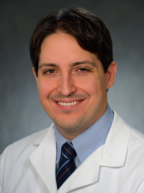 Pasquale Santangeli合作论文数Department of Medicine, Perelman School of Medicine, University of Pennsylvania95
 Gregory E. Supple合作论文数Department of Medicine, Perelman School of Medicine, University of Pennsylvania84
 Michael P. Riley合作论文数Department of Medicine, Perelman School of Medicine, University of Pennsylvania79
 Robert D. Schaller合作论文数Department of Medicine, Perelman School of Medicine, University of Pennsylvania64
 Edward P. Gerstenfeld合作论文数School of Medicine, University of California, San Francisco56
 Mathew D Hutchinson合作论文数52
 Saman Nazarian合作论文数Department of Medicine, Perelman School of Medicine, University of Pennsylvania50
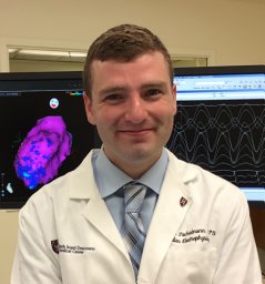 Cory M. Tschabrunn合作论文数Hospital of the University of Pennsylvania, University of Pennsylvania Perelman School of Medicine50
 Joshua M. Cooper合作论文数University of Pennsylvania46
 Jackson J Liang合作论文数41
 Matthew Hyman合作论文数40
 Muser Daniele合作论文数1 Cardiovascular Division, University of Udine;Postgraduate school of Cardiology, University of Triest;2 Cardiovascular Division, Hospital of the University of Pennsylvania;University of Triest, Hospital of the University of Pennsylvania34
 Rupa Bala合作论文数34
 Rajat Deo合作论文数Department of Medicine, Perelman School of Medicine, University of Pennsylvania33
 Jeffrey S. Arkles合作论文数Department of Medicine, Perelman School of Medicine, University of Pennsylvania32
 Ralph J. Verdino合作论文数Perelman School of Medicine, University of Pennsylvania29
 Ramanan Kumareswaran合作论文数Department of Medicine, Perelman School of Medicine, University of Pennsylvania27
 Simon A. Castro合作论文数Cardiovasc Div, Hosp Univ Penn21
 Gustavo S. Guandalini合作论文数Department of Medicine, Perelman School of Medicine, University of Pennsylvania21
 Andres Enriquez合作论文数Department of Cardiology and Cardiovascular Diseases, Pontificia Universidad Católica de Chile20
 Andrew E. Epstein合作论文数Department of Medicine, Perelman School of Medicine, University of Pennsylvania19
 Benoit Desjardins合作论文数Department of Radiology, Perelman School of Medicine, University of Pennsylvania17
 Andrea Russo合作论文数Cooper Medical School, Rowan University;Cooper University Hospital17
 Magnani Silvia合作论文数16
 Tatsuya Hayashi合作论文数15
 Alireza Oraii合作论文数Cardiovascular Diseases Research Institute, Tehran University of Medical Sciences14Rajeev Pathak合作论文数Medical School, College of Health and Medicine, The Australian National University14Arceluz Martín合作论文数Robotic Cardiac Electrophysiology and Arrhythmia Unit, Hospital Universitario La Paz12Maciej Kubala合作论文数Cardiology Department, University Hospital12Wendy S. Tzou合作论文数12Corentin Chaumont合作论文数Dept Cardiol, Rouen Univ Hosp11Betensky Brian P合作论文数11Ioan Liuba合作论文数11Lohit Garg合作论文数10
 Haqqani Haris M合作论文数Cardiovascular Division, Hospital of the University of Pennsylvania10Markman Timothy M合作论文数10David J Callans合作论文数10
 Munveer Thind合作论文数Div Cardiol, Lankenau Heart Inst9William Chik合作论文数School of Medicine, Faculty of Medicine and Health, The University of Sydney9
 Maurie Markman合作论文数Department of Medicine, City of Hope Cancer Centers;Cancer Treatment Centers of America9
 Yasuhiro Shirai合作论文数Advanced Electronic Materials Center, National Institute for Materials Science9Jian-Fang Ren合作论文数9
 William Henry Sauer合作论文数Department of Medicine, Harvard University;Brigham and Women's Hospital9




