BACKGROUND:Patients undergoing first-time atrial fibrillation (AF) ablation can benefit from targeting non-pulmonary vein (PV) triggers. Preprocedural identification of high-risk individuals can guide planning of ablation strategy. This study aimed to create a preprocedural screening tool to identify patients at risk of non-PV triggers during first-time AF ablation.METHODS:All patients who underwent first-time AF ablation at the Hospital of the University of Pennsylvania between 2018 and 2022 were identified. Those who underwent non-PV trigger provocative maneuvers or had spontaneous non-PV trigger firing were included. Non-PV triggers were defined as non-PV ectopic beats triggering AF or sustained focal atrial tachycardia that occurred spontaneously, after AF cardioversion, or after standard provocative maneuvers. The provocative maneuvers included incremental isoproterenol infusion (3, 6, 12, and 20-30 mu g/min) and an atrial burst pacing protocol. Risk factors associated with non-PV triggers in a stepwise multivariable logistic regression model with backward elimination were used to create a risk score.RESULTS:A total of 163 (8.0%) of 2038 patients had non-PV triggers during first-time AF ablation. Based on the multivariable model, we created a risk score using female sex (1 point; odds ratio [OR], 1.90 [95% CI, 1.36-2.67]), sinus node dysfunction (1 point; OR, 1.84 [95% CI, 1.04-3.24]), prior cardiac surgery (1 point; OR, 2.26 [95% CI, 1.45-3.53]), moderate to severe left atrial enlargement (2 points; OR, 3.43 [95% CI, 2.46-4.79]), and cardiac sarcoidosis/amyloidosis (4 points; OR, 7.24 [95% CI, 3.03-17.33]). Internal validation using bootstrap resampling showed an optimism-adjusted C statistic of 0.715 (95% CI, 0.678-0.751). Among all first-time AF ablations, 68.1% of procedures were low-risk for non-PV triggers (scores 0-1, 4.3% risk), 17.8% were intermediate-risk (score 2, 10.5% risk), and 14.1% were high-risk (score >= 3, 22.6% risk).CONCLUSIONS:A preprocedural screening tool can classify patients based on their risk of non-PV triggers during first-time AF ablation. This risk score can guide operators to identify patients who would benefit most from adjunctive non-PV trigger testing. However, further validation is needed to confirm these findings.
Left ventricular (LV) diastolic dysfunction is associated with the development of atrial fibrillation (AF) and risk of recurrence after ablation. The use of an intracardiac echocardiography (ICE) for diastolic function assessment during ablation procedures has not been evaluated. To evaluate the feasibility and utility of ICE obtained measures of LV diastolic function including peak tricuspid regurgitation velocity, trans-mitral flow velocity, mitral annular tissue Doppler velocities, and pulmonary vein flow velocities in patients undergoing AF ablation. We conducted a single-center, prospective evaluation of patients undergoing AF ablation between 2022 and 2024. During sinus rhythm, diastolic parameters were measured with the ICE catheter and direct left atrial pressure (LAP) was recorded prior to AF ablation. Elevated LAP was defined as ≥ 12 mmHg. ICE measured diastolic parameters were compared with those measured on transthoracic echocardiography (TTE). A total of 152 patients (53
Background Left bundle branch area pacing (LBBAP) offers a physiological alternative to traditional pacing and has shown benefit across cardiomyopathy phenotypes. In cardiac sarcoidosis (CS), in which septal inflammation and scarring are common, the performance and clinical impact of LBBAP remain unclear. Objectives The aim of this study was to evaluate procedural success, lead performance, and outcomes of LBBAP in patients with CS. Methods This study retrospectively identified patients with CS who underwent LBBAP from January 2020 to March 2024 at a single tertiary center. CS was defined per Heart Rhythm Society criteria using histologic or imaging-based evidence. Procedural metrics, device parameters, and echocardiographic data were assessed at baseline and follow-up. LBBAP was confirmed by using European Heart Rhythm Association criteria. Results Nineteen patients met inclusion criteria (mean age 59 ± 12 years; 21% female). Atrioventricular block was the most common indication (58%). Septal involvement was seen in 53% (cardiac magnetic resonance imaging) and 61% (positron emission tomography). LBBAP was successful in all patients without acute complications. Baseline paced QRS duration was 139 ± 25 milliseconds; mean threshold was 0.75 ± 0.26 V, R-wave amplitude 15.2 ± 6.3 mV, and impedance 738 ± 208 Ω. Over a 437-day median follow-up, lead parameters remained stable. Among patients with LBBB, QRS >130 milliseconds, and left ventricular ejection fraction (LVEF) <50% (n = 4), LVEF improved from 27% ± 17% to 50% ± 12% (P = 0.06). No differences in pacing parameters were seen based on septal involvement. Conclusions LBBAP in CS is feasible and provided stable long-term lead performance in this cohort. Larger studies are needed to assess its clinical benefit in the presence of septal inflammation and fibrosis.
Background Catheter ablation of mitral annular flutter is challenging, often requiring ablation in the coronary sinus or ethanol infusion into the vein of Marshall (VOM). Intracardiac echocardiography (ICE) from the left atrium provides unobstructed views of the mitral isthmus anatomy. Objective This study investigated the outcomes and safety of mitral isthmus ablation using a stepwise approach guided by left atrial ICE. Methods After transseptal access was achieved, the ICE catheter was advanced into the left atrium. Linear endocardial ablation was performed between the left inferior pulmonary vein and the mitral annulus, followed by coronary sinus ablation and VOM ethanol infusion if there was failure to achieve block. ICE was used to select the ablation plane, to monitor catheter contact, to titrate energy, and to assess lesion formation. Results Thirty patients (age, 70 ± 9 years; 73% male) were included. The mean length of the mitral isthmus was 32.5 ± 5.4 mm, with a depth of 6.0 ± 2.9 mm and a mean thickness of 4.5 ± 0.5 mm. Bidirectional mitral isthmus block was achieved in 29 of 30 cases (97%), with epicardial ablation through the great cardiac vein required in 57% (n = 17 patients) and VOM ethanol infusion in 43% (n = 13 patients). Complications included 1 case of pericardial effusion (3%). During a follow-up of 19 ± 9 months, 13 patients experienced recurrent atrial arrhythmias, with a 12-month survival free of arrhythmias of 60%. Conclusion An anatomic approach to mitral isthmus ablation guided by left atrial ICE is feasible and safe, with high acute procedural success.
BACKGROUND:Targeting non-pulmonary vein triggers (NPVTs) after pulmonary vein isolation may reduce atrial fibrillation (AF) recurrence. Isoproterenol infusion and cardioversion of spontaneous or induced AF can provoke NPVTs but typically require vasopressor support and increased procedural time. OBJECTIVE:The purpose of this study was to identify risk factors for the presence of NPVTs and create a risk score to identify higher-risk subgroups. METHODS:Using the AF ablation registry at the Hospital of the University of Pennsylvania, we included consecutive patients who underwent AF ablation between January 2021 and December 2022. We excluded patients who did not receive NPVT provocation testing after failing to demonstrate spontaneous NPVTs. NPVTs were defined as non-pulmonary vein ectopic beats triggering AF or focal atrial tachycardia. We used risk factors associated with NPVTs with P <.1 in multivariable logistic regression model to create a risk score in a randomly split derivation set (80%) and tested its predictive accuracy in the validation set (20%). RESULTS:In 1530 AF ablations included, NPVTs were observed in 235 (15.4%). In the derivation set, female sex (odds ratio [OR] 1.40; 95% confidence interval [CI] 0.96-2.03; P = .080), sinus node dysfunction (OR 1.67; 95% CI 0.98-2.87; P = .060), previous AF ablation (OR 2.50; 95% CI 1.70-3.65; P <.001), and left atrial scar (OR 2.90; 95% CI 1.94-4.36; P <.001) were risk factors associated with NPVTs. The risk score created from these risk factors (PRE2SSS2 score; [PRE]vious ablation: 2 points, female [S]ex: 1 point, [S]inus node dysfunction: 1 point, left atrial [S]car: 2 points) had good predictive accuracy in the validation cohort (area under the receiver operating characteristic curve 0.728; 95% CI 0.648-0.807). CONCLUSION:A risk score incorporating predictors for NPVTs may allow provocation of triggers to be performed in patients with greatest expected yield.
Background Premature ventricular complexes (PVCs) are common and associated with worse outcomes in patients with heart failure. Class 1C antiarrhythmic drugs (AADs) effectively suppress PVCs, but guidelines currently restrict their use in structural heart disease. Objectives This study aimed to assess the safety and efficacy of class 1C AADs in patients with nonischemic cardiomyopathy (NICM) and implantable cardioverter-defibrillators (ICDs). Methods All patients with NICM and an ICD treated with flecainide or propafenone at the Hospital of the University of Pennsylvania between 2014 and 2022 were identified. PVC burden, left ventricular ejection fraction (LVEF), and biventricular pacing percentage were compared before and during class 1C AAD treatment. Safety outcomes included sustained atrial and ventricular arrhythmias, heart failure admissions, and death. Results We identified 34 patients, 23 receiving flecainide and 11 propafenone. Most patients (62%) had failed other AADs or catheter ablation (68%) prior to class 1C AAD initiation. PVC burden decreased from 20 ± 13% to 6 ± 7% (P < 0.001), LVEF increased from 33 ± 9% to 37 ± 10% (P = 0.01), and biventricular pacing percentage increased from 85 ± 9% to 93 ± 7% (P = 0.01). Sustained ventricular tachycardia (2 vs 9 patients) and admissions for decompensated heart failure (2 vs 3 patients) decreased compared with the 12 months prior to class 1C AAD initiation. Conclusions Class 1C AADs effectively suppressed PVCs in patients with NICM and ICDs, leading to increases in LVEF and biventricular pacing percentage. In this limited sample, their use was safe. Larger studies are needed to confirm the safety of this approach.
 Saman Nazarian合作论文数Department of Medicine, Perelman School of Medicine, University of Pennsylvania59
 David S. Frankel合作论文数Department of Medicine, Perelman School of Medicine, University of Pennsylvania58
 Matthew Hyman合作论文数58
 Francis Edward Marchlinski合作论文数Department of Medicine, Perelman School of Medicine, University of Pennsylvania57
 David J. Callans合作论文数Cardiology Division, Hospital of the University of Pennsylvania55
 Robert D. Schaller合作论文数Department of Medicine, Perelman School of Medicine, University of Pennsylvania54
 Gregory E. Supple合作论文数Department of Medicine, Perelman School of Medicine, University of Pennsylvania52
 Sanjay Dixit合作论文数Department of Medicine, Perelman School of Medicine, University of Pennsylvania47
 David Lin合作论文数Department of Medicine, Perelman School of Medicine, University of Pennsylvania45
 Michael P. Riley合作论文数Department of Medicine, Perelman School of Medicine, University of Pennsylvania36
 Fermin C. Garcia合作论文数Department of Medicine, Perelman School of Medicine, University of Pennsylvania36
 Rajat Deo合作论文数Department of Medicine, Perelman School of Medicine, University of Pennsylvania35
 Pasquale Santangeli合作论文数Department of Medicine, Perelman School of Medicine, University of Pennsylvania32
 Andrew E. Epstein合作论文数Department of Medicine, Perelman School of Medicine, University of Pennsylvania32
 Ramanan Kumareswaran合作论文数Department of Medicine, Perelman School of Medicine, University of Pennsylvania30
 Jeffrey S. Arkles合作论文数Department of Medicine, Perelman School of Medicine, University of Pennsylvania21
 Zado Erica S合作论文数Cardiovascular Division, Hospital of the University of Pennsylvania21
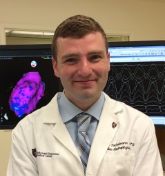 Cory M. Tschabrunn合作论文数Hospital of the University of Pennsylvania, University of Pennsylvania Perelman School of Medicine16Markman Timothy M合作论文数14
 Alireza Oraii合作论文数Cardiovascular Diseases Research Institute, Tehran University of Medical Sciences13Corentin Chaumont合作论文数Dept Cardiol, Rouen Univ Hosp11Arceluz Martín合作论文数Robotic Cardiac Electrophysiology and Arrhythmia Unit, Hospital Universitario La Paz11Poojita Shivamurthy合作论文数Helmsley Electrophysiology Center, Icahn School of Medicine at Mount Sinai10
 Balaram Krishna J Hanumanthu合作论文数Dept Cardiol, Mt Sinai Beth Israel10
 Andres Enriquez合作论文数Department of Cardiology and Cardiovascular Diseases, Pontificia Universidad Católica de Chile10
 Munveer Thind合作论文数Div Cardiol, Lankenau Heart Inst9Katie Walsh合作论文数Consultants Private Clinic, Cork University Hospital9
 Maurie Markman合作论文数Department of Medicine, City of Hope Cancer Centers;Cancer Treatment Centers of America9Weeranun D Bode合作论文数8
 Maiwand Mirwais合作论文数University of Pennsylvania7Lohit Garg合作论文数7Haran Yogasundaram合作论文数6Masahiro Sekigawa合作论文数6Aung Lin合作论文数5Naga Venkata Pothineni合作论文数University of Pennsylvania;Divison of Cardiovascular Medicine, University of Arkansas for Medical Sciences5Irene Lucena-Padros合作论文数4
 Neel Patel合作论文数Dept Internal Med, New York Med Coll4
 Alan M. Sugrue合作论文数Department of Cardiovascular Medicine, Mayo Clinic4Dhakal Bishnu P合作论文数4Maciej Kubala合作论文数Cardiology Department, University Hospital4Ahmed B. Alsalem合作论文数3Jian Liang Tan合作论文数3Maxime Cerantola合作论文数3
 Tarek Zghaib合作论文数University of Pennsylvania Perelman School of Medicine3
 Michael G. Fradley合作论文数Department of Medicine, Perelman School of Medicine, University of Pennsylvania3
 Muser Daniele合作论文数1 Cardiovascular Division, University of Udine;Postgraduate school of Cardiology, University of Triest;2 Cardiovascular Division, Hospital of the University of Pennsylvania;University of Triest, Hospital of the University of Pennsylvania3Qing Lou合作论文数2Christoffel Van Niekerk合作论文数2Jennifer Chee合作论文数2




