Background Ventricular arrhythmias (VAs) near the His-bundle comprise 9% of unexplained VAs and present challenges for ablation caused by the risk of atrioventricular block. Objectives The authors studied the electrocardiographic (ECG) and electrophysiological characteristics of Para-Hisian (PH) VAs, comparing them with septal right ventricular outflow tract VAs. Methods From 210 patients with VAs between 2018 and 2024, 31 (14.7%) with PH-VAs and 23 (10.9%) with septal right ventricular outflow tract VAs were included. ECG characteristics of both were compared, and features differentiating left and right PH and supra- and infra-Hisian VAs were identified. Results Of 31 patients, 15 had VAs from the right PH site and 16 from the left. Median follow-up was 15 months (Q1-Q3: 14-21 months) for left infra-Hisian, 16 months (Q1-Q3: 14-20 months) for left supra-Hisian, and 14 months (Q1-Q3: 14-16 months) for right infra-Hisian and right supra-Hisian VAs (Q1-Q3: 14-15 months). PH-VAs had narrower QRS complexes (134 ± 19.6 ms vs 169 ± 24 ms; P < 0.05), R-wave in lead aVL (100% [31 of 31] vs 4.3% [1 of 23]; P < 0.001), and earlier R-wave transition at or before lead V3 (80.6% [25 of 31] vs 47.8% [11 of 23]; P < 0.05). Left PH-VAs had earlier R-wave transition at lead V2 (50% [8 of 16] vs 20% [3 of 15]; P = 0.036). Right PH VAs had deeper S-wave relative to the preceding sinus beat in lead V1 (73.3% [11 of 15] vs 37.5% [6 of 16]; P = 0.04) and lead aVR (80% [12 of 15] vs 56.3% [9 of 16]; P = 0.01). Postprocedure heart block occurred in 1 patient. Conclusions PH-VAs exhibit unique ECG features based on their origins, and can be effectively treated without affecting atrioventricular conduction.
BACKGROUND:Identifying critical isthmuses in macro-re-entrant atrial tachycardia (MRAT) is challenging. Omnipolar Technology Near Field mapping with the Peak Frequency (PF) tool holds promise for identifying critical regions sustaining re-entry, though PF signal characteristics and the algorithm's clinical value need further exploration. OBJECTIVES:This study sought to analyze properties of PF in MRAT critical isthmuses and evaluate the value of Omnipolar Technology Near Field PF mapping in guiding ablation. METHODS:Consecutive patients undergoing MRAT ablation procedures with fully mapped circuits (using EnSite-X and HD-Grid) and termination during ablation were included. PF values were analyzed globally across the atrium and compared to the ablation termination site (within 5-mm diameter). The absolute maximum PF at and the number of ablation lesions required for termination were recorded. RESULTS:The study involved 21 MRATs (12 left, 9 right atrial) from 17 patients (age 63.9 ± 18.3 years). Termination typically required a median of 1 (Q1-Q3: 1-7) ablation lesion, with 48% terminating after the initial lesion. Maximum PF values at termination sites were significantly higher (615.2 ± 85.7 Hz) than global PF values (317.6 ± 45.4 Hz; P < 0.01) and PF within the 5-mm termination diameter (499.6 ± 115.4 Hz; P < 0.01). Voltage at termination sites (0.39 ± 0.56) was significantly lower than the global average (0.73 ± 0.57; P < 0.01). PF values above 363 Hz differentiated isthmus regions from global atrial PF, with values >530 Hz indicating maximum PF at termination sites. CONCLUSIONS:Peak frequencies at MRAT termination sites are substantially higher than global atrial PFs, supporting the potential of PF mapping as an effective tool for identifying critical isthmuses in MRAT ablation procedures.
Transvenous lead extraction (TLE) has evolved from a high-risk, last-resort procedure to a standardized therapy with expanding indications and improved safety. Although modern electrophysiology (EP) laboratories now rival hybrid operating rooms in capability, many institutions continue to perform TLE exclusively in operating rooms owing to outdated perceptions of risk, logistical barriers, and limited institutional support. This contributes to delays—particularly in infection-related cases—despite clear evidence that early extraction improves outcomes. Contemporary data confirm low rates of surgical complications, especially with advancements such as the vascular occlusion balloon, tandem technique, and ultrasound imaging. With appropriate safeguards, including surgical backup and emergency protocols, EP laboratories can safely accommodate TLE. Broader adoption of this model may enhance efficiency, expedite care, and better align TLE with other routine cardiovascular therapies. A deliberate, phased transition—supported by multidisciplinary collaboration and institutional readiness—is essential to modernizing TLE delivery in a familiar location.
BACKGROUND:Surgical implantation of epicardial implantable cardioverter-defibrillator (ICD) is considered when transvenous ICD placement is not feasible because of congenital anomalies, venous obstruction, or prior infections. Epicardial pacemakers are well-studied but less is known about contemporary epicardial ICD systems in adults. OBJECTIVES:This case series details the indications, techniques, safety, and long-term performance of surgically implanted epicardial high voltage (HV) coils and leads in adults. METHODS:We included all patients who received epicardial HV coils or leads at the Hospital of the University of Pennsylvania between 2014 and 2025. RESULTS:Nineteen patients (14 men, median age 64 years) were identified. Indications included ICD replacement after lead extraction for endocarditis (7), placement during concomitant surgery, including for tricuspid regurgitation (6), left ventricular assist device (3), and lack of venous access (3). HV coils were used in 58%, and 42% received epicardial leads. All patients received epicardial pace-sense leads; 68% received cardiac resynchronization therapy. HV coils or leads were sewn to the epicardium: 48% on the posterior left ventricle, 16% on the anterior right ventricle, 26% on both ventricles, and 11% in the transverse sinus. Two patients had impedances > 200 Ω because of a loose set screw and a damaged connector pin, respectively. Two had impedances < 20 Ω without clinical impact. One coil fractured 1.5 years post-implant. No failed shocks or lead migrations occurred. During a median follow-up of 468 days, 26% died, largely from advanced heart failure, none were attributable to surgery. CONCLUSION:Surgical implantation of epicardial ICD systems with HV coils/leads is safe and feasible. Multidisciplinary planning and long-term monitoring are essential.
BACKGROUND The best approach for ablating ventricular tachycardia (VT) targeting right ventricular (RV) free wall aneurysms in arrhythmogenic right ventricular cardiomyopathy (ARVC) remains undefined. OBJECTIVE We aimed to describe the technical approach, safety, and long-term efficacy of endocardial ablation of VT originating from RV free wall aneurysms in ARVC patients. METHODS We identified ARVC patients with VT mapped to intracardiac echocardiography (ICE)-defined RV free wall aneurysms who underwent endocardial ablation targeting the aneurysmal area. RV free wall aneurysm on ICE was defined as an akinetic or dyskinetic area with diastolic bulging. The primary ablation end point was VT control, defined as freedom from any or multiple (>1) VT recurrences. RESULTS From 2012 to 2023, 14 ARVC patients underwent endocardial VT ablation within ICE-defined RV free wall aneurysms. The median age at first arrhythmia event was 55.5 years (interquartile range [IQR], 32.3-59.8 years). Pathogenic genetic variants were identified in 82% of the patients. Ablation inside the RV aneurysms during ICE monitoring used prolonged radiofrequency applications (median, 111 seconds; IQR, 81-180 seconds), with power titrated up to 29 W (IQR, 29-33 W) to achieve 10%-15% impedance drops. No steam pops occurred. VT noninducibility was achieved in 86% with no complications. During median follow-up of 4.3 years (IQR, 3.1-6.0 years), the primary end point was achieved in 13 patients (93%): 10 VT free and 3 with a single episode of VT. CONCLUSION Endocardial ablation targeting VT from ICE-defined RV free wall aneurysms in ARVC patients using prolonged radiofrequency applications is safe and effective, precluding the need for adjunctive epicardial ablation. Patients with aneurysm-dependent VT were typically older and carried pathogenic genetic variants.
BACKGROUND:Patients undergoing first-time atrial fibrillation (AF) ablation can benefit from targeting non-pulmonary vein (PV) triggers. Preprocedural identification of high-risk individuals can guide planning of ablation strategy. This study aimed to create a preprocedural screening tool to identify patients at risk of non-PV triggers during first-time AF ablation.METHODS:All patients who underwent first-time AF ablation at the Hospital of the University of Pennsylvania between 2018 and 2022 were identified. Those who underwent non-PV trigger provocative maneuvers or had spontaneous non-PV trigger firing were included. Non-PV triggers were defined as non-PV ectopic beats triggering AF or sustained focal atrial tachycardia that occurred spontaneously, after AF cardioversion, or after standard provocative maneuvers. The provocative maneuvers included incremental isoproterenol infusion (3, 6, 12, and 20-30 mu g/min) and an atrial burst pacing protocol. Risk factors associated with non-PV triggers in a stepwise multivariable logistic regression model with backward elimination were used to create a risk score.RESULTS:A total of 163 (8.0%) of 2038 patients had non-PV triggers during first-time AF ablation. Based on the multivariable model, we created a risk score using female sex (1 point; odds ratio [OR], 1.90 [95% CI, 1.36-2.67]), sinus node dysfunction (1 point; OR, 1.84 [95% CI, 1.04-3.24]), prior cardiac surgery (1 point; OR, 2.26 [95% CI, 1.45-3.53]), moderate to severe left atrial enlargement (2 points; OR, 3.43 [95% CI, 2.46-4.79]), and cardiac sarcoidosis/amyloidosis (4 points; OR, 7.24 [95% CI, 3.03-17.33]). Internal validation using bootstrap resampling showed an optimism-adjusted C statistic of 0.715 (95% CI, 0.678-0.751). Among all first-time AF ablations, 68.1% of procedures were low-risk for non-PV triggers (scores 0-1, 4.3% risk), 17.8% were intermediate-risk (score 2, 10.5% risk), and 14.1% were high-risk (score >= 3, 22.6% risk).CONCLUSIONS:A preprocedural screening tool can classify patients based on their risk of non-PV triggers during first-time AF ablation. This risk score can guide operators to identify patients who would benefit most from adjunctive non-PV trigger testing. However, further validation is needed to confirm these findings.
Left ventricular (LV) diastolic dysfunction is associated with the development of atrial fibrillation (AF) and risk of recurrence after ablation. The use of an intracardiac echocardiography (ICE) for diastolic function assessment during ablation procedures has not been evaluated. To evaluate the feasibility and utility of ICE obtained measures of LV diastolic function including peak tricuspid regurgitation velocity, trans-mitral flow velocity, mitral annular tissue Doppler velocities, and pulmonary vein flow velocities in patients undergoing AF ablation. We conducted a single-center, prospective evaluation of patients undergoing AF ablation between 2022 and 2024. During sinus rhythm, diastolic parameters were measured with the ICE catheter and direct left atrial pressure (LAP) was recorded prior to AF ablation. Elevated LAP was defined as ≥ 12 mmHg. ICE measured diastolic parameters were compared with those measured on transthoracic echocardiography (TTE). A total of 152 patients (53
BACKGROUND:Predicting phrenic nerve (PN) location based on right pulmonary vein (RPV) anatomy using preablation imaging may help avoid PN injury. OBJECTIVE:The purpose of this study was to determine the relationship between RPV anatomical variations and PN trajectory. METHODS:One hundred three consecutive patients who underwent preablation computed tomography or magnetic resonance imaging had RPV anatomy identified as typical with separate right superior PV (RSPV) and right inferior PV (RIPV) showing distal branching vs right middle PV (RMPV) or early branching of the RSPV. PN location was identified using high-output pacing (50 mA × 2 ms) over 3 contiguous RPV ostial and paraseptal antral zones: RSPV, RPV carina, and RIPV. The relationship between anatomical variations and the PN trajectory, with the need to adjust planned ablation lines to more distal antral position (greater than additional 10 mm from the ostium), was determined. RESULTS:Early branching of the RSPV occurred in 24%, and an RMPV was present in 21% with anatomical variations more frequent in women (65% vs 38%; P=.01). PN capture extending to the RIPV antrum was significantly more common in patients with an RMPV (59.1%; prevalence ratio [PR] 10.3; 95% confidence interval [CI] 2.5-43.2) or early branching of the RSPV (64%; PR 10.9; 95% CI 2.7-44) compared to typical anatomy (3.6%). Antral ablation line adjustments to avoid PN injury were required in 28% of patients, more frequently in those with an RMPV (50%; PR 5.6; 95% CI 2-15.7) or early branching (56%; PR 5.2; 95% CI 1.3-15.3) compared to typical anatomy (7.1%). CONCLUSION:RMPV or early branching of the RSPV increases the likelihood of PN capture in the RIPV proximal antrum by 10-fold and requires a more distal antral ablation line to avoid phrenic nerve injury.
Background Clinical guidelines recommend cardiac rhythm monitoring in post-stroke patients. Objectives This study sought to assess the prevalence and significance of nonsustained ventricular tachycardia (NSVT) in patients who have had an ischemic stroke or transient ischemic attack (TIA). Methods The CAMPS (Cardiac Ambulatory Monitoring Post Stroke) study was composed of post-stroke or TIA patients who were referred for ambulatory cardiac rhythm monitoring. Between 2019 and 2023, 752 patients completed cardiac monitoring within 1 year of the ischemic event. We evaluated the association between the presence of NSVT and the risk of subsequent stroke, cardiac events, or death. Results Patients were monitored for a mean of 19 ± 7 days, and NSVT was observed in 164 patients (22%). Compared with patients who did not have NSVT, those with NSVT were older, more likely to be male, smoke, and have a higher prevalence of coronary heart disease. Patients with NSVT had a higher risk of subsequent stroke (HR: 2.65; 95% CI: [1.74-4.02]), cardiac events (HR: 2.25; 95% CI: [1.40-3.64]), and death (HR: 1.87; 95% CI: [1.12-3.15]). These estimates remained significant after adjustment for demographics and clinical factors: subsequent stroke (HR: 2.50; 95% CI: [1.59-3.93]), cardiac events (HR: 1.86; 95% CI: [1.11-3.11]), and death (HR: 1.90; 95% CI: [1.09-3.31]). In this exploratory analysis, a higher NSVT burden was associated with increased risk of adverse events. Conclusions NSVT in patients with recent stroke or TIA is independently associated with a 2- to 3-fold increased risk of subsequent stroke, cardiac events, and death after controlling for demographics and clinical factors.
 David J. Callans合作论文数Cardiology Division, Hospital of the University of Pennsylvania439
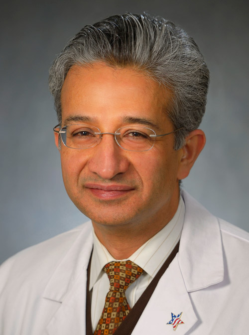 Sanjay Dixit合作论文数Department of Medicine, Perelman School of Medicine, University of Pennsylvania301
 David S. Frankel合作论文数Department of Medicine, Perelman School of Medicine, University of Pennsylvania271
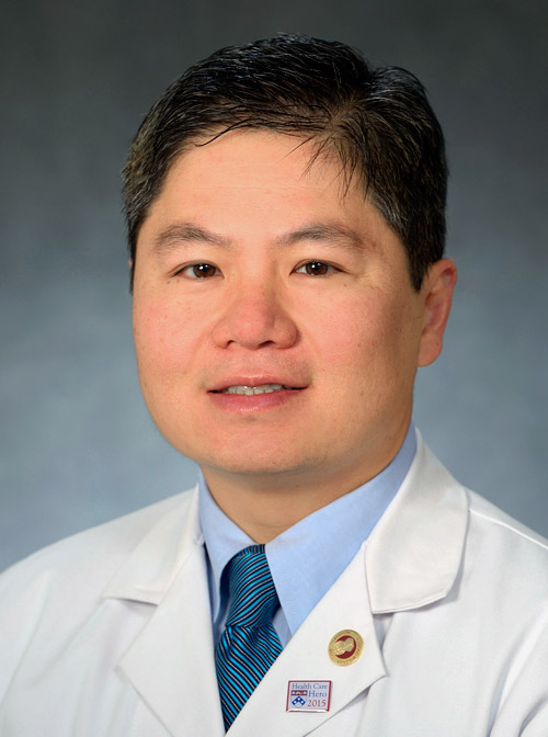 David Lin合作论文数Department of Medicine, Perelman School of Medicine, University of Pennsylvania244
 Fermin C. Garcia合作论文数Department of Medicine, Perelman School of Medicine, University of Pennsylvania234
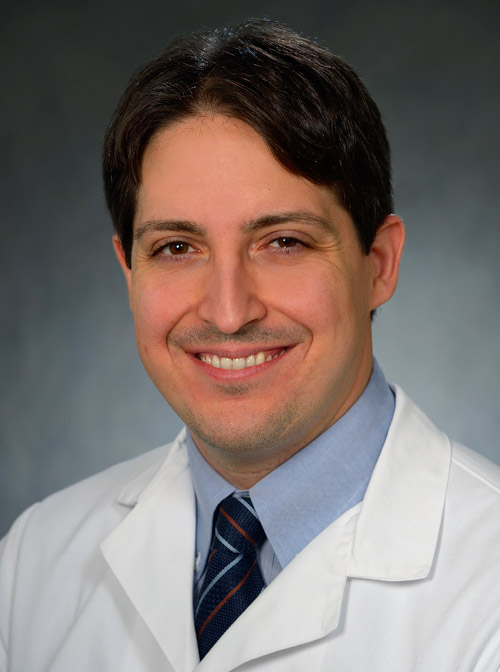 Pasquale Santangeli合作论文数Department of Medicine, Perelman School of Medicine, University of Pennsylvania230
 Zado Erica S合作论文数Cardiovascular Division, Hospital of the University of Pennsylvania230
 Gregory E. Supple合作论文数Department of Medicine, Perelman School of Medicine, University of Pennsylvania189
 Robert D. Schaller合作论文数Department of Medicine, Perelman School of Medicine, University of Pennsylvania169
 Saman Nazarian合作论文数Department of Medicine, Perelman School of Medicine, University of Pennsylvania168
 Edward P. Gerstenfeld合作论文数School of Medicine, University of California, San Francisco154
 Michael P. Riley合作论文数Department of Medicine, Perelman School of Medicine, University of Pennsylvania153
 Mark Josephson合作论文数Harvard Medical School;Beth Israel Deaconess Medical Center126
 Matthew Hyman合作论文数115
 Jeffrey S. Arkles合作论文数Department of Medicine, Perelman School of Medicine, University of Pennsylvania92
 Rajat Deo合作论文数Department of Medicine, Perelman School of Medicine, University of Pennsylvania89
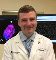 Cory M. Tschabrunn合作论文数Hospital of the University of Pennsylvania, University of Pennsylvania Perelman School of Medicine87
 Jackson J Liang合作论文数86
 Mathew D Hutchinson合作论文数81
 Joshua M. Cooper合作论文数University of Pennsylvania77
 Ramanan Kumareswaran合作论文数Department of Medicine, Perelman School of Medicine, University of Pennsylvania70
 David Schwartzman合作论文数University of Pittsburgh Medical Center68Jian-Fang Ren合作论文数68Rupa Bala合作论文数65
 Muser Daniele合作论文数1 Cardiovascular Division, University of Udine;Postgraduate school of Cardiology, University of Triest;2 Cardiovascular Division, Hospital of the University of Pennsylvania;University of Triest, Hospital of the University of Pennsylvania64Ae Buxton合作论文数63
 Ralph J. Verdino合作论文数Perelman School of Medicine, University of Pennsylvania63
 Gustavo S. Guandalini合作论文数Department of Medicine, Perelman School of Medicine, University of Pennsylvania57
 Andrew E. Epstein合作论文数Department of Medicine, Perelman School of Medicine, University of Pennsylvania57Cd Gottlieb合作论文数56
 Andres Enriquez合作论文数Department of Cardiology and Cardiovascular Diseases, Pontificia Universidad Católica de Chile54Hl Waxman合作论文数53
 William Henry Sauer合作论文数Department of Medicine, Harvard University;Brigham and Women's Hospital53
 Benoit Desjardins合作论文数Department of Radiology, Perelman School of Medicine, University of Pennsylvania45
 Alfred E. Buxton合作论文数Beth Israel Deaconess Medical Center, Harvard Medical School, Harvard University43
 Andrea Natale合作论文数HCA Healthcare;Texas Cardiac Arrhythmia Institute, St. David's Health Care;Scripps Green Hospital40Wendy S. Tzou合作论文数39
 Andrea Russo合作论文数Cooper Medical School, Rowan University;Cooper University Hospital32Ju Doherty合作论文数29
 Vickas V. Patel合作论文数Hospital of the University of Pennsylvania29
 Alireza Oraii合作论文数Cardiovascular Diseases Research Institute, Tehran University of Medical Sciences28
 Luigi Di Biase合作论文数Department of Medicine, Albert Einstein College of Medicine28
 Yasuhiro Shirai合作论文数Advanced Electronic Materials Center, National Institute for Materials Science28
 Henry C. Hsia合作论文数Biomedical Engineering, School of Engineering & Applied Science, Yale University27
 Mouhannad Sadek合作论文数Department of Cardiology, St. Michael's Hospital27David J. Wilber合作论文数27Dm Cassidy合作论文数26Corentin Chaumont合作论文数Dept Cardiol, Rouen Univ Hosp26




