BACKGROUND:Accurate fat redistribution after lacrimal trough ligament release by the conjunctival approach is difficult due to the limited surgical field of view. Therefore, creative methods are urgently needed. AIMS:To explore an innovative technique focused on fat redistribution using a guiding needle in transconjunctival lower eyelid blepharoplasty to correct tear trough deformity. METHODS:Twenty-six patients underwent transconjunctival lower eyelid blepharoplasty. The retroseptal fat was dissected and laid out using a guiding needle for redistribution over the orbital rim. Closing sutures were knotted internally to the periosteum. Patients were followed for 6 months. RESULTS:All patients diagnosed with predominant orbital fat bulge and tear trough deformity without noticeable lower eyelid skin laxity presented impressive correction of the eyelid bags and tear trough deformities at 6 months follow-up. The average score for postoperative effect was 5.692 ± 0.618 on a scale of 1-6 (1 being poor and 6 being good). CONCLUSIONS:This guiding needle is a simple and effective tool for correcting eyelid bags and tear trough deformities by redistributing orbital fat in the transconjunctival lower eyelid blepharoplasty.
Background:Delayed diabetic wound healing is one of the clinical difficulties, the main reason is the limited angiogenesis ability. Deferriamine (DFO) is an iron chelating agent that can induce angiogenesis, but its application is limited due to its short half-life. Increasing the load and slow release performance of desferriamine is beneficial to accelerate diabetic wound healing. Materials and Methods:In this study, we developed collagen (Col)-graphene oxide (GO) and (1% w/w) DFO-loaded nanofiber electrospinning scaffolds (DCG) using the electrospinning technique. We tested the physicochemical properties, drug release performance, and vascularization biological function of the scaffolds, and finally evaluated the promotion of full-thickness wound healing in the diabetic rat models. Results:The results showed that DCG scaffolds have good mechanical properties and water-holding capacity and can release DFO continuously for 14 days. In vitro, the novel DCG scaffold exhibited good biocompatibility, with the up-regulation at the gene level of VEGF and its regulator HIF-1α, promoters of angiogenesis. This was verified in vivo, as the scaffold enhanced granulation tissue formation and improved neovascularization, thereby accelerating wound healing when applied to full-thickness defects on the back of diabetic rats. Conclusion:The DCG nanofiber scaffold prepared in this study has good biocompatibility and vascularization ability, and improves the microenvironment in vivo, and has a good application prospect in diabetic wound repair.
随着年龄增长,面部衰老的问题逐渐出现,衰老的容貌可给求美者带来沉重的心理负担.近年来,各种用于保持年轻容貌、延缓衰老的技术得到了迅速的发展,如外科手术、美塑疗法和注射填充等,但各种方式均存在各自的优缺点.基于能量设备的光声电技术因其简便快捷、见效较快、损伤较小和恢复时间较短等特点受到广大求美者的青睐.现围绕作用机制、临床应用和不良反应等方面,对常见的光声电技术在面部年轻化方面的应用作一综述.
Cutaneous wound healing is complex, requiring a coordinated response by growth factors, drugs, and resident cells of the skin. To simulate native extracellular matrix, electrospun nanofibers offer a favorable microenvironment for biological processes, generating considerable interest in skin tissue regeneration. Furthermore, among the most essential growth factors in wound healing, basic fibroblast growth factor (bFGF) promotes cell migration, proliferation, and differentiation, thus enhancing wound healing. However, its application is limited by a short half-life and loss of bioactivity in normal physiological conditions when used in its naked form and without stabilization. Hence, delivering the growth factor with a controllable releasing speed constitutes a challenge. The aim of this study was to create a growth factor-releasing system that allows for time-controlled release to facilitate skin regeneration. Electrospun collagen-graphene oxide (Col-GO) scaffolds loaded with bFGF were fabricated. The cumulative release rate of the Col-0.2% GO-bFGF group was 30.94 ± 7.77%, which was superior to the other groups. Moreover, core–shell structured Col/GO and polylactic acid (PLA) nanofibers were fabricated by coaxial electrospinning in the attempt of reducing the degradation rate of the scaffolds (Col-GO). The ability of the materials to promoting wound healing in vitro and in vivo was investigated, and the improved skin tissue recovery with the growth factor release system was demonstrated. Importantly, bFGF was sustained-released through the constructed systems, leading to the best wound healing performance when the scaffolds contained the growth factor. The healing rates of Col-GO and core–shell scaffolds loaded with bFGF were 96.39 ± 0.66% and 92.29 ± 0.42%, respectively. Col-GO and PLA through coaxial electrospinning to form core–shell fiber scaffolds for skin tissue engineering applications
鼻基底的骨性标志是上颌骨前壁,包括鼻旁区和梨状孔区。该区域凹陷可产生老态的中面部容貌,称为鼻基底凹陷。肋软骨作为鼻整形常用的自体移植物,材料充分,在肋软骨综合鼻整形中应用肋软骨移植,可较好地改善鼻基底凹陷。对于鼻基底凹陷合并鼻翼肥大患者,如果配合移植物填充,可进一步巩固提升效果,有效改善鼻基底凹陷外观。在没有合适的软骨供区情况下,可考虑用臀部真皮脂肪组织、脂肪颗粒、人造材料等,联合鼻整形手术一起综合改善中面部外观。
Deep full-thickness burn wounds are prone to multi-drug resistant (MDR) infections following injury, which extends the healing time. Thus, providing a bioactive hydrogel dressing with prolonged antimicrobial activity and reduced dressing changes is quite desirable for accelerating burn wound healing and preventing scarring. To achieve this, we developed an injectable hydrogel based on silk sericin (SS), poly(vinyl alcohol) (PVA), and PVA microspheres (MSs) containing vancomycin (VA), gentamicin (GEN), or their association (VG) for the healing of infected burn wounds. The microspheres were prepared by inverse emulsion crosslinking, while the hydrogels were prepared by freeze-thawing cycles. Antibacterial studies showed that gentamicin acts synergistically with vancomycin by increasing the bacterial killing rate and enhancing the biofilm inhibition and eradication effects on methicillin-resistant Staphylococcus aureus more than on Pseudomonas aeruginosa and Escherichia coli. Findings from FESEM images showed that the microspheres were sphere-shaped with a smooth surface and their average diameter ranging from 26.22 to 32.42 μm suitable for parenteral drug delivery. The prepared hydrogel containing 10 ToC. Development of vancomycin/gentamicin-PVA microsphere@PVA/SS hydrogel with sustained drug release, excellent mechanical, and biological properties for burn wound healing.
Mesenchymal stem cell-derived extracellular vesicles (MSC-EVs) have been found to have a great potential for soft tissue repair due to various biological functions, including pro-angiogenesis and low immunogenicity. However, the low yield and heterogeneity of MSC-EVs limited their clinical transformation. This study was designed to develop a novel adipose-derived stem cell engineered nanovesicles (ADSC-NVs) with high production and explore its pro-angiogenetic effect and application in adipose tissue regeneration. Adipose-derived stem cell-derived extracellular vesicles (ADSC-EVs) were isolated from an EVs-free culture medium for human ADSCs (hADSCs). ADSC-NVs were prepared by sequentially extruding ADSCs followed by iodixanol density gradient ultracentrifugation and were compared with ADSC-EVs in morphology, size distribution, protein contents and yield. The pro-angiogenetic effect of ADSC-NVs in different doses (0, 5, 20 and 80 μg/mL) in vitro was determined using transwell assay, tube formation assay, western blot and qRT-PCR. In vivo, BALB/c nude mice were administered injection of a mixture of fat granules and different dose of ADSC-NVs and grafts were harvested at 12 weeks post-transplantation for further analysis. By analyzing the weight and volume of grafts and histological evaluation, we investigated the effect of ADSC-NVs in vessel formation and adipose tissue regeneration. Our results showed yield of purified ADSC-NVs was approximately 20 times more than that of ADSC-EVs secreted by the same number of ADSCs. In vitro, both ADSC-NVs and ADSC-EVs exhibited a dose-dependent pro-angiogenetic effect, despite their distinct miRNA profiles. These effects of ADSC-NVs may be mediated by enriched miR-21-5p via PTEN inhibition and PI3K/p-Akt signaling activation. Furthermore, after a mixed injection of ADSC-NVs, vessel formation and adipose regeneration were observed in vivo in fat implants. Our study developed a potent alternative of ADSC-EVs. ADSC-NVs have a high pro-angiogenesis potential and can be used as cell-free therapeutic biomaterials in soft tissue regeneration.
目的 探讨皮下组织蒂岛状皮瓣修复面部皮肤肿瘤切除术后创面的操作要点及临床效果.方法 回顾自2018年1月至2020年12月,华中科技大学同济医学院附属协和医院整形外科收治的35例面部肿瘤患者,采用皮下组织蒂岛状皮瓣修复术后创面,病损大小1.0 cm×1.0 cm~3.0 cm×3.0 cm.根据肿瘤性质设计切除范围,若疑为恶性者,术中送快速冰冻病理检查,根据病理结果切除组织边缘及基底无肿瘤侵犯再开始创面修复;根据创面位置、大小及面部血管的走行设计皮下组织蒂岛状皮瓣来修复创面,供区直接拉拢缝合.结果 35例受区创面均得到良好的修复,其中2例术后皮瓣尖端出现青紫,经积极干预后恢复,切口均愈合良好;其余患者转移皮瓣成活良好.所有患者术后获随访3~12个月,肿瘤无复发.供区瘢痕大部分隐藏于面部皱褶、发际线或者轮廓边缘,未出现明显眼睑外翻、口角畸形等,皮瓣的颜色、质地与周围皮肤基本相同,局部圆润饱满,外观满意.结论 针对面部良性或低度恶性的皮肤肿瘤,在病变组织切除术后采用皮下组织蒂岛状皮瓣对其皮肤软组织缺损进行创面修复,具有转移灵活、术后外观好、易于操作等优点,是临床值得推荐的方法.
BACKGROUND:There is no consensus regarding the choice of injected drugs for pathological scars. Although the clinical efficacy of different drug treatments was shown in many randomized controlled trials, the efficacies of many drugs are inconsistent. Therefore, this study aimed to determine how different effective drugs are for treating pathological scars. It is anticipated that the study findings may serve as guidelines for plastic surgeons. METHODS:Relevant literature was extracted from the following databases Cochrane Library, Embase, PubMed, Web of Science, CNKI, Weipu, and Wanfang until June 2022, such as randomized clinical trials (RCTs) evaluating different injected drugs for the treatment of pathological scars, including BTA, TAC, 5-Fu, VER, and BLE. RESULTS:This network meta-analysis of 1539 patients from 23 articles revealed that the most effective treatment for a pathological scar was TAC + BTA. The effective rate of TAC + BTA combination therapy was significantly different from that of the BTA, TAC, 5-Fu, VER, and BLM monotherapies. TAC+5-FU was more effective than TAC, 5-FU, VER, or BLM alone, and BTA was more effective than both TAC and 5-Fu. The effectiveness of VER and BLM was the same, but both were better than TAC and 5-Fu. No big differences were found between any of the other local injection therapies. CONCLUSIONS:According to this network meta-analysis, a combination of keloid and hypertrophic scar injection treatment is recommended, especially BTA+TAC. However, this network meta-analysis has some limitations and must be further verified by larger samples and higher quality RCTs. LEVEL OF EVIDENCE III:This journal requires that authors assign a level of evidence to each article. For a full description of these Evidence-Based Medicine ratings, please refer to the Table of Contents or the online Instructions to Authors www.springer.com/00266.
Associated with persistent oxidative stress, altered inflammatory responses, poor angiogenesis and epithelization, wound healing in diabetic patients is impaired. N-acetylcysteine (NAC) is reported to resist excess reactive oxygen species (ROS) production, prompt angiogenesis and maturation of the epidermis. Studies have revealed that graphene oxide (GO) can regulate cellular behavior and form cross-links with naturally biodegradable polymers such as collagen (COL) to construct composite scaffolds. Here, we reported a COL-based implantable scaffold containing a mixture of GO capable of the sustained delivery of NAC to evaluate the wound healing in diabetic rats. The morphological, physical characteristics, biocompatibility and NAC release profile of the GO-COL-NAC (GCN) scaffold were evaluated in vitro. Wound healing studies were performed on a 20mm dorsal full-skin defect of streptozotocin (STZ)-induced diabetic rats. The injured skin tissue was removed at the 18th day post-surgery for histological analysis and determination of glutathione peroxidase (GPx), catalase (CAT) and superoxide dismutase (SOD) activity. In diabetic rats, we confirmed that the GCN scaffold presented a beneficial effect in enhancing the wound healing process. Additionally, due to the sustained release of NAC, the scaffold may potentially induce the antioxidant defense system, upregulating the expression levels of the antioxidant enzymes in the wound tissue. The findings revealed that the antioxidant biocompatible composite collagen dressing could not only deliver NAC in situ for ROS inhibition but also promote the wound healing process. This scaffold with valuable therapy potential might enrich the approaches for surgeon in diabetic wound treatment in the future.
Electrospinning nanofiber scaffolds show great potential in many biomedical applications including regenerative medicine, tissue engineering, drug delivery and wound healing. This article begins with a brief introduction on the wound, and then highlights the application of nanofiber electrospinning scaffolds in wound healing, including sutures, multifunctional dressings, dermal regeneration, epidermalregeneration and full-thickness skin regeneration. Finally, we finish with conclusions and future perspective in this field.
Mesenchymal stem cell-derived extracellular vesicles (MSC-EVs) have been shown to have a high potential for soft tissue repair due to various biological functions, including pro-angiogenesis and low immunogenicity. However, a large scale of MSC amplification for extracellular vesicles preparation may be accompanied by cell senescence, resulting in low yield and heterogeneity of MSC-EVs, limiting their clinical transformation. In search of an alternative, we developed adipose-derived stem cell engineered nanovesicles (ADSC-NVs) through the sequential extrusion of ADSCs and compared them to ADSC-EVs in vitro . Notably, the yield of purified ADSC-NVs was approximately 20 times higher than the yield of ADSC-EVs secreted by the same number of ADSCs. Despite their distinct miRNA profiles, ADSC-NVs and ADSC-EVs demonstrated a dose-dependent pro-angiogenetic effect in vitro . Mechanistically, these effects of ADSC-NVs may be mediated by high miR-21-5p expression levels via PTEN inhibition and PI3K/p-Akt signaling activation. Moreover, in vivo, vessel formation and adipose regeneration were observed in fat implants after mixed injection of ADSC-NVs. Therefore, ADSC-NVs have a high pro-angiogenesis potential and can be used as cell-free therapeutic biomaterials in soft tissue regeneration.
Researches of biomaterials for osteoporotic bone defects focus on the improvement of its anti-osteoporosis ability, due to osteoporosis is a kind of systemic and long-range bone metabolism disorder. Nevertheless, how to steadily deliver anti-osteoporosis drugs in osteoporotic bone defects is rarely studied. Reported evidences have shown that alendronate (Aln) is known to not only restrain osteoclasts from mediating bone resorption but also stimulate osteoblasts to regenerate bone tissue. Here, we developed an engineered implantable scaffold that could sustainably release Aln for osteoporotic bone defects. Briefly, Aln was added into 2% collagen (Col) solution to form a 5 mg/ml mixture. Then the mixture was filled into pre-designed round models (diameter: 5 mm, height: 2 mm) and crosslinked to obtain engineered Col-Aln scaffolds. The release kinetics showed that Aln was released at an average rate of 2.99 μg/d in the initial 8 days and could sustainably release for 1 month. To detect the repair effects of the Col-Aln scaffolds for osteoporotic defects, the Col and Col-Aln scaffolds were implanted into 5 mm cranial defects in ovariectomized rats. After 3 months, the cranial defects implanted with Col-Aln scaffolds achieved more bone regeneration in defect area (11.74 ± 3.82%) than Col scaffold (5.12 ± 1.15%) (p < .05). Moreover, ovariectomized rats in Col-Aln scaffold group possessed more trabecular bone in femur metaphysis than Col scaffold group as analyzed by Micro-CT. This study demonstrated the engineered Col-Aln scaffold has the potential to repair osteoporotic bone defects and resist bone loss in osteoporosis.
Background: Application of distant skin flaps in facial defect reconstruction has limitations such as leaving a patch like appearance and being restricted by the length of the vascular pedicles. Leveraging the abundance of blood supply from superficial muscular aponeurotic system (SMAS), a local skin flap pedicled by SMAS can be used to avoid the aforementioned problems. Herein, we report the clinical application as well as the anatomical study of SMAS-pedicled skin flaps. Methods: This study enrolled patients who underwent facial defect reconstruction surgery between 2013 and 2018 using SMAS-pedicled skin flaps. The flaps were designed according to the size and location of the defect. A follow-up was performed to evaluate the treatment outcomes and incidence of adverse events. In addition, six cadaveric heads were used to perform an anatomical study on the distribution and blood supply of SMAS. Results: Twenty-three cases underwent the defect reconstruction surgery in the frontal regions (three cases), temporal region (four cases), periocular region (four cases), nasal region (seven cases), and other regions (five cases). All the flaps survived well. During the follow-up period up to 12 months, the flaps showed a satisfactory appearance, blood supply, and elasticity. The distribution and blood supply of SMAS at different anatomical regions have been successfully observed. Abundant vascular networks could be found in the SMAS layer. Conclusion: Based on the broad distribution of SMAS and the abundant blood supply, an SMAS-pedicled skin flap could be flexibly designed and versatilely used to reconstruct post-traumatic or post-excisional facial defects. (c) 2020 Published by Elsevier Ltd on behalf of British Association of Plastic, Reconstructive and Aesthetic Surgeons.
PCL (poly-caprolactone) nanofibers have good biocompatibility and high porosity, which are usually utilized for application in wound dressings. However, wound healing could be hindered by the overproduction of reactive oxygen species (ROS) and different factors. Pure nanofibers cannot satisfy these requirements of wound healing. N-acetylcysteine (NAC), as an antioxidant, meets the requirements for wound healing by resisting the overproduction of ROS and by promoting angiogenesis and maturation of the epidermis. In this study, we prepared a sandwich structured PCL-Col/NAC scaffold using the molding method, which consisted of PCL nanofibers at the core and NAC-loaded collagen on both sides. The hydroscopicity and tensile modulus of PCL-Col/NAC scaffolds showed best performance of these properties among groups. Meanwhile, the drug release profiles of PCL-Col/NAC scaffolds were investigated using the HPLC method and the results suggested a sustained drug release of NAC for PCL-Col/NAC scaffolds. In addition, PCL-Col/NAC scaffolds presented better properties than the control groups in cell migration and proliferation. The in vivo wound healing therapy effect was studied using an oval (2 × 1 cm) full-thickness skin defect wound model for SD rats. After 21 days, gross view and histological analysis showed a favorable beneficial therapeutic effect as well as better epidermal maturation compared with the control groups. CD31 immunohistology results revealed relatively more new vessels in the PCL-Col/NAC group than the control groups. This study developed novel PCL-Col/NAC scaffolds with an excellent hydroscopicity, tensile modulus and the ability to promote epidermal maturation and angiogenesis, demonstrating its promising potential in wound healing treatment. © 2019 Wiley Periodicals, Inc. J Biomed Mater Res Part A, 2019.
Wound dressings composed of natural polymers, such as type I collagen, possess good biocompatibility, water holding capacity, air permeability, and degradability, and can be used in wound repair. However, due to the persistent oxidative stress in the wound area, the migration and proliferation of fibroblasts might be suppressed, leading to poor healing. Thus, collagen-containing scaffolds are not suitable for accelerated wound healing. Antioxidant N-acetyl cysteine (NAC) is known to reduce the reactive oxygen species (ROS) and has been widely used in the clinic. Theoretically, the carboxyl group of NAC allows loading of graphene oxide (GO) for sustained release and may also enhance the mechanical properties of the collagen scaffold, making it a better wound-dressing material. Herein, we demonstrated an innovative approach for a potential skin-regenerating hybrid membrane using GO incorporated with collagen I and NAC (N-Col-GO) capable of continuously releasing antioxidant NAC. Methods: The mechanical stability, water holding capacity, and biocompatibility of the N-Col-GO hybrid membrane were measured in vitro. A 20 mm rat full-skin defect model was created to evaluate the repair efficiency of the N-Col-GO hybrid membrane. The vascularization and scar-related genes in the wound area were also examined. Results: Compared to the Col only scaffold, N-Col-GO hybrid membrane exhibited a better mechanical property, stronger water retention capacity, and slower NAC release ability, which likely promote fibroblast migration and proliferation. Treatment with the N-Col-GO hybrid membrane in the rat wound model showed complete healing 14 days after application which was 22% faster than the control group. HE and Masson staining confirmed faster collagen deposition and better epithelization, while CD31 staining revealed a noticeable increase of vascularization. Furthermore, Rt-PCR demonstrated decreased mRNA expression of profibrotic and overexpression of anti-fibrotic factors indicative of the anti-scar effect. Conclusion: These findings suggest that N-Col-GO drug release hybrid membrane serves as a better platform for scarless skin regeneration.
Breast tissue engineering is a promising alternative to standard treatments for breast defects. Although there is a consensus that the mechanical property of the scaffold should best match the reconstructed tissue, the simulation of the soft and elastic tactility of native breast tissues using conventional materials and architecture design requires further study. Previous research has shown that the crystal microstructure-like design can drastically alter the mechanical properties of the constructed scaffolds. In this study, we designed and additive manufactured four kinds of breast scaffolds using polyurethane and termed their architectures as N5S4, N9S8, N7S6 and N4S6. The basic unit cell of each scaffold was similar to a lattice structure from the isometric crystal system. The scaffolds possessed identical porosity but different mechanical properties in which the compressive modulus of the softest scaffolds (N5S4) were similar to that of native breast tissue. When applied in the construction of tissue-engineered breast combining with delayed fat injection technique in nude rat models, the soft scaffolds(N5S4) performed better compared to its stiff counterpart (N4S6), as higher adipose survival, vascularization and milder fibrosis could be observed in N5S4 scaffolds . Lastly, using finite element analysis, we further investigated the influence of the unit cell architectures on the mechanical properties of the scaffolds and simulated the deformation as well as stress distribution patterns of the implanted scaffolds in detail. Thus, a crystal lattice-like architecture design was introduced to tune the mechanical properties of the scaffolds and match the requirements for tissue engineering applications.
Human skin wound repair may result in various outcomes with most of them leading to scar formation. Commonly seen in many cutaneous wound healing cases, hypertrophic scars are considered as phenotypes of abnormal wound repair. To prevent the formation of hypertrophic scars, efforts have been made to understand the mechanism of scarring following wound closure. Numerous in vivo and in vitro models have been created to facilitate investigations into cutaneous scarring and the development of antiscarring treatments. To select the best model for a specific study, background knowledge of the current models of hypertrophic scars is necessary. In this review, we describe in vivo and in vitro models for studying hypertrophic scars, as well as the distinct characteristics of these models. The choice of models for a specific study should be based on the characteristics of the model and the goal of the study. In general, in vivo animal models are often used in phenotypical scar formation analysis, development of antiscarring treatment, and functional analyses of individual genes. In contrast, in vitro models are chosen to pathway identification during scar formation as well as in high-throughput analysis in drug development. Besides helping investigators choose the best scarring model for their research, the goal of this review is to provide knowledge for improving the existing models and development of new models. These will contribute to the progress of scarring studies.
目的研究外涂曲安奈德对预防內眦赘皮矫正术后瘢痕增生的临床效果。方法以自愿参加为前提,将内眦赘皮矫正术后患者分为外用曲安奈德的实验组和外用生理盐水的对照组。分别于术后即刻,术后第1天、第2天、换药时局部涂擦曲安奈德和生理盐水,对瘢痕采用温哥华瘢痕标度法进行评分,对手术效果和瘢痕满意度进行自我评分,比较两组差异性。结果两组各30名患者。患者一般资料有可比性。实验组瘢痕增生发生率明显低于对照组,满意度明显高于对照组。结论外用曲安奈德对预防內眦赘皮矫正术后瘢痕增生的有明显的临床效果。
Background Facial aging is a complex process influencing every layer of the facial structure. Most accepted surgical techniques for facial rejuvenation involve certain manipulation of the superficial musculoaponeurotic system (SMAS). Out of these SMAS-based techniques, SMAS plication or suspension provides excellent outcomes with shorter convalescence and fewer potential complications. Herein, we would like to present our own technique combining SMAS plication, periauricular purse-string, and malar fat pad elevation technique for mid and lower facelift. Methods Through a classical periauricular and temporal incision, a periauricular permanent purse-string suture was woven into the SMAS to suspend sagging soft tissue of the mid and lower face after superficial undermining, then plication of inner and outer SMAS of the purse-string loop was performed to further secure suspension, and at last the malar fat pad was elevated for midface rejuvenation. The shape of the loop varies with patients' age; for younger patients, the loop is more vertical, and for older patients, the loop is more horizontal. Patient-reported outcomes were described using the FACE-Q questionnaire. Results From January 2010 to June 2015, a total of 138 patients were treated with this technique by a same surgeon. Follow-up duration ranged from 1 to 6 years. Preoperative and postoperative photographs were recorded and analyzed. The complications rates were low, and satisfaction rates were high. Patients felt that they appeared 7.3 years younger than their actual age on average and were most satisfied with the appearance of their lower face and jawline. Conclusions Periauricular purse-string reinforced with SMAS plication and malar fat pad elevation technique produces esthetically pleasing outcomes, besides being simple, safe, and personalized.
 Jiaming Sun (孙家明)合作论文数Union Hospital, Tongji Medical College, Huazhong University of Science and Technology22
 Jiecong Wang合作论文数11
 Jie Yang (杨杰)合作论文数Britton Chance Center for Biomedical Photonics, Huazhong University of Science and Technology10
 Muran Zhou合作论文数8
 Thomas A Mustoe合作论文数Department of Surgery, Feinberg School of Medicine, Northwestern University8
 Seok Jong Hong合作论文数Department of Surgery, Feinberg School of Medicine, Northwestern University8
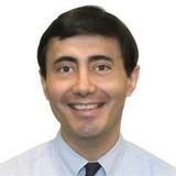 Robert D Galiano合作论文数Department of Dermatology, Feinberg School of Medicine, Northwestern University8
 Jialun Li合作论文数7
 Wang Zhenxing合作论文数7Ke Guo (郭科)合作论文数7Yuan Quan合作论文数7Ping Xie合作论文数7Shengxian Jia合作论文数7ZhengWen Zhang (张正文)合作论文数6Jialong Chen (陈佳龙)合作论文数5Jinfei Hou合作论文数5
 Wei Xu合作论文数5
 Zhenxing Wang (王振兴)合作论文数School of Physics, Huazhong University of Science and Technology;Wuhan National High Magnetic Field Center, Huazhong University of Science and Technology5
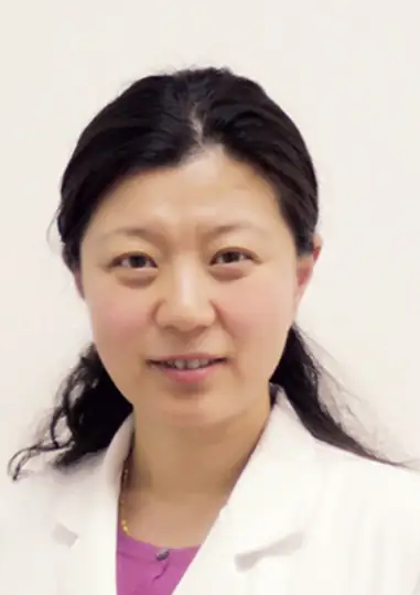 Shan Mou (牟姗)合作论文数Renji Hospital, Shanghai Jiao Tong University School of Medicine5
 Lingyun Xiong合作论文数4Liang Guo合作论文数4
 Rongrong Wang合作论文数4Hongfeng Di (翟弘峰)合作论文数Henan Provincial People's Hospital4Yuyang Zeng合作论文数3Chuchao Zhou合作论文数3
 Chao Luo合作论文数Wuhan Clinical Research Center for Superficial Organ Reconstruction3Jing Tong合作论文数3
 Shaokai Liu合作论文数Union Hosp, Huazhong Univ Sci & Technol3Yang Zhao (赵阳)合作论文数3Feng Xie (谢锋)合作论文数Henan Provincial People's Hospital3Dongyi Zhang (张栋益)合作论文数Henan Provincial People's Hospital3Shensong Kang (康深松)合作论文数Henan Provincial People's Hospital3Zhao Jingling合作论文数3Lifeng Chen合作论文数3
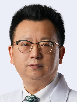 Jiahong Xia (夏家红)合作论文数Union Hospital, Tongji Medical College, Huazhong University of Science and Technology3
 Nengqiang Guo (郭能强)合作论文数Union Hospital, Tongji Medical College, Huazhong University of Science and Technology3
 Guohua Wang (王国华)合作论文数Union Hospital, Tongji Medical College, Huazhong University of Science and Technology3
 Zongquan Sun (孙宗全)合作论文数Union Hospital, Tongji Medical College, Huazhong University of Science and Technology3QIU Chang-hong (邱长虹)合作论文数2WANG Wen-juan (汪文娟)合作论文数2Di Sun合作论文数2Wei Wang合作论文数2Bei Qian合作论文数2Yang Sun合作论文数2
 Haiyan Chou (侴海燕)合作论文数Henan Provincial People's Hospital2王玉荣合作论文数School of Basic Medicine and Clinical Pharmacy, China Pharmaceutical University2
 D James Surmeier合作论文数Department of Neuroscience, Feinberg School of Medicine, Northwestern University;Cavalon Therapeutics, Inc.2Guo Zhang (张果)合作论文数School of Public Health, Tongji Medical College, Huazhong University of Science and Technology2
 D. Marshall Porterfield合作论文数Department of Agricultural & Biological Engineering, College of Engineering, Purdue University;Myco, Inc.;Florence Quantum Labs2




