患者男,72 岁,因摔伤致膝关节肿痛7 h,于2022 年12 月31 日就诊于北京积水潭医院新街口院区创伤骨科.患者于入院16 h前骑自行车时不慎摔倒,伤及左膝,伤后感左膝肿胀、疼痛、活动受限.于急诊行X线及 CT检查,提示左髌骨骨折.予以石膏固定后入院.患者既往患有高血压、糖尿病,口服药物治疗,血压及血糖控制良好.1 个月前因甲状腺癌行甲状腺切除手术.
目的 探讨采用顺行性骨搬移术治疗胫骨骨缺损合并膝内翻畸形的疗效.方法 回顾性分析2012年1月至2020年12月采用顺行性骨搬移术治疗胫骨骨缺损合并膝内翻畸形患者11例,均为男性,年龄(42±16)岁.其中左侧8例、右侧3例;均为骨折术后骨缺损,节段性骨缺损长度为(7.9±2.1)cm;10例为先天性膝内翻、1例为胫骨近段骨折畸形愈合致膝内翻.术前胫骨-股骨解剖轴外侧角为179°(178°,180°),股骨远端解剖轴外侧角为82°(81°,83°),膝关节关节线相交角为0°(0°,1°),胫骨近端解剖轴内侧角为83.1°±3.9°.7例应用单边外固定架、3例应用环形外固定架、1例应用混合式外固定架,采用顺行性骨搬移术治疗骨缺损及内翻畸形.其中3例采用单边外固定架者额外通过摆动夹钳矫正内翻、1例采用环形外固定架者在胫骨近段外侧安装铰链通过延长内侧的螺纹杆矫正内翻.术后测量单边和混合式外固定架近端固定针或环形外固定架的近端环与膝关节面的夹角、下肢机械轴偏向、下肢机械轴通过胫骨平台关节面的位置、胫骨近端机械轴内侧角、畸形矫正的角度、骨愈合时间、骨愈合指数、Paley的功能评分及并发症种类.结果 11例患者均获得随访,拆除外固定架后的随访时间为11(3,25)个月.7例采用单边外固定架和1例采用混合式外固定架者近端固定针与关节面的夹角为0°(-1°,1°),3例采用环形外固定架者近端固定环和关节面的夹角分别为0°、0°、2°.治疗结束时,下肢机械轴偏向(-6±6)mm、下肢机械轴经过胫骨平台的58%±9%、胫骨近端机械轴内侧角为90°(89°,92°)、矫正角度为7.2°± 3.9°.治疗后所有患者骨缺损均达到骨性愈合,骨愈合时间为(12±4)个月(范围7~20个月),骨愈合指数(1.6±0.6)个月/cm(范围1.2~2.9个月/cm);Paley功能评分结果为优7例和良4例.其中3例针道感染和2例软组织感染口服抗生素达到治愈,1例对接端皮肤卡入采用在新鲜化手术中切除卡入的软组织并处理骨折端后达到切口与对接端愈合.结论 顺行性骨搬移术可在治疗骨缺损的同时矫正膝内翻畸形,术后疗效满意.
Objective:To report our experience in using the Ilizarov technique to treat bone defects secondary to Gustilo Ⅲb open tibial fractures with negative clinical signs and serological inflammatory markers.Methods:A retrospective study was performed to analyze the 19 patients with bone defects secondary to Gustilo Ⅲb open tibial fracture who had been treated at Department of Orthopaedics and Traumatology, Beijing Jishuitan Hospital between January of 2010 and June of 2021. They were 15 males and 4 females with an age of (41±11) years. Their inclusion criteria: undergoing treatment with Ilizarov technique (consecutive compression-distraction or bone transport), soft tissue coverage procedures resulting in wound closure and negative clinical infection signs for at least 3 months and normal serological inflammatory markers, and follow-up for more than 3 months after frame removal. Three-phase bone scan was performed for the patients before the present surgery. Debridement, sampling of deep tissues for bacterial culture, and external stabilization with a fixator were performed in the present surgery. Osteotomy for compression-distraction or bone transport technique was carried out at 1 stage or 2 stages. Systemic antibiotic therapy for 6 weeks was continued for those with positive microbiological analysis guided by antibiogram. Recorded were results of intra-operative pus detection around defects, microbiological findings, length of bone defect reconstructed, rate and time of infection recurrence during treatment, fracture union rate, bone healing index, bony and functional results.Results:The interval between primary injury to the present surgery was (10±8) months. The preoperative three-phase bone scan showed infection free in 8 cases, chronic osteomyelitis in 7 cases, and suspicious infection in 4 cases. No pus was found during intra-operative debridement in all. The intra-operative microbiological detection was positive in 1 sample in 1 patient (infection free indicated by bone scan), and in ≥2 samples in 3 patients (bone scan indicating non-infection, infection not excluded and osteomyelitis in 1 case each). The length of bone defect reconstructed was (8±3) cm. The follow-up after the present surgery was (37±15) months. Fracture union was achieved in all cases, with a bone healing index of (1.7±0.5) months/cm. Clinical infection signs were observed 1 (1, 1) month after the present surgery in 6 patients whose microbiological results were all negative. All the 6 patients ended up with no clinical recurrence after empirical use of systemic antibiotics in 5 and radical debridement in one. The bony results showed 16 excellent and 3 good cases while the functional results showed 10 excellent and 9 good cases.Conclusions:In treatment of bone defects secondary to Gustilo Ⅲb open tibial fractures with negative clinical signs and serological inflammatory markers, constant vigilance is needed against low-grade infection. Intra-operative multiple sampling of deep tissues with a standardized protocol and microbiological testing are extremely valuable for diagnosis of fracture-related infections.
OBJECTIVE To study the effect of freshening technique on docking site in tibial bone transport management. METHODS Retrospective cohort study was conducted about the effect of freshening technique on docking site in 20 cases(15 males and 5 females) treated with tibial bone transport from January of 2014 to December of 2019. The age of patients ranged from 19 to 62 years old, with an average of (42.3±11.5)years old. Seven patients had infectious bone defect and 13 patients had non-infectious. Application of freshening technique immediately after docking included resection of invaginated skin or soft tissue, removal of closed sclerotic bone, re-apposition, increasing the contact, acute compression of freshened docking site and grafting from adjacent medullary or bone debris, followed by post-operative gradual compression. RESULTS The amount of segmented bone defect ranged from 5 to 15 cm, with an average of(9.2±2.9) cm. Time required from osteotomy to contact of butt end ranged from 26 to 243 days, with an average of(109.1±51.1) days. The duration needed from 3 to 7 months with an average of(3.7±1.1) months before reaching radiological healing criterion in docking site. Fourteen out of 15 concurrent fibular osteotomy were united. Consolidation time for distracted callus ranged from 5 to 28 months, with an average of (15.0±6.5) months. Bone healing index(BHI) ranged from 0.8 to 2.8 months/cm, with an average of (1.6±0.5) months. One surgical site infection (5%) in tibial was noted. No refractures were found in follow-up ranged from 12 to 73 months, with an average of(37.6±20.3) months after fixator removal. CONCLUSION Freshening technique immediately after docking had advantages of the shorter healing time, avoidance of refracture, and independance of necessity for remote autograft harvest.
目的 观察3D打印技术辅助治疗陈旧髋臼骨盆骨折的临床疗效.方法 2013年2月至2013年12月北京积水潭医院创伤骨科对20例陈旧髋臼骨盆患者进行了3D打印技术辅助的手术治疗.将所有患者骨盆打印为实体模型.使用多枚克氏针钻孔定位,进行截骨.然后进行模拟复位,塑形钢板进行固定.采用视觉疼痛模拟评分(VAS)对患者疼痛情况进行评定,采用Harris评分和EQ-5D评价患者髋部功能及生活质量.结果 3D打印技术可以在术前对骨折截骨位置,复位方式,固定方式进行有效的模拟,显著缩短手术时间,减少手术创伤,减少手术出血;在手术时间手术出血较传统方式均有明显改善.VAS评分术前为(4.3±2.1)分;术后末次随访时降至(1.2±0.9)分(P<0.05).Harris评分术前为(26.8±14.3)分;术后末次随访时为(77.2±11.3)分(P<0.05).EQ-5D 术前为(0.36±0.12)分;术后末次随访为(0.72±0.15)分(P<0.05).8例优4例良;1例差(最终行全髋关节置换术);7例失随访.结论 3D打印技术可以有效的辅助进行陈旧髋臼骨折手术治疗,对手术时间、手术损伤、手术效果均有一定改善.
开放性骨折的治疗对于任何创伤骨科医师都是一个挑战.在诸多问题上仍然需要通过研究提供更高等级证据,随着加速康复外科(ERAS)理念的引入以及循证医学的发展,可以在现有基础上做出多方面的改进,以使患者的主观体验及客观治疗效果得到改善.经过全国创伤骨科专家的讨论后达成本共识,为在急诊情况下处理开放性骨折提供参考.
目的 探讨应用牵拉成骨技术治疗股骨、胫骨肥大型骨不愈合合并畸形患者的疗效.方法 回顾性分析2016年11月至2019年11月在北京积水潭医院创伤骨科应用牵拉成骨技术治疗的3例股骨、胫骨肥大型骨不愈合合并畸形患者的病历资料.3例均为男性.所有骨折不愈合端均不切开.胫骨不愈合者均需经皮截断腓骨,使用Taylor Spatial Frame(施乐辉公司)固定,将13个参数输入计算机,设定牵开速度1 mm/d,生成电子处方.股骨不愈合者使用Orthofix外固定架固定.术后第2天按照1 mm/d的速度牵拉,在牵开恢复长度的过程中逐渐纠正畸形.随访期间,观察患者畸形纠正情况和骨愈合情况.结果 病例A术后20 d畸形完全纠正,术后15个月获得骨性愈合.病例B术后25 d畸形完全纠正,术后15个月获得骨性愈合.病例C术后29 d成角畸形纠正,术后19个月获得骨性愈合.结论 牵拉成骨技术可纠正肥大型骨不愈合的成角畸形及短缩,可作为肥大型骨不愈合的一种微创治疗方法.
目的 探讨采用外固定架尺骨延长治疗遗传性多发性骨软骨瘤(HMO)所致前臂畸形的疗效.方法 回顾分析2014年4月至2018年4月北京积水潭医院创伤骨科采用外固定架尺骨延长治疗HMO所致前臂畸形患者5例.其中男4例,女1例;平均年龄14.5岁(12~16岁);右侧2例,左侧3例;MasadaⅠ型2例,MasadaⅡB型3例.5例患者均接受尺骨延长手术治疗.对3例MasadaⅡB型患者采用环形外固定架,另2例采用单边外固定架.1例合并桡骨骨折患者同时行桡骨骨折切开复位内固定术.截骨术后8~10 d开始行尺骨牵开延长.结果 5例患者均获得随访,平均随访时间16个月(12~30个月).3例MasadaⅡB型患者桡骨头均自行复位.5例患者尺骨平均延长37.5 mm(30~45 mm),平均应用外固定架时间162 d(122~274 d),平均外固定架指数48.9 d/cm.术前和术后平均梅奥肘关节评分分别为36.4分和92.7分,平均肘关节屈曲活动范围分别为118.0°(110° ~130°)和130.0°(120°~150°),平均伸肘活动范围分别为12.7°(10°~20°)和3.5°(0°~10°),平均前臂旋前活动范围分别为18.6°(5°~30°)和44.7°(30°~65°),平均前臂旋后活动范围分别为71.2°(50°~85°)和86.5°(75°~90°).1例桡骨干骨折患者术后3个月骨折愈合.2例出现针道感染;1例尺骨过早愈合,行第2次截骨手术后延长顺利.未见神经血管并发症.5例患者对治疗结果均满意.结论 采用外固定架逐渐延长尺骨治疗HMO患者尺骨短缩畸形和桡骨头脱位安全有效.
Objective To introduce the technique we invented to treat hypertrophic and oligotrophic nonunion of femoral shaft fracture after dynamization of intramedullary nail (IMN) and to report its preliminary results.Methods The data of 2 cases with hypertrophic nonunions of femoral shaft fracture and 1 case with oligotrophic nonunion following IMN dynamization who had been treated by the technique from March 2006 to July 2017 in Beijing Jishuitan hospital were retrospectively studied.There were 2 females and 1 male,aged 50,66 and 24 years old.2 parallel half pins were inserted from anterior to posterior at the trochanteric and condylar zones of the femur for antegrade intramedullary nailing patients,and from lateral to medial sides for retrograde intramedullary nailing patient respectively,then the pins were connected with monolateral limb reconstruction fixator.The fracture nonunion site was compressed by the fixator up to 10-15 mm.The holes at the end of intramedullary nail where their screws were removed for dynamization were locked again with 2 locking screws,and then the frame was removed.The patients are allowed to start their rehabilitation and fully bear their body weight 1 day after the operation.Results The three patients were followed up for 25,22,and 7 months after the surgery,respectively.The X-ray films showed the fracture healed at 7 months in two case,and at 12 months in one.One patient got her nail removed 25 months after the operation,feeling good 37 months after the removal.Conclusion This technique is an option to manage the hypertrophic and oligotrophic fracture nonunion of the femur after dynamization of IMN.It is mini-invasive to the nonunion site,easy to practice and allows early rehabilitation.
To discuss the result of treating tibia deformity with fixator assisted nailing technique (FAN). Methods A total of 5 patients with 7 limbs of tibial deformity?were treated with FAN technique. Etiology: 2 patients with bilater?al tibial deformity suffered from Ricket's disease, 2 patients were malunion after tibial fracture, 1 patient was congenital pseudoar?throsis of tibia. This is a retrospective study. The unilateral external fixator was mounted on the medial side of tibia, and thenthe minimal invasive osteotomy was performed. After the deformity was corrected, the intramedullary nail was inserted to fix the tibia. None of the patients need bone autograft. The pre?operation and post?operation medial proximal tibia angle (MPTA), mechanical axis deviation (MAD) and range of motion (ROM) were measured and analyzed. Results All the 5 patients were followed?up for 12-60 months (average 32 months). The osteotomy site united in 3-5 months (average 4.5 months) post?operatively. According to Paley’s imaging scores, 4 patients were excellent and 1 patient was good. According to Paley's functional result scores, 5 patients were excellent; according to Paley’s bone results evaluation , 4 patients were excellent and 1 patient was good. We achieved de?sired post?operative MAD (from medial 15 mm-lateral 10 mm) in 6 limbs. The MPTA in 7 limbs was corrected to normal (84°-90°). The ROM was not significantly changed before and after operation. The average ROM of knee before operation was 125° (120°-135°),and average postoperative ROM was 120°(115°-130°),No deep infection or neurovascular injury occurred. All pa?tients were satisfied with the method and results of the operation. Conclusion FAN technique combines the advantage of exter?nal fixation and intramedullary nail, and it is a good method to treat tibial deformity, the patient should be carefully evaluated and selected for applying this technique.
目的:探讨保留原锁定接骨板联合Orthofix单边外固定架骨搬移治疗股骨大段骨缺损的临床疗效和安全性.方法:2013年9月至2018年3月收治4例股骨骨折术后骨折不愈合和残留骨缺损患者.男3例,女1例.年龄17~62岁,中位数50岁.均因外伤导致股骨干或股骨远端骨折,前期均采用切开复位锁定接骨板内固定治疗.术后3例患者发生骨折不愈合;1例伤后即存在骨缺损,经多次治疗仍存在大段骨缺损.4例患者均无骨髓炎病史.均一期手术清理不愈合或缺损部位,保留原锁定接骨板,联合Orthofix单边外固定架进行骨搬移.骨搬移速度每天1 mm,分4次完成.待被搬移骨段与股骨远端对合后,二期手术以2枚锁定螺钉通过原锁定接骨板固定被搬移骨段,拆除外固定架.结果:本组患者骨缺损长度6.0~14.0 mm,中位数10.3 mm.一期手术时间116~201 min,中位数170 min.4例患者均获得随访,随访时间16~46个月,中位数25个月.4例均未出现感染,1例出现钉道反应,经局部处理后症状消失;2例患者拆除外固定架6个月后对合端未愈合,其中1例固定被搬移骨段时发生少许偏移,均行对合端清理及自体髂骨植骨术,以1块6孔锁定接骨板辅助固定,4个月后均骨性愈合.最终所有被搬移骨段矿化良好,对合端均愈合.外固定架指数0.360~0.380月·cm-1,中位数0.373月·cm-1.患侧膝关节屈曲活动度,术前30° ~130°,中位数85°;骨搬移完成时30° ~45°,中位数38°;术后16个月60° ~120°,中位数90°.结论:保留原锁定接骨板联合Orthofix单边外固定架骨搬移是治疗股骨大段骨缺损的有效方法,安全性较高,且外固定架放置时间较短.
Objective To evaluate the medial submuscular plating of the femur after limb lengthening and correction with frame in patients with limb length discrepancy and/or angular deformity.Methods A retrospective study was conducted involving 12 patients who had been referred to Department of Orthopaedic Trauma,Beijing Jishuitan Hospital from December 2009 to November 2016 for limb length discrepancy and/or angular deformity.They were 3 males and 9 females,with an average age of 23 years (from 14 to 32 years).Altogether 4 left and 8 right sides were involved.Of them,11 got their length discrepancy and/or angular deformity secondary to injury to distal femoral epiphyseal plate caused by trauma in their childhood and one had length discrepancy and angular deformity following unsuccessful conservative treatment of superior condylar fracture of the femur.The femoral shortening ranged from 3 to 11 cm (average,6.7 cm),and the varus or valgus deformity of the knee ranged from 8° to 18° (average,12°).First,a monolateral fixator was installed on the lateral femur.The limb was lengthened from 7 to 14 days after diaphysis osteotomy.Femoral supracondylar osteotomy was conducted again in patients with knee angular deformity after the lengthening reached the expected length and the deformity was corrected with the aid of external fixator.After medial submuscular plating via the femoral lateral approach using minimally invasive techniques,the external frames were removed.The time for plate removal,limb lengthening and correction of the varus or valgus deformity were documented.Results The mean follow-up was 31 months (from 23 to 43 months) for the 9 patients who had their plates still in situ,and 6.5 months (from 2 to 14 months) for the 3 patients who had subsequently their plates removed.None had blood transfusion.All underwent distraction osteogenesis of the femur to their preoperatively expected length,ranging from 3 to 9 cm (mean,6.2 cm).Both varus and valgus deformities were corrected.None developed a deep infection.The range of motion of the knee joint was similar to that before operation in all but one patient who had a range of motion 40° less than the pre-operative one.The time for external fixation averaged 91 days (from 46 to 113 days),with an external index of 22 d/cm.All patients were satisfied with their outcomes.Conclusion Medial femoral submuscular plating after limb lengthening and angular deformity correction with the external fixator on the lateral side is a useful technique for patients with femoral limb length discrepancy and/or angular deformity,significantly shortening the time for external fixation.
Objective To evaluate the effect of fixator-assisted nailing (FAN) technique for the treatment of femoral deformity,nonunion of femoral shaft fracture with deformity and malunion.Methods The data of 8 patients with nonunion of femoral shaft fracture with deformity,3 patients with femoral deformity caused by Rickets disease and 2 patients with femoral fracture malunion who received treatment FAN technique was retrospectively analyzed.There were 11 males and 2 females,with an average age of 33 years (range,16-50 years).One-stage operation was used in 10 patients:after the fracture was distracted and reduced with external fixator,an intramedullary nail was used for final fixation.Two-stage strategy was used in 3 patients.Unilateral external fixator was used in 12 patients,ring fixator was used in 1 patient.Femoral intramedullary nailing was used in 8 patients,retro-femoral intramedullary nailing was used in 5 patients; autograft from callus of fracture site was used in 1 patient.Results The 12 patients were followed up for 12-48months with an average of 21 months.The fractures of 7 patients united 3-6 months (average,3.9 months)later after the operation.The osteotomy site of 5 patients united 3-5 months (average,3.5 months) later after the operation.The results were evaluated according to the Paley classification of functional results.There were 9 cases rated as excellent,2 as good and 1 as fair.The lower leg discrepancy (LLD) before and after operation was (48.2±23.0) mm and (27.3±24.6) mm,individually; the length of limb increased 4.6-41.0 mm (average,23.3 mm).The pre-and post-operative MAD was (27.5±24.4) mm and (6.3±8.3) mm.There was no neurovascular injury or infection occurred in any of the patients.All the patients were satisfied with the result of treatment.Conclusion FAN technique combines the accuracy,minimal invasiveness and safety of external fixation together with patient compliance of IM nail.It is an effective method to manage nonunion of femoral shaft fracture with deformity,malunion of femur,and femoral deformity cause by rickets.
Objective To report our successful clinical experience of managing tibial metaphyseal bone defects with linear-circular hybrid external fixators to restore limb functions.Methods We managed and fully followed 9 male patients with tibial metaphyseal bone defects from December of 2009 to February of 2013.They were aged from 25 to 66 years (average,45.0 years).Four had a proximal end and 5 a distal end affected;5 cases were infected and 4 non-infected.The average length of tibial shortening was 13.0 mm (from-2 to 30 mm) before operation;the mean length of bone defects was 8.3 cm (from 4 to 13 cm).The non-infected patients were managed with debridement and trimming at the defect end,and primary fixation with linear-circular hybrid external fixators,followed by immediate or delayed osteotomy and bone lengthening after one or two months at the normal metaphyseal end.The infected patients were managed with debridement and fixation before the defects were filled with antibiotics-impregnated bone cement or artificial bone.The osteotomy and bone lengthening was performed secondarily.The fixators were not removed until consolidation was noticed in both the distracted area and the docking site and the patients were able to ambulate without aids.Results On average the 9 were followed up for 22.4 months (from 16 to 35 months).They achieved bony healing in both the distracted area and the docking site.Their average distraction length was 8.9 cm (from 5 to 13 cm).The leg length discrepancy was 4 mm (from1 to 14 mm)shorter on average after operation except in one patient with shortening and equinus deformity who refused correction.The average external fixation index (EFI) was 1.8 month/cm (from 1.2 to 2.6 month/cm).No infection relapsed.According to Paley's criteria,the bony results were excellent in 7 and good in 2 cases,and the functional results were excellent in 4 and good in 5 cases.The total complication rate was 1.4 per patient.Conclusions Bone transport technique with linear-circular hybrid external fixators is effective in management of tibia1 non-union with metaphyseal bone defects because it can preserve functions of the adjacent joints and stabilize securely the small peri-articular segments of cancellous bone.However,surgeons should be prudent in stimulating bone union at the docking site by gradual compression through linear-circular hybrid external fixators.Autograft is strongly recommended at the docking site.
尽管外固定支架为肢体延长最常用器械,但使用其进行肢体延长的也存在以下几个问题:治疗周期长,生活不便,有针道感染、神经血管损伤、邻近关节脱位和马蹄足畸形等并发症发生的可能[1],通常由肢体延长技术本身和长期使用外固定支架所致.因此,有学者[2]一直致力于缩短外固定支架的带架时间和肢体延长方式.目前,肢体延长主要有2种方法:使用内固定辅助外固定支架和全内固定进行肢体延长.本文就这2种方法的特点、优势和缺点作一综述.
Objective To evaluate the effect of a bone transport method using unilateral external fixator in treatment of partial bone defect.Methods Three patients with partial bone defect were reviewed,including 2 males and 1 female,and whose ages were 50,50,and 24 years,respectively.The defects were at medial part of the left proximal tibia in 2 cases.In the first case,the defect was 5 cm in length,1/3-2/3 of transverse diameter in width,with a 5 cm×3 cm skin loss.In the second case,the defect was 6 cm in length and 3 cm in width.For the remaining patient,the bone defect was located in lateral part of the right femur,which was 13 cm in length,1/3-2/3 of transverse diameter in width,with a 15 cm×7 cm scar on it.After debridement of the wound,the Orthofix's limb reconstruction external fixation system was mounted medially,initially with 2-3 HA coated screws in the middle clamp to anchor the near cortex of the segment to be transferred.A partial corticotomy was performed with multiple drill hole technique.The gradual segment transport was started 2 weeks after the operation at a rate of 1 mm/d,4 times/d.Results The follow-up time was 14,28 and 24 months,respectively.The external fixator was removed 8 and 6 months after the osteotomy in 2 patients,when radiographs demonstrated bony union of the docking site as well as mature consolidation of the generated callus.The range of motion of hip,knee,ankle on the injured side was similar to the uninjured side.The segment was unable to be transported successfully in the femur,because the compressiondistraction device was misused.Two months after the first operation,the osteotomy was performed at the same site for the second time.The frame was removed at 10 months after the second osteotomy when the new bone formed well and the fracture healed at the docking site.The patient was able to stand independently and walk with a stick at 17 months after the osteotomy.At the latest follow-up,there was no sign of osteomyelitis.Conclusion Bone transport method using unilateral external fixator is a practical option to treat partial bone defect.In addition to shorten the period with fixator,it can avoid malunion and donor injury.
1 临床资料 患者,男,28岁,主因"车祸后右膝关节屈曲畸形1年"就诊.患者伤后经颅脑外伤抢救,之后发现右股骨内髁骨折畸形愈合合并屈膝畸形.既往体健.查体见右膝屈膝50°畸形,被动屈伸活动度140°~50°(图1),主动屈伸活动130°~50°双下肢感觉、肌力对称存在,血运良好.正侧位X线片示右股骨内髁冠状面骨折畸形愈合(图2).
Objective To evaluate the effectiveness of correction of posttraumatic flexion contracture of knee (FCK) using Ilizarov technique.Methods Six male patients with posttraumatic FCK,aging from 9 to 43 years (mean,24.5 years),treated by using Ilizarov technique from January 2006 to December 2010,were retrospectively analyzed.The preoperative flexion deformity of knee ranged from 35° to 85° (mean,47.6°),and the range of motion ranged from 0° to 70° (mean,15.8°).Among them,five patients had old fracture of knee joint combined with equinus foot deformity (deformity ranging from 25° to 37°,with an average of 31.8°); one patient had femoral supracondylar fracture.We used circular external fixator to correct the FCK and equinus foot deformity gradually,the knee arthrodesis operation was performed in 4 patients because of the damaged hone structure and poor soft tissue condition.Results All patients were followed up for 12-22 months (mean,18 months).The flexion of knee joint was recoverd from preoperative 47.67°±18.63° to postoperative 9.33°±3.50°.Plantar flexion of ankle joint in five equinus foot deformity was recovered from preoperative 31.80°±4.65° to postoperative 3.00°±4.47°.The kuee joint was arthrodesised successfully in 4 patients and the other 2 patients got 30° and 75° ROM.All the patients can walk with a cane,and satisfied with the result of treatment.Pin-site infection was found in 4 patients and was controlled within two weeks.Conclusion Ilizarov technique is an effective treatment method for posttraumatic FCK.Knee arthrodesis should be performed when patients had damaged bone structure and poor soft tissue condition.
患者男,43岁,因"左小腿、踝关节骨折后5个月功能障碍"来就诊.患者就诊前5个月车祸后导致左胫骨干和外踝的闭合骨折,受伤当天行"左外踝骨折切开复位钢板螺钉"固定,术后行长腿石膏前后托固定胫骨干骨折.术后5个月由于发现小腿畸形,伴踝关节负重时疼痛以致不能行走来就诊.
Objective To review gradual correction of proximal tibial varus malunion with a unilateral external fixator for osteogenetic distraction. Methods From July 2004 to August 2010, we treated 5 cases of proximal tibial varus malunion with a unilateral external fixator. They were 2 men and 3 women,with an average age of 32 years (from 18 to 42 years). Two cases were malunion after tibial plateau fracture,2 after proximal tibial fracture, and one after osteogenetic distraction. After fibular osteotomy, a tibial unilateral external fixator was installed before proximal tibial osteotomy. Varus was corrected for 7 to 10 days after surgery by gradual distraction till the same alignment was obtained as the contralateral side. The external fixator was not removed until consolidation and full weight bearing. Results The follow-ups ranged from 5 to 11 months (average, 8. 4 months). Four cases got united after 3 to 6 months (average, 4. 8 months).Time for external fixator ranged from 4 to 8 months (average, 6. 8 months) . One case obtained bone union after bone grafting. All fibulas healed after osteotomy. No other complications were present except mild pin-tract problems. Compared with the contralateral side, the corrected malalignment deviation was laterally displaced by 8 mm on average (from 1 to 13 mm), the corrected medial proximal tibial angle was 90° on average (from 87° to 92°), the limb length discrepancy was -6 to 1 mm, and the tibial length discrepancy was - 2 to 3 mm. Conclusions Proximal tibial varus malunion can be corrected gradually and effectively by a unilateral external fixator. Its advantages over valgus osteotomy and internal fixation are less invasion due to simple transverse osteotomy, accurate correction not only of angulation but also of length discrepancy, and no need of bone grafting or implant removal.
 Xing Teng (滕星)合作论文数22
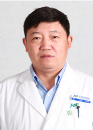 Lei Huang (黄雷)合作论文数 Department of Orthopaedic Trauma,Beijing Jishuitan Hospital,Beijing ,China20
 Manyi Wang (王满宜)合作论文数Beijing Jishuitan Hospital13Gang Zhao (赵刚)合作论文数9Tao Wang (王陶)合作论文数7
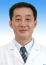 Xinbao Wu (吴新宝)合作论文数Beijing Jishuitan Hospital6
 Xieyuan Jiang (蒋协远)合作论文数Department of Orthopaedic Trauma, Beijing Jishuitan Hospital, Capital Medical University4Wenjie Tang合作论文数3
 Maoqi Gong (公茂琪)合作论文数Beijing Jishuitan Hospital3Weidong Ming (明卫东)合作论文数2GuoWei Rong (荣国威)合作论文数2ZhiLin Xia (夏志林)合作论文数2
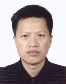 Tiejun Li (李铁军)合作论文数Medical School, Shanghai University2
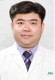 Shiwen Zhu (朱仕文)合作论文数Nanjing Cigarette Factory,Nanjing ,China2bai qiu en *gu ke jia su kang fu lian meng (白求恩·骨科加速康复联盟)合作论文数1bai qiu en gong yi ji jin hui guan jie wai ke zhuan ye wei yuan hui (白求恩公益基金会关节外科专业委员会)合作论文数1Jing-ming DONG (东靖明)合作论文数1Jinyi Tian合作论文数1Shaoliang Li合作论文数1Yuneng Li合作论文数1
 Hangyu Gu合作论文数Univ N Carolina, Dept Appl Phys Sci, Chapel Hill, NC 27515 USA1WANG Xu-yang (王旭洋)合作论文数1GeFeng Han (韩葛峰)合作论文数1HongGu Xiao (肖鸿鹄)合作论文数1Meng Mi (米萌)合作论文数1WenZhi Zhao (赵文志)合作论文数1KaiNan Li (李开南)合作论文数1Wu Wei (魏武)合作论文数1LiJun Liu (刘黎军)合作论文数1Jun Zhang (张军)合作论文数1Jing Wang (王京)合作论文数1Jing Li (李静)合作论文数1Bing Li (李兵)合作论文数1Peng Gao (高鹏)合作论文数1
 Zhijian Sun (孙志坚)合作论文数北京积水潭医院创伤骨科1
 Guanglin Wang (王光林)合作论文数West China School of Medicine, Sichuan University1
 Yimin Chai (柴益民)合作论文数Shanghai Sixth People's Hospital1
 Fang Zhou (周方)合作论文数Peking University Third Hospital1
 Fuxing Pei (裴福兴)合作论文数West China Hospital, Sichuan University1
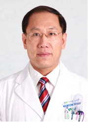 Bosong Zhang (张伯松)合作论文数Beijing Jishuitan Hospital1
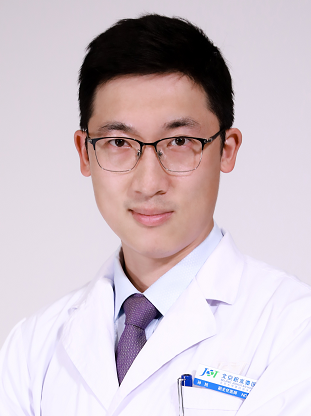 Xu Sun (孙旭)合作论文数Beijing Jishuitan Hospital1
 Yan Zhou (周雁)合作论文数Beijing Jishuitan Hospital1
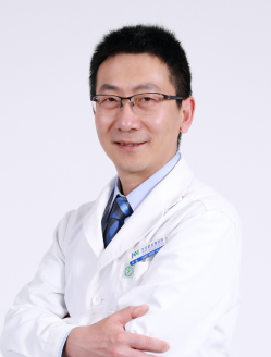 Ting Li (李庭)合作论文数Beijing Jishuitan Hospital/Zhongshan Jishuitan Orthopaedic Hospital1
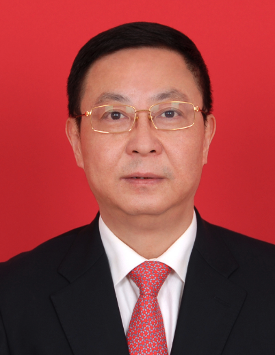 Bin Yu (余斌)合作论文数Nanfang Hospital, Southern Medical University1
 Chunyan Jiang (姜春岩)合作论文数Beijing Jishuitan Hospital;Health Science Center, Peking University1
 Peixun Zhang (张培训)合作论文数Peking University People's Hospital1
 Qiyong Cao (曹奇勇)合作论文数Beijing Jishuitan Hospital1




