目的 探讨老年心脑血管疾病病人颈动脉斑块与BMI、生长激素(growth hormone,GH)之间的关系.方法 选取我院住院的老年心脑血管疾病病人198例,根据颈动脉彩色多普勒超声检查结果是否有颈动脉斑块形成分为斑块组(170例)和非斑块组(28例).观察并比较2组BMI、GH水平及相关临床生化指标的差异,采用多因素Logistic回归分析颈动脉斑块形成的影响因素,采用Pearson相关系数描述GH、BMI及生化指标间的相关性.结果 斑块组与非斑块组在年龄、性别、吸烟史、饮酒史,高血压、糖尿病患病率及BMI、TG、TC、FPG、载脂蛋白A1、GH水平间差异均有统计学意义(P<0.05).Logistic回归分析显示,年龄、吸烟史、饮酒史、高血压、糖尿病、GH、BMI、TG、TC是颈动脉斑块发生的影响因素(P<0.05).Pearson相关分析显示,GH与BMI、TC、TG、FPG呈负相关;BMI与TC、TG、FPG呈正相关.趋势卡方检验提示,随着GH水平的降低和BMI的升高,斑块检出率升高(P<0.01). 结论 老年心脑血管疾病病人颈动脉斑块的发生受多种因素共同作用,年龄、吸烟史、饮酒史、高血压、糖尿病、BMI、TC、TG是其发生的危险因素,GH是其发生的保护因素.老年人应重视合理膳食、适当锻炼、改变生活习惯,降低颈动脉斑块的发生率.
目的 为实现应急救援下脑水肿监测仪的快速连接以判断脑水肿的目的,自行设计了无创脑水肿快速固定装置,并应用于临床.方法 选取2019年3月—2020年2月中国人民解放军总医院第五医学中心神经内科的104例脑水肿患者,应用随机数字表法将患者随机分为观察组(52人)和对照组(52人),观察组实施自行设计的快速电极固定装置进行监测,对照组实施常规电极固定方法监测,分析两组监测的效果.结果 观察组操作时间明显短于对照组,电极移位脱落情况、患者满意度及舒适度明显优于对照组,差异均有统计学意义(P<0.05).结论 应急救援下无创脑水肿监测仪电极快速固定装置大大缩短操作时间且确保电极固定牢固,不易脱落,患者满意度及舒适度明显提高,为应急救援下快速监测赢得了宝贵时间.
焦虑、抑郁是神经内科患者常见的并发症,临床上以易激惹、焦虑、运动阻滞、睡眠障碍及躯体化症状为主要表现[1].在心理健康日益受到人们关注的同时,神经内科患者的心理障碍引起临床医生的高度重视.为了解神经内科住院患者焦虑、抑郁现状,本文对我院神经内科住院患者的焦虑、抑郁状态进行分析,分析其特点及相关危险因素[2].
患者女,66岁,因“口干、双下肢无力2月余”于2013年11月26日入住解放军307医院.患者1个月前无明显诱因出现四肢无力,活动后加重,蹲下后无法自行站起,可自行行走10余米,无晨轻暮重现象,并出现口干、唾液减少,无发热、咳嗽、咳痰、腹胀、腹痛.既往体健,个人史无特殊,家族无遗传病及相关病史.
目的 总结汞中毒所致迟发性周围神经损伤患者的临床特征及诊疗经验.方法 回顾性分析2013年1月至2015年12月解放军总医院第五医学中心收治的37例汞中毒所致迟发性周围神经损伤患者的病历资料.其中男30例,女7例;年龄23~55岁.分析疾病的临床特征、诊治要点,评估临床疗效及患者预后,总结诊疗经验.结果 多数患者为从事工业用汞相关职业者,有长期汞接触史,因慢性汞中毒而损伤神经系统,主要表现为乏力、肢体麻木、肢端肌肉痛,伴头晕、头痛、失眠、记忆力下降、情绪异常、食欲减退等.患者均接受驱汞、营养神经等综合治疗.综合治疗5个月后,27例症状、体征基本消失,10例肢体末梢感觉异常、乏力等明显好转.结论 慢性汞中毒所致周围神经损伤患者有明确的长期汞接触史,合并神经损伤的临床表现主要为肢端感觉神经系统功能的异常.多数患者经驱汞治疗和营养神经等综合治疗可获得良好的效果及近期预后.
目的 了解北京市某医院神经内科患者睡眠障碍、焦虑、抑郁的发生率及其现状,为临床早期干预及治疗提供依据.方法 采用横断面调查方法,对431例神经内科就诊患者使用匹兹堡睡眠质量量表(PSQI)、汉密尔顿焦虑量表14项(HAMA-14)、汉密尔顿抑郁量表24项(HAMD-24)进行他评式问卷调查.结果 被调查的431例患者睡眠障碍的发生率高达84.69%,68.91% 的患者存在不同程度的焦虑或抑郁,其中32.2% 的患者肯定有焦虑,30.6%的患者有明显抑郁,23.9% 的患者焦虑抑郁共病.PSQI平均分为(12.12±4.17)分,HAMA-14的平均分为(10.97±6.95)分,HAMD-24的平均分为(15.29±10.15)分.女性睡眠质量较男性差,更容易患焦虑(P<0.05);住院患者睡眠效率明显低于门诊患者(P<0.05);有焦虑或抑郁的患者睡眠质量显著低于一般患者(P<0.01).结论 神经内科患者睡眠障碍与焦虑抑郁发生率较高,女性高于男性,住院患者睡眠效率更差,应引起医护人员的足够重视,并对患者进行早期预防及治疗.
目的 探讨腔隙性脑梗死患者睡眠与情感障碍及相关性.方法 回顾性研究2014年9月至2017年6月就诊于神经内科的腔隙性脑梗死患者64例,对照组选取同期非脑血管病患者50例.腔隙性脑梗死患者与对照组患者均于就诊1 d~2周内进行匹兹堡睡眠质量指数量表(PSQI)、汉密尔顿焦虑量表(HAMA)、汉密尔顿抑郁量表(HAMD)测定,所有测试均一次完成.比较2组患者PSQI、HAMA、HAMD评分,并对腔隙性脑梗死患者PSQI评分与HAMA、HAMD评分进行相关性分析.结果 腔隙性脑梗死患者PSQI总分及各因子评分除"睡眠效率"外均高于对照组(P<0.05);HAMA总分及各因子评分明显高于对照组(P<0.01);HAMD总分及相关因子中"焦虑/躯体化"、"睡眠障碍"、"迟缓"等因子评分均高于对照组(P<0.05).腔隙性脑梗死合并焦虑抑郁的患者睡眠质量较无焦虑抑郁患者差(P<0.05),且腔隙性脑梗死患者眠障碍评分与情感障碍评分具有明显相关性(P<0.01);焦虑与抑郁评分之间也存在着相关性(P<0.05).结论 腔隙性脑梗死患者比一般患者更容易引起睡眠与情感障碍,且患者睡眠障碍与情感障碍具有明显相关性,应引起临床重视.
气管切开术是用于抢救患者生命的重要治疗手段.在气管切开治疗结束前,需缓慢调整气管套管的末端孔径的大小至最后完全封堵,以保证患者的生命安全和减轻患者的不适感.现在临床上大多采用医用胶布及软木塞等对气管套管末端进行封堵,其操作方法复杂,还存在将胶布线及木屑坠入气管的风险,因此存在许多不足之处.解放军第三○七医院神经内科在气管套管末端加用自行设计的封堵装置,可提高临床治疗中的安全性和有效性.具体介绍如下.
目的 探讨注射肉毒毒素中毒致神经系统损害的临床表现及电生理特征,以利于早期发现和治疗.方法选取2015年4月~2017年11月就诊于解放军307医院神经内科、因注射肉毒毒素中毒致神经系统损害的女性患者42例,选取40例年龄、性别相匹配的正常受试者为对照组,收集中毒患者的临床资料,并对两组周围神经电生理特征进行比较.结果 因注射肉毒毒素中毒致神经系统损害的临床表现主要为视物模糊、视物成双(重影)、四肢无力、部分伴有肢体麻木(全身乏力)、吞咽困难等.神经电生理检查发现,肉毒毒素中毒主要累及运动神经;运动神经检测的结果显示,与正常组相比,中毒组正中神经、腓总神经、胫神经传导速度减慢;正中神经、尺神经波幅降低;正中神经、腓总神经、胫神经潜伏期延长(P<0.05或P<0.01).感觉神经检测的结果显示,中毒组较正常组除正中神经、腓肠神经传导速度减慢(P<0.05),其余无显著性差异.中毒组尺神经、胫神经F波较正常组潜伏期延长(P<0.05).结论 注射A型肉毒毒素易引起中毒,分析其临床表现并结合神经电生理检查可有效反映周围神经损害的程度,并能与其他疾病进行鉴别,有助于临床的早期诊断及治疗.
Mercury and its compounds possess strong neurotoxicity and patients with mercury poisoning often report pain and numbness in the distal extremities that conform to the “stocking–glove” pattern. However, no study has investigated whether damage to small nerve fibers is associated with mercury poisoning. The aims of the present study were to evaluate the effects of different doses of mercury chloride (HgCl2) on intraepidermal nerve fibers density (IENFD) and Langerhans cells (LCs) in the plantar skin of rats and to assess the possible relationship between changes in IENFD and sensory testing. Male Sprague–Dawley rats were divided into three experimental groups and administered HgCl2 solutions via gavage at three different doses (4.25, 8.5, and 17 mg/kg/day) for 21 days. Subsequently, behavioral tests and pathological changes in IENFD and LCs were assessed at three different time points (1, 2, and 3 weeks). Rats in all three HgCl2 groups exhibited varying degrees of weight and hair loss. Thermal hypersensitivity was evident in all the HgCl2 groups (for middle‐2w subgroup, p < 0.05). Mechanical sensitivity tests revealed hyposensitivity in all the HgCl2 groups except the high‐1w subgroup. Significant decreases in IENFD (for the high‐1w, middle‐1w, low‐2w, and low‐3w subgroups, p < 0.05) and significant increases in the density of LCs (except for the low‐1w and high‐2w subgroups, all p < 0.05) were found in all groups after HgCl2 exposure. An association analysis revealed a significant correlation between the decrease in IENFD and the increase in LCs densities (r = −0.573, p < 0.01). The present study demonstrated a decrease in IENFD and an increase in LCs density in the plantar skin of rats after HgCl2 poisoning, indicating that damage of the small nerve fibers occurs after mercury poisoning.
Objective To investigate the clinical and electrophysiological features and treatments for thallium poisoning. Methods Twelve cases of thallium poisoning patients were from hospital 307 of PLA between June 2012 and October 2017 and their data were retrospectively analyzed. Twelve sex-and age-matched healthy subjects were selected as control group. Result The clinical manifestations of thallium poisoning were mainly symptoms of nervous and gastrointestinal systems as well as hair loss. Thallium poisoning compromised function of motor nerves including the prolonged distal latency of ulnar and common peroneal nerve, the decreased amplitude and slowed nerve conduction velocity of common peroneal nerve and tibial nerve, which were statistically different from control group (P<0.05). Thallium poisoning also impaired function of sensory nerve including the prolonged distal latency and decreased amplitude of median , ulnar and sural nerve, the slowed nerve conduction velocity of median , ulnar, radial and sural nerve which were statistically different from control group (P<0.05 or P<0.01 ). Electroencephalogram (EEG) of 7 cases revealed mild abnormality EEG in 6 cases and moderate abnormality EEG in one case. Patients received potassium supplementation, diuresis, oral Prussian blue, intramuscular injection of sodium dimercaptopropanesulfonate and other treatment. Severe cases had good outcome after hemoperfusion and plasma exchange. Conclusions Thallium poisoning is rare in clinic and typical clinical features and electrophysiological examination are helpful to the diagnosis and differential diagnosis of diseases. Timely increasing thallium excretion and symptomatic support treatment can effectively improve the prognosis of the patients.
目的 探讨重症脑血管病患者脑电图变化,并分析其预后.方法 选取本院收治的60例重症脑血管病、且均行连续脑电图检测(continuous electroencephalogram monitoring,CEEG)的患者,分析CEEG分级与预后的关系.结果 本组60例患者中19例CEEG表现为弥散性慢活动,3例死亡;4例表现为间歇性δ节律,无1例死亡;12例癫痫样活动,3例死亡;4例三相波,2例死亡;5例单节律活动,均死亡;9例低波幅活动,均死亡;6例局灶性异常,无1例死亡;1例爆发-抑制性类型,死亡.CEEG中Ⅰ~Ⅱ级存活率为92.31%(12/13)、Ⅲ级为60.71%(17/28)、Ⅳ~V级为31.58%(6/19),差异有统计学意义(P<0.05).Spearman等级相关性分析结果显示,CEEG分级与预后呈正相关(r2=0.76,P<0.05).结论 CEEG可作为判断重症脑血管病患者预后的重要方式.
失眠是脑卒中患者常见的并发症状,不仅影响患者疾病的预后,还严重影响患者的情绪及生活质量,因此治疗脑卒中后的失眠极其关键.目前脑卒中后失眠的治疗方法种类繁多、效果不一,临床上结合实际情况,采用各种方法联合治疗,可以更加安全有效的解决脑卒中患者由失眠带来的困扰.本文将对中西医治疗、心理治疗、物理治疗脑卒中后失眠等方面的进展进行综述.
目的 分析急性重度中毒性脑病EEG特征,评价EEG诊断和预后的预测价值.方法 选取2013年4月至2016年2月军事医学科学院附属医院急诊科收治的急性重度中毒脑病患者60例,作为病例组,体检中心接待的健康志愿者50例作为对照组,均进行EEG检测.结果 治疗后24 h EEG分级低于治疗前,差异有统计学意义(P<0.05);病死率为20%,Logistic分析显示,EEG分级与院内死亡有关(OR=3.3,95% CI:1.4~7.645),差异有统计学意义(P<0.05);EEG分级预测患者病死率的AUC为0.778(P< 0.05);存活患者24h后,EEG分级与其认知功能量表评分(mini-mental state examination,MMSE)不存在相关性(r=-0.216,P=0.156);θ功率与δ功率Fp1、F3、C3水平病例组低于对照组,差异有统计学意义(P<0.05).结论 急性重度中毒性脑病患者EEG与正常人存在显著差异,EEG分级随着治疗而逐渐改善,EEG分级可作为死亡风险预测指标,EEG异常等级越高,死亡风险越高,存活者24 h后EEG分级不能预测认知功能.
DOI: 10.3969/j.issn.1672-6731.2018.01.012
Objective To explore the clinical features and therapeutic outcomes of positive leueine-rich glioma inactived-1 antibody (Anti-LGI1) associated 1 imbic encephalitis.Methods One case with anti-LGI1 1imbic encephalitis was analyzed retrospectively,and the literature was reviewed.Results A young woman was admitted to the hospital due to subacute onset of declination of recent-memory function,epilepsy,mood disorder and amenorrhea.LGI1 antibody both in blood and cerebrospinal fluid was positive.T2 or flair MRI scans revealed abnormal signals in bilateral medial temporal lobes and hippocampus.While PET-CT showed the decreased metabolism of bilateral medial temporal lobes and hippocampus.And glucocorticoid combined intravenous immunoglobulins (IVIg) showed remarkable outcome.Conclusion Anti-LGI1 1imbic encephalitis was characterized by memory function impairment,epilepsy,mood disorder with specific LGI1 antibody.Bilateral medial temporal lobes and hippocampus were major affected while immune therapeutic effect was significant.Prompt diagnosis and treatment might be helpful for patients in recovery.
With the development of modern industry,heavy metal pollution in China is becoming increasingly serious.Large-scale water sources and farmland have been severely contaminated.Frequent contact with heavy metal in life or at work has caused a marked increase in heavy metal poisoning in recent years.Nervous system damage caused by heavy metal poisoning is extremely common in clinic,and poses a threat to a patient's life,bringing heavy burden to society and families.Therefore,early prevention and treatment are crucial.This article takes commonpoisoning several common heavy metals as an example and combine the domestic and foreign research to review the damage to the nervous system caused by heavy metal poisoning.We aim to make patients and medical staff more alert to such poisoning and provide the basis for clinical prevention,diagnosis,treatment and research.
Objective To study the injury of SD rats' vascular endothelial cell poisoned with different doses of mercuric chloride (HgCl2). Methods The SD rat animal models were created by gavaging different doses of HgCl2. A total of 54 male rats were randomized into 4 groups:high, medium, low dose HgCl2 group (n=15 in each, poisoned with 17 mg·kg-1·d-1, 8.5 mg·kg-1·d-1, 4.25 mg·kg-1·d-1 HgCl2, respectively), and control group (n=9). The levels of Hg in blood cell, NO and endothelin-1 (ET-1) in serum were measured on the 7th day, 14th day and 21th day, and the number of circulating endothelial cells (CEC) was counted at the same time. Results Level of Hg:The levels of Hg in control group were close to zero, and there were statistically significant differences between high, medium, low dose HgCl2 groups and control group. Levels of serum ET-1:Between-group comparison showed significant differences between high dose HgCl2 group and control group, and within-group comparison revealed significant differences during different time. Lev-els of serum NO:Between-group comparison showed that there were significant differences between high, medium, low dose HgCl2 groups and control group, and also among the high, medium, low dose HgCl2 groups at the same time, and with-in-group comparison revealed showed that there are significant differences during different time. CEC counts:Between-group comparison showed significant differences between high, medium, low dose HgCl2 groups and control group, and with-in-group comparison revealed significant differences during different time. Conclusion HgCl2 can cause direct injury and dysfunction to vascular endothelial cells, and its injury has the same changing trend with dose and time.
Some patients with cardiocerebral vascular diseases still have recurrent ischemic events after clopidogrel administration,suggesting clopidogrel may not play an expected antiplatelet effect.Studies demonstrated that clopidogrel resistance was seen in some patients.The mechanism of clopidogrel resistance is still unclear.This article reviews the relationship between clopidogrel resistance and gene polymorphism and drug interactions.
中毒性周围神经损伤在临床中明确诊断后,将毒性暴露因素降低或消除,患者病情多可好转或恢复[1]。然而多数中毒性周围神经病变患者临床表现及辅助检查缺乏特异性,尤其在早期,往往较难诊断。因此,该类疾病的确诊需要对相关疾病较高水平的了解和掌握。基于上述原因,本文拟总结中毒性周围神经病的临床和电生理特点,对不同形式的中毒性周围神经病进行分类,以尽可能提高临床诊断的准确率。
 BinBin Sun (孙彬彬)合作论文数20ZhiFang Li (李志方)合作论文数17
 Penglin Ma (马朋林)合作论文数Guiqian International Hospital14
 Dongning Wei (魏东宁)合作论文数The 8th Medical Center of Chinese PLA General Hospital10LiXue Liu (刘力学)合作论文数9TianHao Wang (王天昊)合作论文数8Juan LAN (兰娟)合作论文数7LeShi Zhang (张乐石)合作论文数7Chun Chi (池春)合作论文数6Hao WEN (文浩)合作论文数5XueLin Xia (夏学林)合作论文数5XiaoFei Geng (耿晓非)合作论文数5Gang Lin (林刚)合作论文数5Fan YANG (杨帆)合作论文数4Hao-ming XU (徐浩明)合作论文数4Youping Zhou合作论文数4Weining Xue (薛伟宁)合作论文数4Yujia Liu (刘宇佳)合作论文数3Jing Wang (王菁)合作论文数3Haomin Xu (徐浩民)合作论文数3Weixi Yang (杨维晰)合作论文数3Jingfeng Liu (刘景峰)合作论文数3
 Luyan Ji (季琭妍)合作论文数School of Education, Guangzhou University3Nana Ni (倪娜娜)合作论文数2Wanshen Guo合作论文数2liu lixue合作论文数2xin yao (姚欣)合作论文数2Yan Jie (闫洁)合作论文数2ChengWen Sun (孙成文)合作论文数2LvHua Gai (盖绿华)合作论文数2GuoGang Bai (白国刚)合作论文数2WeiLi Liu (刘伟丽)合作论文数2JunShan Li (李俊山)合作论文数2Yuan Chen (陈远)合作论文数2Sugang Liu (刘素刚)合作论文数中国人民解放军军事科学院军事医学研究院2Zuosen Yang (杨作森)合作论文数2
 Xusheng Huang (黄旭升)合作论文数Chinese People's Liberation Army General Hospital2dehai yu (余德海)合作论文数2Kaili Leng (冷凯利)合作论文数2
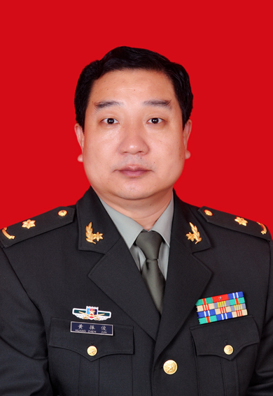 Zhenjun Huang (黄振俊)合作论文数The 8th Medical Center of Chinese PLA General Hospital2WU WAN-nan (吴皖南)合作论文数1Kai Yao合作论文数1Guangda Yao合作论文数1shuyan liu (刘树艳)合作论文数1honglin wei (魏洪林)合作论文数1wei li (李玮)合作论文数1
 Wanlu Wu合作论文数1zhifang li合作论文数1




