Triple-negative breast cancer (TNBC) presents formidable therapeutic challenges due to its triple defense system including antioxidant capacity, glycolytic metabolism, and immunosuppressive microenvironment. To overcome these interconnected resistance mechanisms, we developed a multifunctional nanogel (3-in-1 NG) consisting of Fe2+-crosslinked boronate-conjugated mannose-alginate with encapsulated D-alpha-tocopheryl polyethylene glycol succinate (TPGS), which enables efficient tumor delivery. 3-in-1 NG achieved a 52.9% tumor growth inhibition rate and significantly impeded metastatic progression in 4T1 models. Mechanistically, the pH-triggered mannose release led to intracellular accumulation of mannose-6-phosphate. This effectively blocked glycolytic activity and reversed immunosuppressive lactate accumulation, priming tumors for ferroptosis. The nanoplatform simultaneously executes therapeutic functions including metabolic disruption via mannose-mediated glycolysis inhibition, TPGS-induced immunogenic cell death triggering dendritic cell maturation and cytotoxic T-cell infiltration, and Fe2+- dependent lipid peroxidation initiating ferroptosis cascade. These synergistic mechanisms established self-reinforcing therapeutic actions where metabolic inhibition enhanced both immune recognition and ferroptosis susceptibility, creating a feed-forward cycle that progressively dismantled tumor defenses. Our work pioneers a nanomedicine strategy that simultaneously exploits the metabolic plasticity, redox adaptability, and immune escape of TNBC, providing a unified synergistic solution for refractory malignancies.
Cancer cells evade conventional therapies by dynamically switching between glycolysis and oxidative phosphorylation (OXPHOS). Simultaneous blockade of both pathways could overcome this plasticity but requires coordinated delivery of two inhibitors to the same cell. Here we design a glycyrrhizic acid (GA) nanogel that functions as both a structural carrier and a prodrug. GA itself serves as the structural carrier. It hydrolyzes in the acidic tumor microenvironment to become glycyrrhetinic acid (GRA), an OXPHOS inhibitor. The nanogel also encapsulates mannose, a glycolysis inhibitor. A crisis-inducing nanogel (CNG) is fabricated via Fe2+-crosslinked borate networks using microfluidic assembly, enabling pH-responsive disassembly. Fe2+ serves as both a structural crosslinker and a therapeutic component for ferroptosis. Upon exposure to acidic pH, CNG synchronously releases mannose and converts GA to GRA. Mannose suppresses glycolysis, GRA inhibits OXPHOS, and together they induce severe ATP depletion and bioenergetic collapse, which directly activates transient receptor potential cation channel subfamily M member 4 (TRPM4) ion channels. This activation triggers pathological Na+ influx and a cell death modality referred to as necrosis induced by sodium overload (NECSO). Simultaneously, liberated Fe2+ catalyzes the Fenton reaction, driven lipid peroxidation, and triggers ferroptosis. The convergence of NECSO and ferroptosis amplifies immunogenic cell death. In a 4T1 breast cancer model, CNG suppresses tumor growth and remodels the immunosuppressive microenvironment without systemic toxicity. This work establishes a "carrier-as-prodrug" nanoplatform that co-delivers two metabolic inhibitors with spatiotemporal precision. It also identifies TRPM4 as a mechanistically linked ion channel target in energy-stressed tumors.
Ischemia-reperfusion (I/R) injury is prevalent in the medical field and significantly limits the therapeutic outcomes of various ischemic diseases, adversely affecting patient prognosis. The pathogenesis of I/R injury is highly complex, involving intricate interactions among oxidative stress, inflammatory responses, mitochondrial dysfunction, and multiple cell death pathways. Once the mitochondrial respiratory chain is impaired, it triggers oxidative stress responses, leading to the excessive production of reactive oxygen species (ROS). Excessive ROS not only directly damage cells but also activate inflammatory responses and initiate multiple cell death signalling pathways, such as necroptosis, pyroptosis, and ferroptosis, thereby exacerbating tissue damage. Moreover, the clinical manifestations of I/R injury vary significantly across different organs, such as the heart, brain, kidneys, liver, and lungs, and are further influenced by patients' underlying conditions, posing challenges for clinical diagnosis and treatment. Therefore, constructing a comprehensive assessment system based on individual patient characteristics (such as genetic polymorphisms and comorbidities) to accurately predict the risk of I/R injury is particularly important. Currently, there are diverse strategies for the prevention and treatment of I/R injury, but translating basic research into clinical application remains challenging. Developing personalized treatment plans tailored to different cell types holds promise for overcoming existing therapeutic bottlenecks, significantly improving patient outcomes, and providing new directions for addressing the challenges of I/R injury.
OBJECTIVE:Endothelial-mesenchymal transition (EndMT) is a key contributor to the progression of sepsis-induced myocardial injury (SIMI). Defective mitophagy can result in oxidative stress and mitochondrial dysfunction, both of which play a critical role in EndMT. Sirtuin 3 (SIRT3), a major deacetylase responsible for mitochondrial quality control, has the potential to regulate EndMT, although the exact mechanism remains unclear. Therefore, this study aims to investigate the role of SIRT3 in mediating EndMT and cardiac remodeling during SIMI. METHODS:Wild-type and SIRT3 knockout mice were induced with lipopolysaccharide (LPS) for 24-h to mimic SIMI. Human cardiac microvascular endothelial cells were treated with LPS for in vitro experiments. Cardiac function was measured by echocardiography. Cardiac fibrosis was determined by Sirus red and Masson's trichrome staining. The expression of endothelial biomarkers and mesenchymal biomarkers was detected using immunofluorescence and western blot to determine EndMT. Mitochondria function and mitophagy were determined by transmission electron microscopy (TEM) and protein biomarkers. The interaction of SIRT3 with PINK1/Parkin was detected by immunoprecipitation (IP) and co-IP. RESULTS:Following endotoxin exposure, SIRT3 knockout mice exhibited a more severe EndMT phenotype and increased collagen deposition in cardiac tissues, along with mitochondrial dysfunction and impaired mitophagy. Similarly, LPS treatment induced mitochondrial oxidative stress and disrupted mitophagy flux during EndMT in CMECs, effects that were partially rescued by either rapamycin treatment or SIRT3 upregulation. Furthermore, SIRT3 overexpression enhanced deacetylation of the PINK1/Parkin pathway, thereby promoting mitophagy. CONCLUSION:Our findings suggest that SIRT3 suppresses EndMT-mediated cardiac fibrosis by promoting PINK1/Parkin-dependent mitophagy, offering novel insights for the treatment of SIMI.
Lysosomes are best known for their roles in inflammatory responses by engaging in autophagy to remove inflammasomes. Here, we describe an unrecognized role for the lysosome, showing that it finely controls macrophage inflammatory function by manipulating the lysosomal Fe2+—prolyl hydroxylase domain enzymes (PHDs)—NF-κB—interleukin 1 beta (IL1B) transcription pathway that directly links lysosomes with inflammatory responses. TRPML1, a lysosomal cationic channel, is activated secondarily to ROS elevation upon inflammatory stimuli, which in turn suppresses IL1B transcription, thus limiting the excessive production of IL-1β in macrophages. Mechanistically, the suppression of IL1B transcription caused by TRPML1 activation results from its modulation on the release of lysosomal Fe2+, which subsequently activates PHDs. The activated PHDs then represses transcriptional activity of NF-κB, ultimately resulting in suppressed IL1B transcription. More importantly, in vivo stimulation of TRPML1 ameliorates multiple clinical signs of Dextran sulfate sodium-induced colitis in mice, suggesting TRPML1 has potential in treating inflammatory bowel disease. Lysosomes are best known for their roles in inflammatory responses via autophagy. Here, Xing et al., find that lysosomes regulate macrophage inflammatory responses by finely controlling interleukin-1β production, altering lysosomal Fe2+ release through the TRPML1 channel.
Drug transmission through the blood-brain barrier (BBB) is considered an arduous challenge for brain injury treatment following the return of spontaneous circulation after cardiac arrest (CA-ROSC). Inspired by the propensity of melanoma metastasis to the brain, B16F10 cell membranes are camouflaged on 2-methoxyestradiol (2ME2)-loaded reactive oxygen species (ROS)-triggered "Padlock" nanoparticles that are constructed by phenylboronic acid pinacol esters conjugated D-a-tocopheryl polyethylene glycol succinate (TPGS-PBAP). The biomimetic nanoparticles (BM@TP/2ME2) can be internalized, mainly mediated by the mutual recognition and interaction between CD44v6 expressed on B16F10 cell membranes and hyaluronic acid on cerebral vascular endothelial cells, and they responsively release 2ME2 by the oxidative stress microenvironment. Notably, BM@TP/2ME2 can scavenge excessive ROS to reestablish redox balance, reverse neuroinflammation, and restore autophagic flux in damaged neurons, eventually exerting a remarkable neuroprotective effect after CA-ROSC in vitro and in vivo. This biomimetic drug delivery system is a novel and promising strategy for the treatment of cerebral ischemia-reperfusion injury after CA-ROSC.
MCOLN1/TRPML1 is a nonselective cationic channel specifically localized to the late endosome and lysosome. With its property of mediating the release of several divalent cations such as Ca2+, Zn2+ and Fe2+ from the lysosome to the cytosol, MCOLN1 plays a pivotal role in regulating a variety of cellular events including endocytosis, exocytosis, lysosomal biogenesis, lysosome reformation, and especially in Macroautophagy/autophagy. Autophagy is a highly conserved catabolic process that maintains cytoplasmic integrity by removing superfluous proteins and damaged organelles. Acting as the terminal compartments, lysosomes are crucial for the completion of the autophagy process. This review delves into the emerging role of MCOLN1 in controlling the autophagic process by regulating lysosomal ionic homeostasis, thereby governing the fundamental functions of lysosomes. Furthermore, this review summarizes the physiological relevance as well as molecular mechanisms through which MCOLN1 orchestrates autophagy, consequently influencing mitochondria turnover, cell apoptosis and migration. In addition, we have illustrated the implications of MCOLN1-regulated autophagy in the pathological process of cancer and myocardial ischemia-reperfusion (I/R) injury. In summary, given the involvement of MCOLN1-mediated autophagy in the pathogenesis of cancer and myocardial I/R injury, targeting MCOLN1 May provide clues for developing new therapeutic strategies for the treatment of these diseases. Exploring the regulation of MCOLN1-mediated autophagy in diverse diseases contexts will surely broaden our understanding of this pathway and offer its potential as a promising drug target.Abbreviation: CCCP:carbonyl cyanide3-chlorophenylhydrazone; CQ:chloroquine; HCQ: hydroxychloroquine;I/R: ischemia-reperfusion; MAP1LC3/LC3:microtubule associated protein 1 light chain 3; MCOLN1/TRPML1:mucolipin TRP cation channel 1; MLIV: mucolipidosis type IV; MTORC1:MTOR complex 1; ROS: reactive oxygenspecies; SQSTM1/p62: sequestosome 1.
中枢神经系统疾病是近年来全球疾病负担的主要原因,已成为全球重大的公共卫生问题.血-脑脊液屏障限制了某些药物直接向大脑的传递,阻碍了中枢神经系统疾病的治疗.随着纳米技术的发展,脑靶向纳米递药系统被证实可提高穿越血-脑脊液屏障的效率,实现药物在大脑的高效释放.本文综述近年来纳米递药系统在治疗中枢神经系统疾病中的进展,为提高中枢神经系统疾病药物疗效提供更多、更有价值的思路.
Targeted co-delivery and co-release of multi-drugs is essential to have an integrative collaborative effect on treating cancer. It is valuable to use few drug carriers for multi-drug delivery. Herein, we develop cRGD-modified nanoparticles (cRGD-TDA) of a conjugate of doxorubicin as cytotoxic agent, adjudin as an anti-metastasis agent and D-α-tocopherol polyethylene glycol 1000 succinate (TPGS) as a reactive oxygen species inducer linked with pH-sensitive bonds, and then combine the nanoparticles with PD-L1 antagonist to treat 4T1 triple-negative breast cancer. cRGD-TDA NPs present tumor-targeted co-delivery and pH-sensitive co-release of triple agents. cRGD-TDA NPs combined with PD-L1 antagonist much more significantly inhibit tumor growth and metastasis than single-drug treatment, which is due to their integrative collaborative effect. It is found that TPGS elicits a powerful immunogenic cell death effect. Meanwhile, PD-L1 antagonist mitigates the immunosuppressive environment and has a synergistic effect with the cRGD-TDA NPs. The study provides a new strategy to treat refractory cancer integratively and collaboratively.
目的:探讨窒息性心搏骤停/心肺复苏(CA/CPR)后的不同时间点,大鼠皮质神经元中黑色素瘤缺乏因子2(AIM2)炎症小体的表达变化以及对神经元死亡的影响.方法:健康雄性SD大鼠60只,随机分为假手术组(sham)和心肺复苏组(CPR).CPR组实施心搏骤停/心肺复苏(CA/CPR),即5min窒息法建立心搏骤停模型后行CPR,根据复苏后时间点的不同分为五组,即1.5、3、6、12、24 h组.采用Western Blot检测皮质AIM2、凋亡相关斑点样蛋白(ASC)、含半胱氨酸的天冬氨酸蛋白水解酶-1(caspase-1)、白细胞介素-1 β(IL-1 β)、IL-18表达情况,免疫荧光法观察AIM2和caspase-1分别在神经元中表达情况,尼氏染色观察大鼠皮质神经元的形态变化.结果:与sham组相比,CPR组各时间点大鼠皮质AIM2、ASC、caspase-1、IL-1β、IL-18表达均增多,在12 h达到高峰(P<0.05).免疫荧光结果显示,CPR组各时间点大鼠皮质神经元上均有AIM2、caspase-1阳性表达,其中12 h表达最多(P<0.05).尼氏染色结果显示,CPR组各时间点皮质神经元受损严重且存活数量明显减少(P<0.01),其中12h较6h神经元受损数量增加(P<0.05).结论:CA/CPR后大鼠皮质AIM2及相关炎性因子表达上调并造成神经元损伤,该作用可能是缺血性脑损伤的主要机制之一.
BackgroundCombined anesthesia can be a promising option for hip surgery when neuraxial anesthesia is contraindicated. Lumbar and sacral plexus blocks, and femoral nerve and lateral femoral cutaneous (LFC) nerve blocks in combination with general anesthesia (GA) are commonly used in elderly patients undergoing arthroplasty for hip fracture surgery. However, no study has compared these two anesthetic strategies in the perioperative period. MethodsA total of 41 elderly patients scheduled for arthroplasty for hip fracture surgery were randomized into group A (n = 20) and group B (n = 21). Group A received femoral nerve block, LFC nerve blocks, and GA, and group B received lumbar plexus block, sacral plexus block, and GA. Primary outcomes were incidences of hemodynamic events and changes in blood pressure (BP) and heart rate (HR). Secondary outcomes included time and drug consumption, infusion and bleeding volume, eyes opening time after surgery, and postoperative quality recovery rate. ResultsCompared with group B, group A showed a lower incidence of intraoperative hypotension (p < 0.001), higher BP [including mean arterial pressure (MAP), systolic BP (SBP), and diastolic BP (DBP)] following induction (IN), and higher HR from mid-surgery. Time required for nerve blockade (p < 0.001) and ephedrine consumption was significantly shorter in group A (p < 0.001), while sufentanil consumption was higher as compared to group B (p = 0.002). No significant differences in other intraoperative parameters and postoperative quality recovery rate were reported during the observation. ConclusionOur pilot data indicate that compared with lumbar and sacral plexus blocks, femoral nerve and LFC nerve blocks may provide more stable intraoperative hemodynamics and a comparable postoperative recovery for elderly patients undergoing arthroplasty for hip fracture under GA. Further studies with a larger sample size are needed to derive stronger evidence.
多种药物被研究证实对中枢神经系统疾病有显著疗效,然而其临床应用受到血脑屏障这一解剖结构的限制.近年来,纳米药物递送系统被证明可保证药物在递送过程中的结构完整性;同时,研究发现聚焦超声联合微泡技术能够安全、无创、可逆地开放血脑屏障并递送药物.这两项技术具有良好的临床应用前景.本文对跨血脑屏障纳米药物递送系统和聚焦超声开放血脑屏障的研究进展进行综述,以期为促进药物透过血脑屏障研究的开展提供思路.
自噬是一种细胞内降解途径,可将大量细胞质、受损细胞器等底物包裹后运至溶酶体降解从而维持细胞内稳态。在自噬过程中,自噬流的完整性是必需的。影响自噬流通畅的因素很多,如广受关注的内体分拣转运复合物(endosomal sorting complexes required for transport,ESCRT)与自噬的相互关系。ESCRT主要负责膜的剪切,介导细胞膜重构和分裂,在细胞内吞和胞质分裂过程中发挥重要作用。近年来,大量研究表明ESCRT突变体内常积累自噬小体,从而使自噬流受到影响。深入研究提示ESCRT参与调节不同类型自噬的多个过程,而且其亚单位与自噬相关蛋白之间也有交互作用。该文对近年来的相关研究进行综述。
Neonatal hypoxic–ischemic (H‐I) injury, which mainly causes neuronal damage and white matter injury (WMI), is among the predominant causes of infant morbidity (cerebral palsy, cognitive and persistent motor disabilities) and mortality. Disruptions to the oxygen and blood supply in the perinatal brain affect the cerebral microenvironment and may affect microglial activation, excitotoxicity, and oxidative stress. Microglia are significantly associated with axonal damage and myelinating oligodendrocytes, which are major pathological components of WMI. However, the effects of H‐I injury on microglial functions and underlying transformation mechanisms remain poorly understood. The historical perception that these cells are major risk factors for ischemic stroke has been questioned due to our improved understanding of the diversity of microglial phenotypes and their alterable functions, which exacerbate or attenuate injuries in different regions in response to environmental instability. Unfortunately, although therapeutic hypothermia is an efficient treatment, death and disability remain the prognosis for a large proportion of neonates with H‐I injury. Hence, novel neuroprotective therapies to treat WMI following H‐I injury are urgently needed. Here, we review microglial mechanisms that might occur in the developing brain due to neonatal H‐I injury and discuss whether microglia function as a double‐edged sword in WMI. Then, we emphasize microglial heterogeneity, notably at the single‐cell level, and sex‐specific effects on the etiology of neurological diseases. Finally, we discuss current knowledge of strategies aiming to improve microglia modulation and remyelination following neonatal H‐I injury. Overall, microglia‐targeted therapy might provide novel and valuable insights into the treatment of neonatal H‐I insult.
Background: Hypoxic/ischaemic encephalopathy (HIE) is a severe condition leading to high neonatal mortality and long-term brain damage. Autophagy and apoptosis are involved in hypoxic/ischaemic (HI) neuronal injury. Previous reports have shown that CHMP2A, an essential component of the endosomal sorting complexes required for transport (ESCRT)-III complex, is required for double-membrane autophagosome formation and is an anti-apoptotic protein. However, whether CHMP2A affects autophagy and apoptosis triggered by HIE remains unknown.Methods: The levels of CHMP2A, autophagy, and apoptosis were assessed by immunoblotting and immunofluorescence in HIE mice and primary cultured neuron (oxygen/glucose deprivation, OGD model). CHMP2A was overexpressed or knockdown in vivo by intracerebroventricular viral injection. Immunofluorescence, western blot, TUNEL staining, Nissl staining, and behavioral testing were performed to test the function of CHMP2A in regulating autophagy, apoptosis, and cognitive function .Findings: CHMP2A expression was reduced in hippocampal neurons after HI injury. Downregulating CHMP2A expression in HI mice caused autophagy deficits along with autophagosome accumulation, exacerbation of apoptosis, and cognitive impairment, whereas upregulating CHMP2A expression rescued these autophagy defects and reduced apoptosis and ameliorated cognitive dysfunction. Moreover, upregulating CHMP2A did not significantly reverse the aggravation of apoptosis induced by chloroquine treatment, indicating that enhanced autophagic flux could partly prevent apoptosis and that CHMP2A regulated apoptosis via autophagy after OGD injury.Interpretation: These findings show that CHMP2A exerts an anti-apoptotic function through the positive regulation of autophagy following neuronal injury, which may open new avenues for therapeutic targeting of autophagy in HIE.Funding: This work was supported by the National Natural Science Foundation of China (No. 81974284), the Shanghai Municipal Commission of Health (grant no. 201740118) and Medicine Engineering joint foundation at Shanghai Jiao Tong University (No. YG2016MS17).Declaration of Interest: The authors declare that they have no conflicts of interest.Ethical Approval: The C57BL/6 mice were supplied by the Experimental Animal Center of Shanghai Jiaotong University [SYXK (Shanghai, China) 2011-0128, 1 January 2011]. The Animal Research Committee of Shanghai Jiaotong University approved all experiments. All procedures were carried out in accordance with the National Institute of Health Guide for the Care and Use of Laboratory Animals (no. 2016-0056) and the ARRIVE guidelines. Furthermore, all efforts were made to minimize pain and to reduce the number of animals used.
Abstract Background An increase in blood flow in the forearm arteries has been reported after brachial plexus block (BPB). However, few studies have quantitatively analysed the blood flow of the forearm arteries after BPB or have studied only partial haemodynamic parameters. The purpose of the present study was to comprehensively assess blood flow changes in the distal radial artery (RA) and ulnar artery (UA) after BPB performed via a new costoclavicular space (CCS) approach using colour Doppler ultrasound. Methods Thirty patients who underwent amputated finger replantation and received ultrasound-guided costoclavicular BPB were included in the study. The haemodynamic parameters of the RA and UA were recorded before the block and 10 min, 20 min, and 30 min after the block using colour Doppler ultrasound to determine the peak systolic velocity (PSV), end-diastolic velocity (EDV), mean velocity (Vmean), pulsatility index (PI), resistance index (RI) and area. The volumetric flow rate (VFR) was calculated using the formula Q = area×Vmean. The aforementioned parameters were compared not only before and after the BPB but also between the RA and UA. Results Compared with those of the respective baselines, there was a significant increase in the PSV, EDV, Vmean, area, and VFR and a significant decrease in the PI and RI of the RA and UA 10 min, 20 min, and 30 min post-block. The increase 30 min post-block in EDV (258.68 % in the RA, 279.63 % in the UA) was the most notable, followed by that in the Vmean (183.36 % in the RA, 235.24 % in the UA), and the PSV (139.11 % in the RA, 153.15 % in the UA) changed minimally. The Vmean and VFR of the RA were significantly greater than those of the UA before the BPB; however, there was no significant difference in the VFR between the RA and UA after the BPB. Conclusions A costoclavicular BPB can increase blood flow in the forearm arteries. The RA had a higher volumetric flow rate than the UA before the BPB; however, the potential blood supply capacity of the UA was similar to that of the RA after a BPB. Trial registration This study was registered at the Chinese Clinical Trial Registry (http://www.chictr.org.cn/searchproj.aspx, clinical trial number: ChiCTR 1900023796, date of registration: June 12, 2019)
Abstract Background Activation of the absent in melanoma 2 (AIM2) inflammasome and impaired autophagosome clearance in neurons contribute significantly to cardiac arrest and return of spontaneous circulation (CA-ROSC) injury, while the mechanism by which the AIM2 inflammasome is regulated and relationship between the processes remain poorly understood. Recently, charged multivesicular body protein 2A (CHMP2A), a subunit of endosomal sorting complex required for transport (ESCRT), was shown to regulate phagophore closure, and its depletion led to the accumulation of autophagosomes and induced cell death. Here, we investigated whether CHMP2A-mediated autophagy was an underlying mechanism of AIM2-associated inflammation after CA-ROSC and explored the potential link between the AIM2 inflammasome and autophagy under ischemic conditions. Methods AIM2 inflammasome activation and autophagic flux in the cortex were assessed in the CA-ROSC rat model. We injected LV-Vector or LV-CHMP2A virus into the motor cortex with stereotaxic coordinates and divided the rats into four groups: Sham, CA, CA+LV-Vector, and CA+LV-CHMP2A. Neurologic deficit scores (NDSs), balance beam tests, histopathological injury of the brain, and expression of the AIM2 inflammasome and proinflammatory cytokines were analyzed. Results AIM2 inflammasome activation and increased interleukin 1 beta (IL-1β) and IL-18 release were concurrent with reduced levels of CHMP2A-induced autophagy in CA-ROSC rat neurons. In addition, silencing CHMP2A resulted in autophagosome accumulation and decreased autophagic degradation of the AIM2 inflammasome. In parallel, a reduction in AIM2 contributed to autophagy activation and mitigated oxygen–glucose deprivation and reperfusion (OGD-Rep)-induced inflammation. Notably, CHMP2A overexpression in the cortex hindered neuroinflammation, protected against ischemic brain damage, and improved neurologic outcomes after CA. Conclusions Our results support a potential link between autophagy and AIM2 signaling, and targeting CHMP2A may provide new insights into neuroinflammation in the early phase during CA-ROSC.
BACKGROUND:Carotid artery stenosis (CAS) is one of the leading causes of ischemic stroke. However, knowledge of the changes in the plaque itself is lacking. Information about the ultrasound and clinical features of CAS will help elucidate the changes in prognostic and risk factors.METHODS:We evaluated 736 patients with carotid stenosis for an average 18-month follow-up. According to their degree of CAS stenosis, patients were allocated to one of three groups: regression (n=125), stable (n=443), or progression (n=168). An ordinal regression analysis was used to determine the risk factors for atherosclerosis progression. A logistic regression was subsequently applied to investigate the effects of CAS stenosis on cerebrovascular events after adjusting for various factors.RESULTS:The progression group had more male patients (P=0.02), hypoechoic plaque (P<0.01), high-risk high sensitivity C-reactive protein (hs-CRP) (P=0.02), ulcerative plaque (P=0.05), and hyperlipidemia (P=0.05) than the other two groups. There were no significant differences in residual ultrasound and clinical features among the three groups, including age, high-density lipoprotein cholesterol (HDL-C), low-density lipoprotein cholesterol (LDL-C), intima-media thickness (IMT), body mass index (BMI), diabetes mellitus (DM), hypertension (HTN), coronary heart disease (CHD), statin use, ulcerative plaque. The ordinal regression analysis identified hypoechoic plaque (OR, 1.53; 95% CI: 1.14-2.05; P<0.01) and high-risk hs-CRP (OR, 1.75; 95% CI: 1.17-2.61; P<0.01) as independent risk factors for CAS progression. Logistic regression analysis revealed that the stroke/transient ischemic attack adjusted odds ratio was 1.80 (95% CI: 1.03-3.13) in the progression group.CONCLUSIONS:High-risk hs-CRP and hypoechoic plaque are independently associated with CAS progression. The progression of carotid stenosis is associated with a high risk of cerebrovascular events.
 Wei Jiang (江伟)合作论文数Shanghai Sixth People's Hospital Affiliated to Shanghai Jiao Tong University School of Medicine13
 Xintao Wang合作论文数9
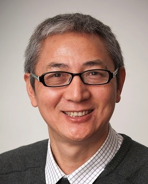 Shengrong Guo (郭圣荣)合作论文数School of Pharmaceutical Sciences, Shanghai Jiao Tong University7Yue Hu合作论文数6
 Sun Dawei合作论文数6Rongjiao Shao合作论文数5
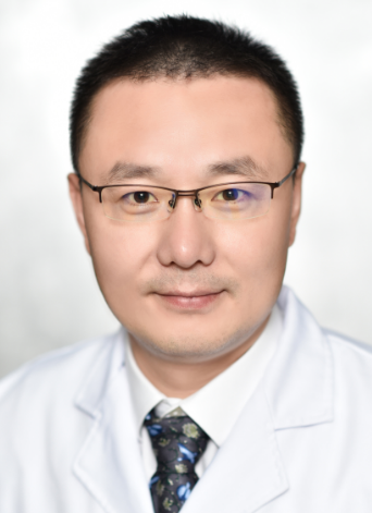 Hanbing Shang (尚寒冰)合作论文数Ruijin Hospital, Shanghai Jiao Tong University School of Medicine5
 Quanhong Zhou (周全红)合作论文数Shanghai Sixth People’s Hospital Affiliated to Shanghai Jiao Tong University School of Medicine4
 Li Wang (王莉)合作论文数Shanghai Sixth People's Hospital4Dawei Sun (孙大伟)合作论文数3
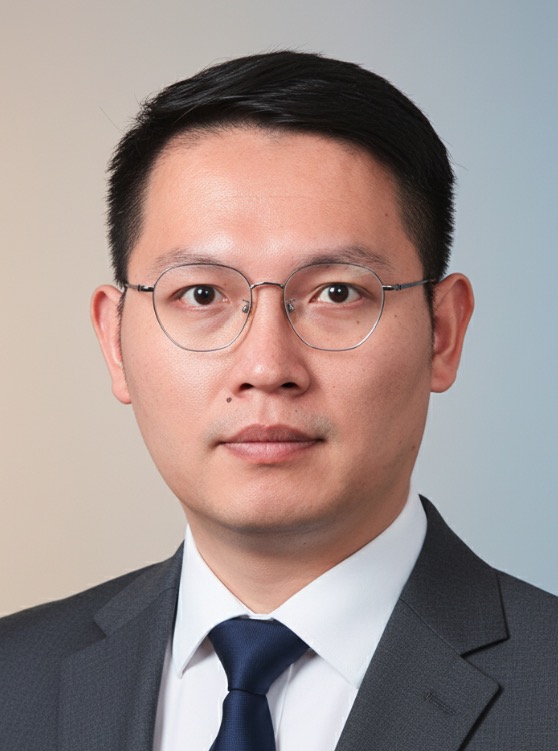 Zhu Jin (金竹)合作论文数School of Pharmaceutical Sciences, Shanghai Jiao Tong University3
 Yuanyuan Shen (沈圆圆)合作论文数Shenzhen University,Health Science Center3Yiyang Xia (夏艺洋)合作论文数2Qixia Lu合作论文数2Chenming Zou合作论文数2Yongxu Wei合作论文数2Xu Tianhua合作论文数2Ping Zeng合作论文数2
 Tao Xu (许涛)合作论文数Shanghai Sixth People's Hospital Affiliated to Shanghai Jiaotong University2Mingjian Liu合作论文数2
 Yijun Cheng合作论文数Cent South Univ, Sch Informat Sci & Engn, Changsha, Hunan, Peoples R China2Xiaotao Xu合作论文数2Zhenghong Chen合作论文数2Fei-Fei Zhang合作论文数2LI Qingqing (李晴晴)合作论文数2Aihua Qi (祁爱花)合作论文数2Meng-Meng Wang合作论文数2Shaohui Xu合作论文数2YiXia Yang (杨懿霞)合作论文数2
 Wang Wuyang合作论文数Department of Physiology and Biophysics, Dalhousie University2Guo Aijie合作论文数2
 Yi Li (李轶)合作论文数Shanghai Ninth People’s Hospital, Shanghai Jiao Tong University School of Medicine2
 Yan Wang (王彦)合作论文数School of Pharmaceutical Sciences, Shanghai Jiao Tong University2
 Aizhong Wang (王爱忠)合作论文数Shanghai Sixth People's Hospital2Ying Xue合作论文数2Yanhong Xing合作论文数2Yang Zhang合作论文数1Jiansong Qi合作论文数1Huijie Han合作论文数1Majdi Al Amili合作论文数1Zhongheng Sui合作论文数1Lijun Hu合作论文数1Qi-Hui Shao合作论文数1Liye Yi合作论文数1Xiaorong Gao合作论文数1Ziqian Zhang合作论文数1Sheng Liang合作论文数1Bin Chen合作论文数1




