Manual processing of spinal CTA images for detecting dural arteriovenous fistulas (SDAVF) is laborious and operator-dependent. We developed an automated AI system (SDAVFdoc) that integrates a 3D convolutional neural network with anatomical prior knowledge to both identify SDAVF and localize the fistula site in a multicenter study of 718 patients. The system sequentially segments spinal structures, localizes the fistula region using anatomical priors, and finally classifies SDAVF likelihood via DenseNet within the foramina. The draining vein cluster segmentation model, using a threshold of 42.5, achieved high accuracy in distinguishing SDAVF cases, with F1-scores ranging from 0.932 to 0.960 across test sets. The DenseNet-based fistula detection model showed a high AUC of 0.928-0.954 across test sets. Compared to technicians, the system reduced processing time from 40.75 ± 11.57 min to 1.05 ± 0.29 min (P < 0.001) and clicks from 761.80 ± 202.05 to 9.68 ± 2.12 (P < 0.001), greatly streamlining clinical workflows. This AI-driven approach enables fast, accurate screening and localization of SDAVF.
The widespread adoption of computed tomography has increased the detection of lung nodules. However, deep learning methods for classification of benign and malignant nodules often fail to comprehensively integrate global and local features, and most of these methods have not been validated through clinical trials. Here we developed DeepFAN, a transformer-based model trained on more than 10,000 pathology-confirmed nodules, and conducted a multireader, multicase clinical trial (Chinese Clinical Trial Registry: ChiCTR2400084624) to evaluate its efficacy in assisting junior radiologists. DeepFAN achieved diagnostic area under the curve (AUC) values of 0.939 (95% CI 0.930-0.948) on an internal test set and 0.954 (95% CI 0.934-0.973) on a clinical trial dataset involving 400 cases across three independent medical institutions. Explainability analysis indicated higher contributions from global than local features. The average performance of 12 readers improved significantly: by 10.9% (95% CI 8.3-13.5%) for AUC, 10.0% (95% CI 8.9-11.1%) for accuracy, 7.6% (95% CI 6.1-9.2%) for sensitivity and 12.6% (95% CI 10.9-14.3%) for specificity (all P < 0.001). Nodule-level interreader diagnostic consistency improved from fair to moderate (overall κ: 0.313 versus 0.421; P = 0.019). These results indicate that DeepFAN can effectively assist junior radiologists and could help to homogenize diagnostic quality and reduce unnecessary follow-up of patients with indeterminate pulmonary nodules.
OBJECTIVE:Radiologists often face challenges in differentiating benign from malignant sacral bone lesions due to their similar imaging characteristics. This study aimed to develop an ensemble deep learning (DL) model that can preoperatively distinguish between benign and malignant sacral tumors using noncontrast computed tomography images. MATERIALS AND METHODS:Preoperative sacral CT scans from 569 patients with confirmed sacral lesions were analyzed. Data from Center 1 were utilized in model development and internal test via fivefold cross-validation, and those from Centers 2 and 3 were employed in external test. Various ensemble models combining human-readable interpretation and DL were developed. The diagnostic performance of the models and radiologists was assessed using metrics such as precision, recall, accuracy, area under the curve (AUC), F1 score, and confusion matrix. Furthermore, the clinical benefits derived from radiologists' interpretations and supported by the DL model were evaluated. RESULTS:The ensemble model, which integrates 3D-DenseNet121 with human interpretation, exhibited the most robust performance. The ensemble model demonstrated high performance on the internal and external test sets and achieved AUCs of 0.9139 and 0.8713, F1 scores of 0.9054 and 0.8571, precision of 0.9041 and 0.8824, recall of 0.9136 and 0.8333, and accuracy of 0.8630 and 0.8182, respectively. Across the external test cohort, all radiologists experienced improvements in AUC, accuracy, sensitivity, and specificity. Notably, junior radiologists demonstrated significant improvements compared with senior radiologists. CONCLUSION:The potential clinical application of the DL model lies in its capacity to considerably enhance the diagnostic efficiency of radiologists. CRITICAL RELEVANCE STATEMENT:This study presents the first ensemble deep learning model integrating 3D-DenseNet121 with radiologists' interpretation for preoperative differentiation of sacral tumors on noncontrast CT that improved diagnostic performance across all experience levels, particularly for junior radiologists. KEY POINTS:First artificial intelligence-radiologist ensemble for noncontrast computed tomography (NCCT)-based sacral tumor classification. Boosts all radiologists' performance, with the greatest gains for juniors, potentially reducing referrals. Enables reliable NCCT diagnosis, overcoming contrast/magnetic resonance imaging dependency in musculoskeletal oncology.
This study aimed to investigate the application of T2-based MRI delta-radiomics as a novel predictive tool for neoadjuvant chemotherapy (NACT) response in patients with osteosarcoma. We retrospectively analyzed data from 152 patients with pathologically confirmed osteosarcoma who underwent NACT at our institution. Axial T2-weighted MRI sequences were acquired both at baseline (pre-NACT) and after NACT (post-NACT). After image segmentation and preprocessing, 1158 radiomic features were extracted from the T2-weighted images. We developed and compared four models: the conventional quantitative imaging features-based model (CQIF model), the pre-NACT radiomics model, the post-NACT radiomics model, and the Delta-Radiomics model. Model performance was assessed using the area under the receiver operating characteristic curve (AUC) and accuracy (ACC). Based on histopathological assessment, patients were divided into two groups: good responders (n = 57) and poor responders (n = 95). Significant differences in change rates for tumor diameter and volume were observed between the two groups (P < 0.001). The Delta-Radiomics model demonstrated superior predictive performance compared to other models, achieving an AUC of 0.796, ACC of 0.756, sensitivity of 0.529, specificity of 0.893, PPV of 0.750, and NPV of 0.758 in the test set. However, the Delong test revealed no significant differences among these models, except between the Post-NACT and Delta-Radiomics models (P < 0.05). T2-based MRI delta-radiomics showed strong predictive value for NACT response in patients with osteosarcoma. This model holds potential for guiding clinical decision-making and improving patient management by identifying responders early in the treatment course.
Thyroid-associated ophthalmopathy (TAO), the most common orbital disease in adults, is a specific autoimmune condition closely associated with thyroid dysfunction, primarily affecting orbital fat and extraocular muscles. Multiparametric magnetic resonance imaging (MRI) holds unique value in diagnosing TAO, staging disease activity and progression evaluation by providing multidimensional information on tissue morphology, edema, fat infiltration, and fibrosis. This review examines conventional and advanced MRI techniques—such as T1WI, T2WI, diffusion-weighted imaging, dynamic contrast-enhanced MRI and magnetization transfer imaging—for their research progress and clinical utility. Recent advances in artificial intelligence (AI) applications for orbital MRI in TAO are also discussed. AI-enhanced multiparametric orbital MRI facilitates the comprehensive evaluation of TAO, covering image acquisition, quantification of metrics, and the establishment of predictive assessment models. Despite these advancements, challenges persist, such as the need to standardize quantitative parameters, validate findings through larger multicenter studies, and develop more efficient scanning protocols. Future research should prioritize parameter standardization, automated analysis workflows, and integration of multiparametric data to advance personalized TAO management.
Purpose To develop a fully automated hybrid approach to predict sacral tumor types from preoperative noncontrast CT (NCCT ) images. Materials and Methods In this retrospective, multicenter study, scans were available in 690 patients who had histopathologically confirmed preoperative sacral NCCT performed between January 2011 and May 2024. A fully automated hybrid model integrated two deep convolutional neural network models (model 1 and model 2) through a fully automated pipeline. Model 1 segments tumors and hip bones automatically from NCCT images, producing masks that are used by model 2. For the first time, the hip bone was used as a reference frame for tumor localization. The second model, CL-MedImageNet, is an innovative six-classification model that allows the simultaneous input of tumor images, clinical data, and location information. This streamlined, automated system ensures efficient data integration and processing between the two models. The efficacy of the model was assessed in comparison to that of radiologists, using metrics including the area under the curve (AUC), F1 score, and confusion matrix. Results In all, 690 patients (mean age, 46 years ± 17 [SD]; 377 male patients) were included. Segmentation achieved mean Dice coefficients of 0.82 ± 0.11 (validation), 0.81 ± 0.12 (internal test), and 0.81 ± 0.12 (external test) after postprocessing; interobserver Dice coefficient was 0.96. The CL-MedImageNet classifier attained macro average AUCs of 0.89 (95% CI: 0.83, 0.93), 0.88 (95% CI: 0.84, 0.92), and 0.87 (95% CI: 0.79, 0.92) in validation, internal, and external test sets, respectively, with macro average F1 scores of 0.63, 0.63, and 0.56. The highest achieved precision and sensitivity were both 0.66 across all sets. CL-MedImageNet outperformed radiologists (macro average AUCs, 0.87 vs 0.80, P = .002; 0.87 vs 0.83, P = .45). Conclusion The fully automated NCCT-based CL-MedImageNet pipeline demonstrated high segmentation accuracy and robust six-class classification, outperforming expert radiologists. Keywords: Applications - CT, Deep Learning, Radiomics, Segmentation, Skeletal-Axial, Pelvis, Sacral Tumors Supplemental material is available for this article. © The Author(s) 2026. Published by the Radiological Society of North America under a CC BY 4.0 license.
To address the limited availability of genetic testing, this study aimed to develop lumbar MRI-radiomics models to predict International Staging System/Revised International Staging System (ISS/R-ISS) stages in newly diagnosed multiple myeloma (ndMM). This two-center retrospective study analyzed 164 ndMM patients. Radiomics models were developed based on single or dual sequence multi-mobility features from T1-weighted imaging (T1-WI) and T2-weighted fat-suppressed (T2-FS) images. A clinical model was also constructed as the baseline for comparison. A fusion model combining optimal radiomics features and peripheral blood biomarkers was subsequently compared against both the clinical model and the radiomics models. Model performance was evaluated using area under the receiver operating characteristic curve (AUC), accuracy, sensitivity, and specificity across training, internal, and external test sets. Independent risk factors were identified via two-step logistic regression. Differences in AUC values were compared using the DeLong test, while the net reclassification improvement (NRI) was applied to assess reclassification performance. The T1_WL model proved to be the most effective for ISS stratification (AUCs: 0.743 internal, 0.707 external), while the cross-region model showed superior predictive power for R-ISS stratification (AUCs: 0.814 internal, 0.763 external). The fusion model demonstrated significantly superior performance compared to both the radiomics model (P < 0.001) and clinical model (P < 0.001), achieving the highest AUCs of 0.869 (internal) and 0.825 (external). Significant net reclassification improvements were also observed (NRI = 1.536 internal, 1.296 external; all P < 0.001). A lumbar MRI-radiomics strategy enables practical, non-invasive risk stratification of nd-MM in resource-constrained environments.
Cancer remains a leading cause of global mortality, with therapeutic resistance posing a formidable clinical obstacle. Inorganic nanotheranostics-multifunctional platforms integrating diagnostic and therapeutic capabilities-offer a promising strategy to overcome this barrier. This review systematically examines how distinct classes of inorganic nanomaterials (including iron, gadolinium, titanium, gold, manganese, carbon, and silicon-based systems) are engineered to counteract specific resistance mechanisms through unique physicochemical properties and biological interactions. We highlight their roles in enhancing drug delivery, modulating the tumor microenvironment, enabling precise imaging, and synergizing with various treatment modalities such as chemotherapy, radiotherapy, and immunotherapy. Advances in stimulus-responsive design, targeted delivery, and combinatorial therapies are discussed. Finally, we critically evaluate the translational challenges-including pharmacokinetics, long-term safety, and manufacturing scalability-and outline future directions toward clinically effective, personalized nanotheranostic interventions in oncology.
RATIONALE AND OBJECTIVES:Accurate proptosis measurement change is critical for managing thyroid-associated ophthalmopathy (TAO), yet traditional Hertel exophthalmometry suffers from inter-observer inconsistency. This study aimed to investigate the efficacy of magnetic resonance imaging (MRI)-based proptosis measurement and developed an automated tool to enhance diagnostic reproducibility. MATERIALS AND METHODS:We retrospectively analyzed data from 250 patients with TAO prospectively enrolled at Center 1 between December 2023 and June 2025. Proptosis was independently measured by three radiologists (R1-R3) using both 2D and 3D MRI sequences; clinical measurements via Hertel exophthalmometry were obtained within one week for comparison. An automated measurement tool was developed and internally validated using manual annotations from the senior radiologist (R1) as the ground truth. To assess generalizability, the tool was externally validated on axial 2D MRI datasets from two independent cohorts: Center 2 (n = 65) and Center 3 (n = 48). RESULTS:Orbital MRI-based measurements demonstrated excellent inter-observer agreement for both 2D (ICC: 0.948; 95% CI: 0.938-0.957) and 3D sequences (ICC: 0.954; 95% CI: 0.939-0.966). MRI findings showed a strong correlation with Hertel exophthalmometry (Spearman's ρ = 0.76-0.80). The automated tool maintained high performance across all cohorts, achieving concordance correlation coefficients (CCC) of 0.928-0.973 and intraclass correlation coefficients (ICC) of 0.916-0.973 in validation. CONCLUSION:This MRI-based automated tool provides a reliable, complementary, and standardized alternative to Hertel exophthalmometry, particularly useful when clinical measurements are discordant or demand high precision for longitudinal monitoring. It demonstrated excellent accuracy, efficiency, and cross-center generalizability.
OBJECTIVE:Cervical cancer remains a major public health challenge among women in China. This study aimed to comprehensively assess long-term trends and future projections of cervical cancer burden in China. METHODS:Data on cervical cancer in China from 1990 to 2023 were extracted from Global Burden of Disease Study2023. Prevalence, incidence, deaths, years lived with disability (YLDs), years of life lost (YLLs), disability-adjusted life years (DALYs), and age-standardized rates (ASRs) were analyzed. Temporal trends were evaluated using estimated annual percentage change (EAPC) and Joinpoint regression. Das Gupta decomposition quantified the contributions of population growth, aging, and epidemiological change. Nordpred was used to project deaths and DALYs from 2024 to 2045. RESULTS:From 1990 to 2023, prevalent cervical cancer cases increased from 445,795 (95% uncertainty interval [UI]=301,749-642,609) to 752,306 (95% UI=481,273 to 1,011,544). YLDs increased by 62%, whereas YLLs decreased by 4%. Overall DALYs decreased slightly from 1,457,115 (95% UI=1,070,025 to 2,064,940) to 1,430,054 (95% UI=896,628 to 1,874,757), while deaths increased by 16%. Age-standardized DALYs rate (ASDR) declined from 294.14 to 132.72 per 100,000 population, with an EAPC of -2.43 (95% confidence interval=-2.54 to -2.33). Decomposition showed that population aging and growth offset favorable epidemiological changes, and projections suggested continued declines in ASDR with relatively stable deaths through 2045. CONCLUSION:Despite sustained declines in ASRs, the absolute burden of cervical cancer in China remains considerable due to demographic shifts. These findings underscore the urgent need to strengthen human papillomavirus vaccination coverage and expand equitable cervical cancer screening programs.
PURPOSE:To develop and validate a non-invasive magnetic resonance imaging (MRI)-based deep learning and radiomics approach for the preoperative differentiation of p53 abnormal (P53abn) endometrial cancer, facilitating refined risk stratification for personalized treatment planning. METHODS:In this retrospective multi-institutional analysis, we examined data from 920 patients with histologically confirmed endometrial cancer who underwent preoperative MRI. A two-stage deep learning architecture (V-Net followed by VB-Net) was developed to automate tumor delineation across three participating centers. Extracted radiomic features from these segmented regions were leveraged to build machine learning classifiers-support vector machines (SVM), random forests (RF), logistic regression (LR), and decision trees (DT)-aimed at distinguishing p53-abnormal tumors from other molecular subtypes. Model efficacy was assessed using the Dice similarity coefficient (DSC) for segmentation accuracy and the area under the receiver operating characteristic curve (AUC) for classification performance. RESULTS:The automated segmentation achieved Dice similarity coefficients (DSC) of 77.4%, 84.9%, and 80.1% on T2-weighted imaging (T2WI), diffusion-weighted imaging (DWI), and contrast-enhanced T1-weighted imaging (CE-T1WI) sequences, respectively. Among the four classification models developed, the RF classifier demonstrated the highest AUC values in both internal CV cohort (0.924) and external test cohort (0.863). No statistically significant differences were observed between automated and manual segmentation results across all models (P = 0.109-0.454). CONCLUSION:The integrated deep learning and radiomics pipeline developed in this study provides a promising non-invasive approach for preoperative risk stratification of endometrial cancer. The model has demonstrated high performance in identifying the P53abn subtype, offering a valuable tool to support personalized treatment planning.
BACKGROUND:Accurate prediction of early recurrence (ER) after radical resection remains a critical challenge in pancreatic ductal adenocarcinoma (PDAC). This study aimed to develop and validate an integrated radiomic-pathology (Rad-Path) model for ER prediction and to elucidate its underlying biological mechanisms. METHODS:A retrospective cohort of 225 PDAC patients who underwent R0 resection was included. Preoperative CT images and whole-slide images (WSI) were collected for the extraction of radiomic features and computational pathology features. Selected features were used to develop 11 distinct machine learning models. The SHapley Additive exPlanations (SHAP) algorithm was employed to evaluate feature importance. Single-cell RNA sequencing (scRNA-seq) and spatial transcriptomics (ST) were performed on prospectively collected specimens. RESULTS:The final Rad-Path model achieved AUCs of 0.851 and 0.814 in the internal and external validation cohorts, respectively. The predicted ER group was specifically linked to the enrichment of fibroblasts and pancreatic stellate cells, as well as dysregulation in extracellular matrix (ECM)-related pathways. This finding was validated histopathologically, as predicted ER patients predominantly displayed a "reactive-dominant" phenotype marked by abundant activated fibroblasts and ECM deposition. CONCLUSION:Our study offers a high-performance predictive model for ER in PDAC and establishes ECM remodeling as a key biological mechanism underlying the predictions.
BACKGROUND:Accurate segmentation of pelvic and sacral tumors (PSTs) in multi-sequence magnetic resonance imaging (MRI) is essential for effective treatment and surgical planning. PURPOSE:To develop a deep learning (DL) framework for efficient segmentation of PSTs from multi-sequence MRI. MATERIALS AND METHODS:This study included a total of 616 patients with pathologically confirmed PSTs between April 2011 to May 2022. We proposed a practical DL framework that integrates a 2.5D U-net and MobileNetV2 for automatic PST segmentation with a fast annotation strategy across multiple MRI sequences, including T1-weighted (T1-w), T2-weighted (T2-w), diffusion-weighted imaging (DWI), and contrast-enhanced T1-weighted (CET1-w). Two distinct models, the All-sequence segmentation model and the T2-fusion segmentation model, were developed. During the implementation of our DL models, all regions of interest (ROIs) in the training set were coarse labeled, and ROIs in the test set were fine labeled. Dice score and intersection over union (IoU) were used to evaluate model performance. RESULTS:The 2.5D MobileNetV2 architecture demonstrated improved segmentation performance compared to 2D and 3D U-Net models, with a Dice score of 0.741 and an IoU of 0.615. The All-sequence model, which was trained using a fusion of four MRI sequences (T1-w, CET1-w, T2-w, and DWI), exhibited superior performance with Dice scores of 0.659 for T1-w, 0.763 for CET1-w, 0.819 for T2-w, and 0.723 for DWI as inputs. In contrast, the T2-fusion segmentation model, which used T2-w and CET1-w sequences as inputs, achieved a Dice score of 0.833 and an IoU value of 0.719. CONCLUSIONS:In this study, we developed a practical DL framework for PST segmentation via multi-sequence MRI, which reduces the dependence on data annotation. These models offer solutions for various clinical scenarios and have significant potential for wide-ranging applications.
To investigate the knowledge and expectations of abdominopelvic MR elastography (MRE) among radiologists from multiple institutions, and to rate the image quality of MRE maps. Radiologists from Beijing and Tianjin were invited to participate in an online survey. Full MRE maps of the uterus, prostate, pancreas, liver, and kidney from 93 research papers published between 2017 and 2024 were displayed for image quality rating, blinded to the MRE systems. Before and after the image-review session, the participants’ knowledge and expectations about abdominopelvic MRE were investigated. Eighty-one radiologists finalized the survey, all but one from tertiary hospitals. Their knowledge of MRE mainly came from the literature and conferences, and only 29.6
OBJECTIVE:To compare the relative safety and efficacy of the Chiba needle and the Trocar needle in CT-guided microcoil localization of pulmonary nodules. METHODS:A retrospective study was conducted on 118 patients who underwent CT-guided microcoil localization and subsequent video-assisted thoracoscopic surgery (VATS) resection from September to November 2023. Patients were divided into the Chiba needle group (n = 75) and the Trocar needle group (n = 43). Characteristics of patients, lesions, procedures, and surgeries were statistically analyzed. Univariate and multivariate logistic regression analyses were used to determine potential risk factors for technical failure and complications. RESULTS:The success rate of localization was 97.3 % for the Chiba needle group and 100 % for the Trocar needle group, with no significant difference (p = 0.533). Complications included pneumothorax in 16 % of the Chiba group and 18.6 % of the Trocar group (p = 0.914), and parenchymal hemorrhage in 25.3 % and 41.9 % respectively (p = 0.098). There were no significant differences in puncture depth, procedure duration, or interval between procedure and surgery. Multivariate logistic regression analyses identified longer puncture depth as a risk factor both for pneumothorax (p = 0.027) and parenchymal hemorrhage (p = 0.006), while the Trocar needle was identified as a risk factor for parenchymal hemorrhage (p = 0.035). CONCLUSION:This study found no significant difference in the effectiveness of Chiba and Trocar needles for preoperative CT-guided lung nodule localization. Both needles showed high success rates and comparable pneumothorax profiles. However, the Trocar needle was found associated with a higher incidence of parenchymal hemorrhage.
This study developed an end-to-end deep learning (DL) model using non-enhanced MRI to diagnose benign and malignant pelvic and sacral tumors (PSTs). Retrospective data from 835 patients across four hospitals were employed to train, validate, and test the models. Six diagnostic models with varied input sources were compared. Performance (AUC, accuracy/ACC) and reading times of three radiologists were compared. The proposed Model SEG-CL-NC achieved AUC/ACC of 0.823/0.776 (Internal Test Set 1) and 0.836/0.781 (Internal Test Set 2). In External Dataset Centers 2, 3, and 4, its ACC was 0.714, 0.740, and 0.756, comparable to contrast-enhanced models and radiologists (P > 0.05), while its diagnosis time was significantly shorter than radiologists (P < 0.01). Our results suggested that the proposed Model SEG-CL-NC could achieve comparable performance to contrast-enhanced models and radiologists in diagnosing benign and malignant PSTs, offering an accurate, efficient, and cost-effective tool for clinical practice.
BackgroundOsteosarcoma (OS) is the most common primary malignant bone tumor. Exploring quantitative parameters that reflect the outcome of neoadjuvant chemotherapy (NACT) in patients with OS can help advance the treatment of patients.PurposeTo explore the role of T2-weighted (T2W) magnetic resonance imaging (MRI) radiogenomic features in characterizing changes in patients with OS and on NACT.Material and MethodsA total of 21 patients with OS were examined retrospectively and divided into a poor-response group (n = 13) and a good-response group (n = 8). A total of 98 radiomic features and 31 gene expression profiles were analyzed for each patient. Age, sex, alkaline phosphatase, pathologic type, tumor size, and tumor location were also analyzed. Comparisons between the good- and poor-response groups were made using the t-test, Mann-Whitney U test, or Fisher's exact test. The relationships between radiomic features and gene expression profiles were conducted using Spearman's correlative analyses.ResultsStatistical differences in 19 radiomics features and glutathione-s-transferase 1 were found between the good- and poor-response groups (P < 0.05). The receiver operating characteristic curve showed that four NGTDM busyness features had the best performance in predicting the NACT of patients with OS, with an area under the curve of 0.788, sensitivity of 0.750, and specificity of 0.923. Correlation analysis showed that the HLA_I, CD274, GSTP1, and CCND3 were significantly correlated with one or more radiomics features (P < 0.05).ConclusionThe T2W MRI radiogenomic features can be used as biomarkers for the early response evaluation of NACT in OS. This is the first study to analyze the association of T2 radiogenomic features with NACT in patients with OS to assist in the assessment of NACT.
The increasing complexity of lung surgeries necessitates the need for enhanced imaging support to improve the precision and efficiency of preoperative planning. Despite the promise of 3D reconstruction, clinical adoption remains limited due to time constraints and insufficient validation. To address this, we evaluate an artificial intelligence-driven 3D reconstruction system for pulmonary vessels and bronchi in a retrospective, multi-center multi-reader multi-case study. Using a two-stage crossover design, ten thoracic surgeons assess 140 cases with and without the system's assistance. The system significantly improves the accuracy of anatomical variant identification by 8% (p < 0.01), reducing errors by 41%. Improvements in secondary endpoints are also observed. Operation procedure selection accuracy is improved by 8%, with a 35% decrease in errors. Preoperative planning time is decreased by 25%, and user satisfaction is high at 99%. These benefits are consistent across surgeons of varying experience. In conclusion, the artificial intelligence-driven 3D reconstruction system significantly improves the identification of anatomical variants, addressing a critical need in preoperative planning for thoracic surgery.
Early prediction of chemotherapy efficacy continues to be a significant challenge in osteosarcoma patients. This study aims to investigate the impact of habitat analysis based on Intravoxel Incoherent Motion (IVIM) and Dynamic Contrast-Enhanced magnetic resonance imaging (DCE-MRI) for predicting neoadjuvant chemotherapy (NACT) response in osteosarcoma patients. We prospectively analyzed 71 patients diagnosed with osteosarcoma who underwent pre-treatment IVIM and DCE-MRI. IVIM metrics, including pseudo-diffusion coefficient (ADC_fast), diffusion coefficient (ADC_slow), and fractions of ADC_fast (f_ADC_fast), were evaluated alongside DCE-MRI parameters such as Ktrans, Ve, and Kep. We assessed the predictive value of habitat regions’ features from these functional images for NACT outcomes. Model performance was measured using the area under the receiver operating characteristic curve (AUC). Among the data, 39 patients were classified as poor responders and 32 as good responders. Demographic factors, including age, sex, and lesion location, showed no significant differences between groups (P > 0.05). Our findings indicated that an increase in volume in habitat 3, coupled with decreases in f_ADC_fast in habitat 1, ADC_slow in habitat 5, and f_ADC_fast in habitat 5 was valuable indicators for predicting the response of osteosarcoma to NACT. The Habitat Feature-Based IVIM-Bi (HAB-IVIM-Bi) model demonstrated the best performance, as indicated by the Delong test, which yielded all P values < 0.05. It achieved an AUC of 0.797, accuracy of 0.761, sensitivity of 0.719, and specificity of 0.795. Habitat analysis based on multiparametric MRI demonstrates promising potential for non-invasive prediction of NACT response in osteosarcoma. The HAB-IVIM-Bi model may serve as a valuable tool for treatment stratification and personalized therapy planning.
 Lei Chen (陈雷)合作论文数Department of Radiology,Peking University People's Hospital,Beijing ,China45
 Ping Yin合作论文数35Cheng Jin合作论文数28
 Xiangke Du (杜湘珂)合作论文数Peking University People's Hospital24Zhuo Liu (刘卓)合作论文数State Key Laboratory of Synthetical Automation for Process Industries, Northeastern University23
 Yi Wang (王义)合作论文数Peking University Shougang Hospital22
 Lai Yunyao (赖云耀)合作论文数20ZHANG Zhuolu (张卓璐)合作论文数17WeiWei Qi (綦维维)合作论文数17
 Chao Sun (孙超)合作论文数北京协和医学院北京医院17
 Wanqing Liao (廖万清)合作论文数Naval Medical University13LiBao Hu (胡立宝)合作论文数12ChuanXi Hao (郝传玺)合作论文数12Jin Cheng (程瑾)合作论文数12Caizhen Feng (冯彩珍)合作论文数11Yinli Zhang合作论文数11
 Jianliu Wang (王建六)合作论文数Peking University People's Hospital11
 Yingjiang Ye (叶颖江)合作论文数Peking University People's Hospital11
 Ning Mao合作论文数10Sicong Wang合作论文数9Fei Zheng合作论文数9WANG Xu (王煦)合作论文数9XiaoYi Liu (刘晓翌)合作论文数9Chen Chen (陈尘)合作论文数9
 Jianping Xu合作论文数Origins Institute, Faculty of Science, McMaster University;Microbiology and Molecular Genetics, Duke University;Institute of Microbiology, Jiangxi Academy of Sciences9Xinyi Gou合作论文数8Fan Chai合作论文数8Xin Zhi (郅新)合作论文数8Jing Wu (武靖)合作论文数8Jian Gao (高健)合作论文数8
 Xin Yu (于欣)合作论文数Alberta Institute, Wenzhou Medical University8
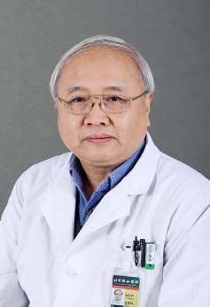 Zhengyu Jin (金征宇)合作论文数Peking Union Medical College;Peking Union Medical College Hospital8Xia Liu (刘霞)合作论文数8Ling FU (付玲)合作论文数7
 Weihua Pan (潘炜华)合作论文数The First Affiliated Hospital of Naval Medical University7
 Jie Tian (田捷)合作论文数School of Medical Science and Engineering, Beihang University7
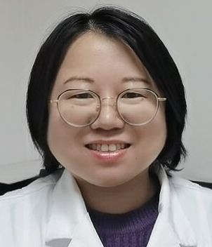 Ying Liu (刘颖)合作论文数Radiology Department, Peking University Third Hospital7Shengcai Wei合作论文数6Haihong LONG (隆海红)合作论文数6HAO Qi (郝琪)合作论文数5Qingyi Qi合作论文数5Xun Yao合作论文数5Xiaoxuan Jia合作论文数5Jingjing Cui合作论文数Department of Research and Development, Shanghai United Imaging Intelligence, Co., Ltd5Hao CHEN (陈皓)合作论文数5Yu Kang (康钰)合作论文数5Ye Sun (孙烨)合作论文数5




