目的 探讨携带免疫检查点VISTA抗体的溶瘤病毒HSV-1-ab-VISTA在小鼠(C57BL/6J)移植瘤模型中的抗肿瘤效果.方法 利用CRIPSR-Cas9基因编辑技术制备携带VISTA分子抗体的HSV-1-ab-VISTA溶瘤病毒;琼脂糖凝胶电泳检测VISTA抗体的基因组片段大小;蛋白免疫印迹法检测VISTA抗体表达情况;构建小鼠移植瘤模型,游标卡尺测量溶瘤病毒治疗后瘤体大小的变化,且进行分析.结果 成功构建了携带免疫检查点VISTA抗体的溶瘤病毒HSV-1-ab-VISTA,和野生病毒相比,病毒增殖和裂解癌细胞的能力没有差别,HSV-1-ab-VISTA溶瘤病毒治疗显著缩小了小鼠的肿瘤大小.结论 HSV-1-ab-VISTA溶瘤病毒可抑制小鼠结直肠移植瘤的生长.
目的 探讨T细胞活化V结构域Ig抑制因子(VISTA)和程序性死亡受体-1(PD-1)信号通路联合阻断在大剂量单次X射线照射白化小鼠(BALB/C)小鼠双移植瘤中诱导远隔效应可能的机制.方法 构建BALB/C小鼠双移植瘤模型,采用液相悬浮微球蛋白分析技术检测治疗前后细胞因子表达变化;流式细胞术检测小鼠移植瘤模型治疗前后瘤内和脾脏免疫细胞比例的变化;游标卡尺测量瘤体大小的变化.统计学分析以上指标.结果 X射线大剂量照射联合VISTA和PD-1拮抗剂(CA170)治疗显著缩小小鼠第1肿瘤和第2肿瘤瘤体,提高了细胞因子IFN-γ表达,并增加了肿瘤内CD8+T细胞的浸润和扩增.结论 大剂量X射线照射联合CA170治疗可以显著增强X射线大剂量照射对小鼠结肠癌的抑制,并有效地改善肿瘤微环境.
BACKGROUND:Intrahepatic cholangiocarcinoma (ICC) is the second most common primary liver cancer in humans after hepatocellular carcinoma and a rare epithelial malignancy that results in a poor prognosis. According to the Liver Cancer Study Group of Japan classification, ICC can be divided into three types: Mass-forming (MF) type, periductal-infiltrating (PI) type, and intraductal-growth type. The MF type is the most common, accounting for 57.1-83.6% of ICCs. Nevertheless, little is known about the epidemiology and treatment of MF ICC.AIM:To examine the prognostic factors for patients with MF ICC.METHODS:We carried out a retrospective analysis of consecutive patients with MF ICC treated at the Faculty of Hepato-Pancreato-Biliary Surgery of Chinese PLA General Hospital between January 2008 and December 2018. According to the treatment received, the patients were divided into either a resection group or an exploration group.RESULTS:The pooled 1-, 3-, and 5-year survival rates in the 68 patients with MF ICC were 66.5%, 36.3%, and 9.3%, respectively. Univariate analysis revealed that surgical resection (P < 0.001), nodal metastasis (P < 0.001), tumor location (P = 0.039), vascular invasion (P < 0.001), ascites (P < 0.001), and differentiation (P = 0.009) were significantly associated with the prognosis and survival of MF ICC. Multivariate analysis revealed that ascites (hazard ratio [HR] = 5.6, 95% confidence interval [CI]: 1.6-18.9, P = 0.006) and vascular invasion (HR = 2.5, 95%CI: 1.0-6.1, P = 0.045) were independent risk factors for MF ICC. The pooled 1-, 3-, and 5-year survival rates in the 19 patients of the exploration group were 5.3%, 5.3%, and 0, respectively. Among the 49 patients who underwent surgical resection, the pooled 1-, 3-, and 5-year survival rates were 93.5%, 49.7%, and 14.4%, respectively. Univariate and multivariate analyses revealed that vascular invasion (HR = 3.1, 95%CI: 1.2-8.5, P = 0.024) and nodal metastasis (HR = 3.2, 95%CI: 1.4-7.6, P = 0.008) were independent prognostic risk factors for surgical resection patients.CONCLUSION:The prognosis of MF ICC patients is dismal, especially those with ascites or vascular invasion. Surgical resection is a key factor in improving overall survival in patients with MF ICC, and vascular invasion and lymph node metastasis affect the efficacy of surgical resection.
Objective:To evaluate the surgical efficacy and to explore the prognostic factors of gallbladder cancer.Methods:Clinical data of 162 patients with gallbladder cancer admitted to the First Medical Center of Chinese PLA General Hospital from January 2013 to January 2015 were retrospectively analyzed. Among them, 77 patients were male and 85 female, aged (61±11) years on average. The informed consents of all patients were obtained and the local ethical committee approval was received. 9 cases were classified as stage T1a, 10 cases of stage T1b, 28 cases of stage T2, 103 cases of stage T3 and 12 cases of stage T4, respectively. Intraoperative and postoperative conditions of all patients were observed. The factors affecting survival and prognosis were analyzed. Survival analysis was performed by Kaplan-Meier method and Log-rank test. The independent prognostic factors were identified by Cox proportional hazards regression model.Results:Simple cholecystectomy was performed in 9 cases with stage T1a. Among the 153 cases with stage T1b and above, radical resection was conducted in 81 cases, palliative surgery in 45 cases and abdominal exploration in 27 cases. 140 patients were followed up after surgery. The median survival time was 12(9-15) months. The 1-, 3- and 5-year cumulative survival rates were 51.4%, 25.0% and 22.1%, respectively. The 5-year survival rates of patients with stage T2b undergoing wedge resection and patients receiving segment Ⅳb+Ⅴresection were 44.4% and 100.0%, where significant difference was observed between two groups (χ2=9.00, P<0.05). The 5-year survival rate of patients with stage T3 after radical resection and more than 6 lymph nodes dissection was 33.3%, significantly higher than 4.5% in their counterparts with less than 6 lymph nodes dissection (χ2=4.17, P<0.05). Cox proportional hazards regression model analysis showed that TNM staging and R0 resection were the independent influencing factors for the survival of patients with stage T1b and above gallbladder cancer after radical resection (HR=1.08, 3.23; P<0.05).Conclusions:Segment Ⅳb+Ⅴresection can bring clinical benefits to patients with stage T2b gallbladder cancer. Local dissection of more than 6 lymph nodes can benefit patients with stage T3 gallbladder cancer. TNM staging and R0 resection are the independent prognostic factors of patients with stage T1b and above gallbladder cancer after radical resection.
中晚期肝癌手术风险大,预后差。随着精准医学的发展,术前新辅助及转化治疗或将改变肝癌围手术期患者的治疗模式。本文报道1例累及肝后腔静脉的左肝巨大肝细胞肝癌,经过术前联合应用抗血管生成的多靶点激酶抑制剂及免疫检查点阻断剂,在两次局部血管栓塞治疗后,成功开展了根治性扩大左半肝切除的病例。
Objective To investigate the risk factors of clinical prognosis of mass-forming intrahepatic cholangiocarcinoma (ICC) patients after surgical resection. Methods Clinical data of 48 patients with mass-forming ICC who underwent surgical resection in PLA General Hospital from January 2010 to October 2016 were retrospectively analyzed. The informed consents of all patients were obtained and the local ethical committee approval was received. Among them, 33 patients were male and 15 were female, aged from 29 to 73 years with a median age of 53 years. Tumors were located in the right lobe of the liver in 37 cases and left lobe in 11 cases. The mean tumor diameter was (6.8±2.6) cm. All patients underwent R0 resection. The clinicopathological parameters were included in the analysis of prognostic factors. Kaplan-Meier survival curve and Log-rank test were conducted for univariate analysis. Cox's proportional hazard model was conducted for multivariate analysis. Results During the postoperative follow-up, 28 cases died and 20 cases survived. The survival time was 3-82 months with a median survival time of 20 months. The 1-, 3- and 5-year survival rate was 95.7%, 50.9% and 14.8%, respectively. Univariate analysis indicated that HBsAg, degree of tumor differentiation, lymph node metastasis and vascular invasion were the impact factors for the survival prognosis (χ2=4.097, 6.415, 11.069, 9.117; P<0.05). Multivariate analysis revealed that lymph node metastasis was an independent risk factor for the prognosis of patients undergoing surgical resection (HR=3.306, 95%CI: 1.184-9.233; P<0.05). Conclusion Lymph node metastasis is an independent risk factor for the clinical prognosis of mass-forming ICC patients after surgical resection. Key words: Intrahepatic cholangiocarcinoma; Hepatectomy; Prognosis; Risk factors
Objective To analyze the clinical features and CT imaging findings of85 patients with drug-induced liver injury (DILI) to provide references for clinical prevention, diagnosis and treatment. Methods A total of 85 patients with DILI diagnosed in the hospital from January 2010 to July 2018 were included in the study. Gender, age, medical history, medication history, clinical features, clinical classification, biochemical indicators of liver function and CT findings were analyzed retrospectively. Results The ratio of male to female in this study was 1:1.18, and most patients were between 41 and60 years old, accounting for 73.75%. The most common clinical manifestations included poor appetite, fatigue, upper abdominal discomfort and loss of appetite. Among the patients with DILI, the proportion of three or more combinedmedication was the highest, followed by traditional Chinese medicines, antibiotics, antiviral drugs and non-steroidal anti-inflammatory drugs. The main clinical type of DILI was hepatocyte type, followed by cholestasis typeand the mixed type. The levels of alanine aminotransferase (ALT) and aspartate aminotransferase (AST) in hepatocytetype and mixed type were significantly higher than those in cholestasis type (P<0.05), but there was no significant difference in the level of glutamyl transferase (GGT) among the three types (P>0.05). CT showed that main types of DILI were diffuse liver injury and focal liver injury, accounting for 52.94%and 43.53%, respectively. Multifocal liver injury was rare, accounting for only 3.53%.The CT number of the them were 31~42 Hu, 41~49 Hu and 45~51 Hu, respectively. CT showed decreased liver parenchyma density, multiple small patchy low-density lesions and multifocal low-density patchy shadow. Conclusion DILI occur in all ages, and the clinical manifestations are not specific. The drugs causing DILI are various. Therefore, clinical use of drugs should focus on medication safety when treating diseases. The main clinical type of DILI is hepatocyte injury, and the CT image is characteristic, which is of certain reference value for clinical diagnosis.
Objectives The study aimed to evaluate the prognostic value of systemic inflammation markers [neutrophil-to-lymphocyte ratio (NLR), platelet-to-lymphocyte ratio (PLR), Prognostic Nutritional Index (PNI)] and hepatic inflammation markers [aspartate aminotransferase-to-platelet ratio index (APRI), γ-glutamyl transferase (γ-GT)/alanine aminotransferase (ALT)] in patients with hepatitis B virus (HBV)-associated hepatocellular carcinoma (HCC) and further to develop a novel prognostic score model.Methods A total of 401 cases with HBV-associated HCC who underwent hepatectomy as initial therapy were included in the analysis. Kaplan–Meier was performed to construct survival curves and receiver operating characteristic (ROC) analysis was used to detect the optimal cut-off value of markers. The prognostic score model was constructed using significant inflammation markers in the Cox model. Each factor was given a score of 1 and patients were stratified according to the scores.Results In the Cox model, α-fetoprotein (AFP), ALT, tumour differentiation, maximum size of tumours, TNM stage, PNI and γ-GT/ALT were independently prognostic factors. We established a preoperative inflammation-based prognostic scoring model combining PNI and γ-GT/ALT. The novel preoperative inflammation-based prognostic score was superior (area under the curve [AUC], 0.659) to 7th tumour-node-metastasis (TNM) stage (AUC, 0.600) despite no statistical significance (p = .1036).Conclusion PNI and γ-GT/ALT are independent predictors for prognosis. The novel prognostic score model based on systemic and hepatic inflammation markers is suitable for the prognosis evaluation in patients with HBV-associated HCC.
目的:分析研讨恩替卡韦与前列地尔联合治疗乙型肝炎肝硬化腹水的临床疗效.方法:选择我院2016年3月—2017年9月收治的乙型肝炎肝硬化腹水患者80例,用1:1随机数字法分为两组,每组40例,对照组接受恩替卡韦治疗,研究组接受恩替卡韦联合前列地尔治疗,观察对比两组患者治疗效果.结果:研究组治疗总有效率高于对照组(P<0.05).治疗前两组患者肝功能指标(AST、Alb、TBil)比较,差异无统计学意义(P>0.05);治疗后,研究组各指标均低于对照组(P<0.05).结论:临床治疗乙型肝炎肝硬化腹水采用恩替卡韦联合前列地尔药物,肝功能改善更为明显,疗效突出,应用价值大.
目的 分析吉西他滨联合奥沙利铂经肝动脉化疗栓塞治疗中晚期肝癌的临床疗效.方法 选取2014年1月~2016年1月我院收治的中晚期肝癌患者86例,随机分为观察组和对照组各43例.对照组给予阿霉素经肝动脉化疗栓塞进行治疗,观察组给予吉西他滨联合奥沙利铂经肝动脉化疗栓塞进行治疗,以28天为1个疗程,2个疗程后对两组患者疗效、治疗前后甲胎蛋白(AFP)水平及癌胚抗原(CEA)水平进行比较,观察两组患者化疗毒副反应的发生情况,患者治疗出院后随访1年,观察两组患者癌症的复发率及死亡率.结果 观察组患者治疗有效率为72.09%,对照组患者治疗有效率为51.16%,两组比较,差异有统计学意义(P<0.05);治疗后,两组患者AFP、CEA水平均明显降低,且观察组较对照组下降更明显,差异有统计学意义(P<0.05);两组患者化疗毒副反应发生率比较,差异无统计学意义(P>0.05);随访1年,两组患者复发率比较,差异有统计学意义(P<0.05),死亡率比较,差异无统计学意义(P>0.05).结论 吉西他滨联合奥沙利铂经肝动脉化疗栓塞治疗中晚期肝癌的临床疗效好,化疗毒副反应轻,治疗后复发率低.
Objectives: To propose a novel clinical classification system of gallbladder cancer, and to investigate the differences of clinicopathological characteristics and prognosis based on patients who underwent radical resection with different types of gallbladder cancer. Methods: The clinical data of 1 059 patients with gallbladder cancer underwent radical resection in 12 institutions in China from January 2013 to December 2017 were retrospectively collected and analyzed.There were 389 males and 670 females, aged (62.0±10.5)years(range:22-88 years).According to the location of tumor and the mode of invasion,the tumors were divided into peritoneal type, hepatic type, hepatic hilum type and mixed type, the surgical procedures were divided into regional radical resection and extended radical resection.The correlation between different types and T stage, N stage, vascular invasion, neural invasion, median survival time and surgical procedures were analyzed.Rates were compared by χ(2) test, survival analysis was carried by Kaplan-Meier and Log-rank test. Results: Regional radical resection was performed in 940 cases,including 81 cases in T1 stage,859 cases in T2-T4 stage,119 cases underwent extended radical resection;R0 resection was achieved in 990 cases(93.5%).The overall median survival time was 28 months.There were 81 patients in Tis-T1 stage and 978 patients in T2-T4 stage.The classification of gallbladder cancer in patients with T2-T4 stage: 345 cases(35.3%)of peritoneal type, 331 cases(33.8%) of hepatic type, 122 cases(12.5%) of hepatic hilum type and 180 cases(18.4%) of mixed type.T stage(χ(2)=288.60,P<0.01),N stage(χ(2)=68.10, P<0.01), vascular invasion(χ(2)=128.70, P<0.01)and neural invasion(χ(2)=54.30, P<0.01)were significantly correlated with the classification.The median survival time of peritoneal type,hepatic type,hepatic hilum type and mixed type was 48 months,21 months,16 months and 11 months,respectively(χ(2)=80.60,P<0.01).There was no significant difference in median survival time between regional radical resection and extended radical resection in the peritoneal type,hepatic type,hepatic hilum type and mixed type(all P>0.05). Conclusion: With application of new clinical classification, different types of gallbladder cancer are proved to be correlated with TNM stage, malignant biological behavior and prognosis, which will facilitate us in preoperative evaluation,surgical planning and prognosis evaluation.
目的:分析研讨骨髓细胞形态学诊断不明原因发热(FUO)的临床价值.方法:随机从解放军第152中心医院2012年3月至2017年10月收治的经病理科确诊的FUO的患者中抽取100例纳入到讨论中,患者均接受实验室常规检查和骨髓细胞形态学检查,本研究以病理检查的诊断结果为准,将检查结果与病理科结果进行比较.结秉:实验室检查的准确率为25.00%,低于骨髓细胞形态学检查的95.00%,差异具有统计学意义(P<0.05);骨髓细胞形态学检查的准确率与病理检查比较,差异无统计学意义(P>0.05).结论:临床采用骨髓细胞形态的方法诊断FUO的诊断准确率高,该方法可为临床诊治FUO提供一定依据.
Objective To investigate the diagnosis and treatment for severe hemorrhage of portal vein system after pancreaticoduodenectomy (PD).Methods Clinical data of 6 patients with severe portal vein hemorrhage after PD in Chinese PLA General Hospital from January 2000 to December 2017 were retrospectively analyzed.All patients were male,aged 50-70 years with a median age of 56 years.The informed consents of all patients were obtained and the local ethical committee approval was received.The primary diseases were 2 cases of distal bile duct carcinoma,2 cases of pancreatic head ductal adenocarcinoma,1 case of duodenal carcinoma and 1 case of duodenal papilla carcinoma.3 patients underwent pylorus-preserving PD and 3 underwent classic PD.Results Among the 2 149 cases undergoing PD,6 suffered from portal vein system hemorrhage after operation with an incidence of 0.28%.Portal vein hemorrhage occurred from 6 to 38 d after PD with a median of 20 d.All 6 cases were complicated with pancreatic fistula,with symptom of abdominal bleeding or hematochezia.Portal vein or superior mesenteric vein hemorrhage was confirmed by reoperation or angiography.3 patients received portal vein stent implantation and 3 underwent reoperation.After operation,4 cases survived and 2 died of hemorrhagic shock.Conclusions Massive portal vein system hemorrhage after PD is rare.Its diagnosis depends on clinical manifestations,surgical exploration and angiography.The treatments include surgical suture and interventional therapy.Portal vein stent implantation has been proven a safe and effective treatment and can be the preferred alternative treatment for the complication.
The clinical data of 32 patients with giant hepatic hemangioma (GHH) who underwent radiofrequency ablation(RFA)treatment in the PLA General Hospital and Affiliated Peace Hospital of Changzhi Medical College from June 2011 to May 2016 were retrospectively analyzed, including 26 cases treated with laparoscopy-guided RFA and 6 cases treated with ultrasound-guided percutaneous transhepatic RFA+laparoscopy. A Total of 41 lesions were ablated, the diameters of lesions ranged from 2.0 to 12.0 cm. The RFA time ranged from 18 to 72 min and the volume of intraoperative bleeding varied from 5 ml to 150 ml. The incidence of postoperative complications was 56%, which mainly were fever and hemoglobinuria. Patients were followed up for 12 to 60 months; the lesions were incompletely ablated in 3 cases. Radiofrequency ablation is an effective and safe method for the treatment of giant hepatic hemangiomas .
OBJECTIVE: MiR-655-3p has been reported to play important roles in tumor initiation, development, and metastasis in several cancers. This study aimed to assess the potential role of miR-655-3p in the pathogenesis of hepatocellular carcinoma (HCC).PATIENTS AND METHODS: The expression levels of miR-655-3p in HCC tissues were detected by qPCR. The relationship between clinicopathologic characteristics and miR-655-3p was analyzed by chi-square test. Kaplan-Meier curves and multivariate Cox proportional models were used to study the impact on clinical outcome.RESULTS: miR-655-3p was significantly down-regulated in HCC tissues compared to normal adjacent liver tissues (p < 0.01). Low miR-655-3p expression was observed to be closely correlated with positive microvascular invasion, advanced tumor stage and lymph node metastasis (p < 0.05, respectively). The results of Kaplan-Meier analysis showed that patients with high miR-655-3p expression lived shorter than those with low miR655-3p expression (Log-rank test, p = 0.0002). Multivariate analysis revealed that miR-655-3p was an independent risk factor for HCC (HR=1.533, 95% CI: 0.988-3.891; p = 0.002).CONCLUSIONS: Our data showed that low expression of miR-655-3p was associated with significant characteristics of patients with HCC, and it could function as a potential unfavorable prognostic biomarker.
Objective To investigate the application value of radiofrequency ablation (RFA) for primary liver cancer complicated with bile duct tumor thrombus (BDTT). Methods Clinical data of 14 patients with primary liver cancer and BDTT undergoing RFA in Chinese PLA General Hospital between December 2009 and December 2014 were retrospectively analyzed. There were 11 males and 3 females, aged from 45 to 68 years old with a median age of 54 years old. All patients were complicated with liver cirrhosis. The informed consents of all patients were obtained and the local ethical committee approval was received. The patients first underwent ultrasound-guided RFA through open surgery for the tumor, and then underwent choledochotomy + removal of BDTT, and finally ablation of the root of BDTT. Perioperative status, incidence of postoperative complications and clinical prognosis of the patients were observed. Results All patients underwent surgery successfully. The intraoperative blood loss was ≤50 ml and the postoperative length of hospital stay ranged from 8 to 34 d. The incidence of postoperative complications was 71%(10/14), including 10 cases with pleural effusion, 8 with exacerbation of ascites, 6 with elevated bilirubin and 3 with renal dysfunction. All complications were alleviated after liver-protecting and nutrition supportive therapy. No death was observed during the perioperative period. At 1 months after surgery, residual tumor was found in 2 cases and they were treated with transcatheter arterial chemoembolization (TACE). Ten patients died during the follow-up. The survival time ranged from 9 to 22 months with a median of 17 months and the 1-year survival rate 85%. Conclusions RFA is a safe, effective and ideal treatment for patients with unresectable primary liver cancer and BDTT, and it can significantly reduce the intraoperative blood loss during BDTT removal. Key words: Liver neoplasms; Bile duct tumor thrombus; Catheter ablation; Prognosis
We have investigated the effects of agaricoglycerides (AG) in a mouse model of hepatic I/R injury. I/R triggered increases/changes in markers of liver injury, hepatic oxidative stress, tumor necrosis factor-α (TNF-α), interleukin-1β (IL-1β), and nuclear factor κB (NF-κB). AG significantly reduced the extent of liver inflammation and oxidative stress and also attenuated the NF-κB activation as well as TNF-α and IL-1β production. Our results indicate that AG may represent a novel protective strategy against I/R-induced injury and inflammatory diseases.
The objective of this study was to investigate the effects of different preoperative biliary drainage (PBD) methods on complications in jaundiced patients following pancreaticoduodenectomy. We retrospectively analyzed 270 extrahepatic bile duct cancer patients who underwent pancreaticoduodenectomy. A total of 170 patients without PBD treatment were defined as the non-PBD group. According to different PBD methods, 45, 18, and 37 patients were classified into the percutaneous transhepatic biliary drainage (PTBD), endoscopic nasobiliary drainage (ENBD), and endoscopic retrograde biliary stent (ERBS) groups, respectively. Clinical characteristics and complications were compared among the 4 groups. Preoperative cholangitis occurred in 14 (8.2%) and 8 (21.6%) patients in the non-PBD and ERBS group, respectively (P = 0.04). Compared with the non-PBD group, delayed gastric emptying (DGE) and wound infection occurred significantly more often in the ERBS group. The incidence of severe complications was significantly lower in the PTBD group than the non-PBD group (P = 0.03). Postoperative hospital stay and complication rates were significantly higher in the ERBS group than the PTBD group. There were no significant differences in complications between ENBD and other groups. In conclusion, PTBD can improve surgical outcomes by reducing severe complication rate in jaundiced patients following pancreaticoduodenectomy. ERBS increased the rates of DGE and wound infection due to high incidence of cholangitis before operative intervention and should be avoided. ENBD carried no special effect on complications and needs further analysis.
Objective To examine the expression of Ki-67 and topoisomeraseⅡα( TopoⅡα) in ampullary carcinoma, and investigate the correlation of Ki-67, TopoⅡα with pathological data and prognosis. Methods The SP immunohistochemical method was used to examine the expression of Ki-67, TopoⅡα in 71 ampullary carcinomas. The correlation of the two factors with pathological data and prognosis was analyzed statistically. Results The positive rate of Ki-67 and TopoⅡα expression was 71.8%(51/71), 45.1%(31/71), respectively. The expression of Ki-67, TopoⅡαand their coexpression were associated with tumor diameter (P=0.008, P=0.029, P=0.004). Ki-67 expression was significantly correlated with TopoⅡαexpression (r=0.378, P=0.001).The expression of Ki-67, TopoⅡαhad significant negative correlation with survival period (P=0.018, P=0.001). The survival period of patients with coexpression of Ki-67 and TopoⅡα was obviously lower than those without Ki-67 and/or TopoⅡαexpression (P=0.001, P=0.018). Conclusion Ki-67 and TopoⅡα are important prognosis factors for ampullary carcinoma patients. Combined detection of Ki-67 and TopoⅡαmay play a more important role in prognostic prediction of ampullary carcinoma.
Xanthohumol may prevent and cure diabetes and atherosis, have oxidation resistance and antiviral function as well as anticancer effect preventing cancer cell metastasis. We investigate whether the anticancer effect of xanthohumol induces growth inhibition and apoptosis of human liver cancer through NF-κB/p53-apoptosis signaling pathway. Human liver cancer HepG2 cell were treated with 10, 20, 30 and 40 µM xanthohumol for 48 h. The present study showed that the anticancer effect of xanthohumol was effective in inhibiting proliferation and inducing apoptosis of human liver cancer HepG2 cells. Furthermore, the caspase-3 activity of human liver cancer HepG2 cells was increased by xanthohumol. In addition, 48-h treatment with xanthohumol suppressed NF-κB expression and promoted p53, cleaved PARP, AIF and cytochrome c expression and downregulated XIAP and Bcl-2/Bax expression in human liver cancer HepG2 cells. Therefore, the anticancer effect of xanthohumol induces growth inhibition and apoptosis of human liver cancer through the NF-κB/p53-apoptosis signaling pathway.
 Wenzhi Zhang (张文智)合作论文数Hainan Hospital of Chinese PLA General Hospital22
 Jiahong Dong (董家鸿)合作论文数Beijing Tsinghua Changgung Hospital21
 Xiaoqiang Huang (黄晓强)合作论文数Chinese PLA General Hospital15YuQuan Feng (冯玉泉)合作论文数11
 Zhiqiang Huang (黄志强)合作论文数Chinese People's Liberation Army General Hospital11Zhe Liu (刘哲)合作论文数9Ming Su (苏明)合作论文数8
 Jing Wang (王敬)合作论文数Chinese PLA General Hospital7HangYu Zhang (张航宇)合作论文数6NingXin Zhou (周宁新)合作论文数6
 Rong Liu (刘荣)合作论文数Chinese PLA General Hospital;School of Medicine, Nankai University6Jian Feng (冯健)合作论文数5
 Bin Liang (梁斌)合作论文数Peking University People's Hospital5YongWei Chen (陈永卫)合作论文数4
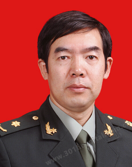 Shouwang Cai (蔡守旺)合作论文数Chinese PLA General Hospital4lin zhao (赵霖)合作论文数3Zhi-Qiang Huang合作论文数2quanwen chen (陈全文)合作论文数2LIU Yang (刘洋)合作论文数2WANG Xin (王欣)合作论文数2Jing DONG (董菁)合作论文数2Mingxing Lin (林明星)合作论文数2HuiMin Cheng (程惠民)合作论文数2LingBo Huo (霍凌波)合作论文数2WanQing Gu (顾万清)合作论文数2HuiHuang Xiao (肖辉煌)合作论文数2XiangFei Meng (孟翔飞)合作论文数2ZhiWei Liu (刘志伟)合作论文数2
 Shichun Lu (卢实春)合作论文数School of Medicine, Nankai University2
 Xianjie Shi (史宪杰)合作论文数The Eighth Affiliated Hospital Sun Yat-Sen University2
 Minggen Hu (胡明根)合作论文数Chinese PLA General Hospital2
 Yongliang Chen (陈永亮)合作论文数Chinese PLA General Hospital2
 Yang Liu (刘阳)合作论文数Chinese PLA General Hospital2X H Mao合作论文数1Zhaojun Chen合作论文数1
 Guodong Zhao (赵国栋)合作论文数Chinese People's Liberation Army General Hospital1Y D Qiu (裘一丹)合作论文数1Yong-Wei Chen合作论文数1lijie huang (黄丽洁)合作论文数1Jia-Hong Dong合作论文数1Yang LIU (刘洋)合作论文数1Yingdi Liu合作论文数1M Su合作论文数1Haoyun Zhang合作论文数1hongying meng (孟红英)合作论文数1Li Bai合作论文数1Kai Jiang合作论文数1




