Objective To explore the clinical factors influencing CSF in elderly patients and association and coronary artery diameter.Methods A retrospective cohort study was conducted on 236 consecutive elderly patients undergoing coronary angiography in the First Medical Center of Chinese PLA General Hospital between May 2018 and December 2021.Based on CTFC,115 patients were identified as CSF group and 121 as NCA group.Their clinical data and coronary artery diameter were analyzed.Results The CSF group had significantly larger proportions of chest pain history, taller height, larger body surface area, higher white blood cell count and neutrophil count, and larger left anterior descending diameter, left circumflex artery diameter, and right larger coronary artery diameter, while lower lymphocyte count and HDL-C level when compared with the NCA group(P<0.05,P<0.01).HLD-C was an independent protective factor for CSF(OR=0.899,95%CI:0.819-0.986,P=0.024) and neutrophil was an independent risk factor for CSF(OR=1.775,95%CI:1.294-2.435,P=0.000).The diameters of left anterior descending, left circumflex artery and right coronary artery were positively correlated with their corresponding CTFC(P=0.000),neutrophil was positively correlated with average CTFC(P=0.017),while lymphocyte negatively(P=0.018).Conclusion Increased coronary artery diameter and high neutrophil are observed in the CSF elderly, who are in a “sign of macrovessel”.Neutrophil is an independent risk factor for CSF in the elderly patients.
目的 全球急性冠状动脉事件注册(GRACE)评分与B型钠尿肽(BNP)、C反应蛋白、D-二聚体对老年急性前壁心肌梗死患者猝死风险评估的价值.方法 选择2008年8月~2020年4月解放军总医院第一医学中心诊治的首次急性前壁心肌梗死患者97例,其中院内猝死40例(猝死组),未发生院内猝死57例(对照组).比较2组BNP、C反应蛋白、D-二聚体及GRACE评分的差异,并分析BNP、C反应蛋白、D-二聚体及GRACE评分用于评估老年急性前壁心肌梗死患者猝死发生的风险.结果 猝死组GRACE评分、C反应蛋白、D-二聚体和BNP水平明显高于对照组,差异有统计学意义(P<0.01).BNP、C反应蛋白、D-二聚体和GRACE评分对老年急性前壁心肌梗死患者猝死风险评估的敏感性分别为77.14%、88.00%、89.19%、85.00%,特异性分别为77.36%、74.47%、55.36%、82.46%,AUC 分别为0.810、0.850、0.776、0.870.BNP、C 反应蛋白、D-二聚体与 GRACE 评分联合评估老年急性前壁心肌梗死患者猝死风险敏感性为82.60%,特异性为84.40%,AUC为0.902.各指标联合检测的特异性明显高于各指标单独检测(P<0.05).结论 BNP、C反应蛋白、D-二聚体与GRACE评分对老年急性前壁心肌梗死患者猝死风险评估有较高的临床价值.
早在1938年,Burstein J等报道9例心电图标准肢体导联QRS波均以S波为主的病例,患者有冠心病和/或慢性肺病.1942年,Schwartz SP等总结7例有上述心电图表现的患者,其中6例尸检发现右心室肥大.1969年,Te-chuan Chou等提出S1S2S3综合征(S1S2S3 syndrome)的具体诊断标准.
[定义] 2018年11月,丹麦医生Henning Bundgaard与来自荷兰和英国的研究人员报告了一种新型心电图综合征,其特征性表现为持续性非缺血性广泛ST段压低.患者可进展为房颤、室速/室颤、心脏性猝死,属于常染色体显性遗传心律失常综合征,被称为"家族性ST段压低综合征",其心电图特征性改变也被称为"Bundgaard征"(Bundgaard Sign).
目的 探讨正加速度适应性训练对大鼠胃黏膜损伤指数及PGE2含量的影响.方法 40只雄性SD大鼠随机分为5组,每组8只,分别标记为A、B、C、D、E组.A组为空白对照组;B组大鼠每天+5 Gz值暴露5 min,连续暴露5d;C组大鼠每天+10 Gz值暴露5 min,连续暴露5d;D组大鼠适应性训练(+4 Gz值每天暴露3 min,连续暴露5 d)后+5 Gz值每天暴露5 min,连续暴露5d;E组大鼠适应性训练(+4 Gz值每天暴露3 min,连续暴露5 d)后+10 Gz值每天暴露5 min,连续暴露5d.实试验结束后肉眼观察胃黏膜损伤情况,计算损伤指数(lesion index,LI),ELISA法检测胃黏膜PGE2的含量.结果 肉眼观察,除A组外,其余各组胃黏膜均可见不同程度损伤.C组损伤指数(5.625±1.767)高于B组(1.750±0.707).适应性训练后,D组损伤指数(0.875±0.641)低于B组(1.750±0.707),E组损伤指数(2.250±1.035)低于C组(5.625±1.767),差异有统计学意义(P<0.05).C组PGE2含量[(24.598±6.017) pg/ml]低于B组[(60.468±9.697) pg/ml],适应性训练后,E组PGE2含量[(46.165±13.996) pg/ml]高于C组的[(24.598±6.017) pg/ml](P<0.05).结论 低速正加速度适应性训练可减轻高速正加速度带来的胃黏膜损伤,其机制与胃黏膜内PGE2分泌增多有关.
目的 示指无名指长度比(2D∶4D)和某些心脏疾病之间的关联有所报道,其与冠心病(CAD)的关系尚不明确.我们调查2D∶4D是否可以作为预测CAD的一个指标.方法 本研究包括了884人,分为4组,221例CAD患者和221名健康对照.使用电子卡尺测量手指长度.t检验和Spearman相关系数用于检测2D∶4D组间的差异以及2D∶4D比值和CAD相关性.结果 仅在男性观察到右手2D∶4D比例高于左手.在男性右手对照组和双手CAD组,2D∶4D比例均高于女性(P <0.001).在男性CAD组,双手2D∶4D比值均高于男性对照组(P <0.001),但在女性没有观察到此趋势.各组2D∶4D比例和年龄之间没有显著关系(P>0.05,),在男性双手2D∶4D比例和CAD呈显著的正相关(P <0.001).结论 本研究表明在男性的双手2D∶4D比值和CAD之间存在显著相关.
Objective To analyze the medication with β blocker in elderly patients with stable angina.Methods From June 1,2010 to December 1,2010,investigations were made among the patients with coronary heart diseases in our hospital.Among them,186 elderly patients with stable angina were selected out and their clinical data were statistically analyzed.Results The usage rate of β blocker was 54.8%(102/186) among these patients.There was no significant difference in the usage rate(P>0.05) among ones with different gender and age.Conclusion The usage rate of β blocker is low among senile patients with stable angina and the dosage is insufficient,which indicates the insufficient usage of this kind of medicine.
Objective To observe the effects of CpG oligonucleotide(ODN)on the angiogenesis of subcutaneous implanted tumor of lung carcinoma in the nude mice.Methods The models of subcutaneous implanted tumor in nude mice was established and randomly divided into 4 groups:group A receiving radiation and medical administration[CpG ODN 1826(1 μg/μl)+ β ray(RT:8 Gy)],group B receiving simple medical administration[CpG ODN 1826(1 μg/μl)],group C receiving irradiation[saline(100 μl/one)+β ray(RT:8 Gy)]and group D[saline(100 μl/one)].Seven days after the implantation of lung cancer cell,all those groups received the intratumoral injection of medicine and irradiation for 5 d.The expression of VEGF-C mRNA and NRP-1 mRNA in the tumor tissue was detected by RT-PCR.Results The relative expression levels of VEGF-C mRNA in the tumor tissue of the four groups above were respectively 18.34±3.19,29.62±3.14,51.13±2.81 and 53.46±5.67.The relative expression levels of NRP-1 mRNA in those groups were respectively 23.57±5.73,34.72±5.13,59.95±4.76 and 62.49±6.34.The expression level of group A decreased very significantly compared with those of the other three groups(P<0.01).Compared with the expression levels of group C and group D,that of group B decreased very significantly(P<0.01).Conclusion CpG ODN1826 can remarkably inhibit the angiogenesis of nude mice tumor,and its mechanism may be correlated with the inhibition of VEGF-C and NRP-1 expression.
<正>静脉输液是最常应用的临床给药方法之一,而上肢静脉为首选部位。为了保护病人的静脉血管,减少因静脉穿刺后护理不当造成的问题发生,笔者在静脉输液完毕后采用改良拔针方法,不按压穿刺部位,抬高穿刺侧肢体,并与传统方法进行对比,取得较好效果,现报告如下。
Objective To investigate the influence of oligodeoxynucleotide containing CpG motif(CpG ODN) on the radiosensitivity of human lung cancer cell line A549.Methods A549 cells were exposed to CpG ODN at different concentration(1,3,10,30 μg/ml) and β-ray irradiation(5 Gy).The cell proliferation and colony formation were measured afterward.The secretion levels of TNF-α,IL-12 and INF-γ were detected by ELISA method.The NO level was detected by Griess method.Results CpG ODN inhibited the proliferation of A549 cells,and presented a dose-effect relationship.CpG ODN(10 μg/ml) combined with β-ray irradiation had the best effects on inhibiting the proliferation of cells,and it increased the secretion of TNF-α,IL-12,INF-γ and NO.Conclusion CpG ODN has an obvious radiosensitizing effect on A549 cells,which may be related to its inhibition of A549 cell proliferation and enhancement of the secretion of TNF-α,IL-12,INF-γ and NO.
Objective Objective To evaluate the efficacy and safety of domestic oseltamivir in the treatment of influenza A(H1N1)virus infection.Methods A randomized and control clinical trial was conducted among 245 individuals with H1N1 virus infection.They were divided into two groups at random:oseltamivir group(123 cases,oseltamivir 75 mg,twice a day for 5 d)and control group(122 cases,no oseltamivir treatment).Both groups were intravenously given Double Rhizoma Coptidis Injection(3.6 g,once a day for 5 d).The clinical features and the disappearance time of their symptoms were observed,and the blood routine,urine routine as well as liver and kidney function were examined.Results 75.61% of patients in oseltamivir group showed a significant improvement versus 9.84% in control group(P< 0.01).The effective rate was 95.12% in oseltamivir group versus 54.92% in control group(P<0.01).The disappearance time of fever in oseltamivir group and control group was(28.67±15.92)h versus(52.12±20.70)h(P< 0.01).The remission time in oseltamivir group was significantly shorter than that in control group(P<0.01).The usage rates of antibiotics and paracetamol tablets were much less in oseltamivir group than those in control group(P<0.05).No evident adverse reactions were observed in both groups.Conclusion Oseltamivir is effective and of good tolerance in the treatment of influenza A(H1N1)virus infection.It can apparently reduce the duration and severity of influenzal symptoms.
Objective To assess the efficacy and safety of blood vessel suture device, Starclose, in coronary interventional procedure for senile patients. Methods Among the 204 senile patients who underwent coronary interventional procedure, Starclose was used in 110 who patients served as a suture group, and manual compression hemostasis was used in 93 patients who served as a control group. Hemostatic time, immobilization time of the lower extremities, and total incidence rate of operation-related complications in both groups were recorded, respectively. Results No difference was found in the success rate for hemostasis between the two groups (P>0.05). The hemostatic time and immobilization time of the lower extremities were shorter in suture group than in control group (P<0.01) while the operation-related complication rate was significantly higher in control group than in suture group (P<0.05). Conclusion Starclose has a rather good efficacy and safety when applied in the interventional diagnosis and treatment of coronary heat disease of senile patients.
临床教学的过程管理是一项非常复杂的系统工程,随着医学模式的转变和现代医学的快速发展,对医学人才的培养提出了更高的要求,同时也为临床理论教学和实践教学提供了发展的良机.
AIM: To evaluate the influence of postdilatation with noncompliance balloon on subacute thrombosis after implanting intracoronary drug-eluting stents(DES).METHODS: A cohort of 305 consecutive coronary heart disease patients with DES implanted from January 2006 to September 2008 were divided into two groups: postdilatation group and non-postdilatation group.Clinical features,pathological changes of coronary arteries,implantation of drug-eluting stents and formation of subacute thrombosis were recorded and compared.Effects of postdilatation on subacute thrombosis were observed and analyzed.RESULTS: No statistical differences were observed in clinical features,pathological changes of coronary arteries and the number of DES implanted between groups.The rates of subacute thrombosis in the two groups were 1.2% and 0%,respectively,with no significant difference.CONCLUSION: Postdilatation with noncompliance balloon after DES deployment did not significantly reduce the rate of subacute thrombosis.
Objective To evaluate the effect of post -dilatation with highpressure for reducing subacute thrombosis after intracoronary drug-eluting stent(DES) implantation. Methods The cohort of consecutive coronary heart disease patients with implanted DES from January 2004 to June 2009 were studied. The 838 patients were divided into 2 groups depending on whether they were post-dilatated with high-pressure. There were 501 patients in non-post-dilatation group,male 353,female 148,aged 40 -82 years,mean age 61.4 years,and 337 patients in post-dilatation group,male 235,female 102,aged 37 -86 years,mean age 62.9 years. The clinical features,pathological changes of coronary artery,profiles of DES implantation and subacute thrombosis were recorded. Besides,the effects of post-dilatation for reducing subacute thrombosis of 2 groups were observed and analyzed. Results There were no statistical differences in the aspects of clinical features,pathological changes of coronary artery and profile of DES implantation between 2 stages. The rates of subacute thrombosis in 2 stages were 0 % and 1.62 % respectively,which were significantly decreased,there was statistical difference between 2 stages(P 0.05). Conclusion The post-dilatation with high-pressure after DES deployment could reduce the rate of subacute thrombosis.
Objective To evaluate the impact of optical coherence tomography(OCT) for high pressure post-dilatation after coronary artery stent implantation.Methods A cohort of 29 consecutive coronary heart disease patients with an average age of(59.2±4.5) years(40-77 years) old who had finished percutaneous coronary intervention from July 2007 to July 2009 were retrospectively analyzed.Coronary angiography and stent implantation were done according to standard methods,and OCT examination was done after stents expanded with named pressure and high pressure balloon post-dilatation respectively,as well as the stent struts apposition,intimal prolapse and micro-dissection were analyzed.Results Twenty-nine patients finished OCT examination successfully without angina and heart failure complications in the surrounding operation stage.The distance between the vessel wall and stent struts of high pressure balloon post-dilatation(94.00±22.42) μm was significantly shorter than that of the stent named expansion(137.38±26.80) μm,with statistically significant difference(P0.01).The incidence of intimal prolapse and micro-dissection with high-pressure balloon post-dilatation(respectively 14 and 0) were significantly less than that of stent named expansion(respectively 32 and 5),and both of them had statistic difference(P0.05).Conclusion There is a certain guide role of OCT for high pressure balloon post-dilatation after stent implantation.
Several kinds of noninvasive cardiovascular imaging techniques and noninvasive coronary angiography have become ma- jor methods in the diagnosis and treatment of coronary heart disease.But there are still a lot of difficulties and disputs on the order to assess borderline coranary lesion severity,the necessity of interventional revasculorization,the identification of vulnerable plaques and the evaluation of the instant and long-term effects of revasculorization.The advantages of intracoronary optical coherence tomography (OCT)are the discrimination of vessel stenosis degrees and extent of stenosis.Resolving power of OCT is approximately 10-fold higher than that of IVUS,and it is similar with that of pathologic findings.But the application of OCT in the elderly patients with coro- nary heart disease is rarely reported.This paper reviews the progress in the diagnosis of borderline coranary lesion using OCT so that borderline coranary lesion can be treated effectively.
OBJECTIVE:To investigate the constituent expression of PH domain leucine-rich repeat protein phosphatase 1 (PHLPP1) in human umbilical vein endothelial cells (HUVECs) and the effect of PHLPP1 gene transfer on the proliferation of the cells in vitro. METHODS:Cultured HUVECs were transfected with pcDNA3-GFP or pcDNA3HA-PHLPP1 via lipofectamine 2000. The cell proliferation ability was determined by cell counting and MTT colorimetric assay, and Western blotting was used to detect the protein expression of PHLPP1 in the cells. RESULTS:No PHLPP1 protein was detected in the non-transfected cells or pcDNA3-GFP-transfected cells. pcDNA3HA-PHLPP1 gene transfection significantly increased PHLPP1 expression in the HUVECs (P<0.01), but the cell proliferation status remained unchanged (P>0.05). The absorbance of the cells measured by MTT assay was 0.134-/+0.0152, 0133-/+0.014 and 0.137-/+0.016, with cell counts of (8.293-/+0.962)x10(5), (7.937-/+0.101)x10(5) and (8.127-/+0.112)x10(5), respectively, showing no significant differences between the 3 groups (P>0.05). CONCLUSIONS:Phosphatase PHLPP1 may not be the most important signal protein in the regulation of HUVEC proliferation.
AIM: To investigate the serum level of IL-6,IL-8 and TNF-α and the effect of salvianolate in patients with unstable angina pectoris(UAP) after stent implantation.METHODS: Sixty-eight patients undergoing stent implantation were divided randomly into conventional therapy group and salvianolate group.The levels of IL-6,IL-8 and TNF-α were evaluated by ELISA before and 24 h and 1 week after stent implantation,and the levels of the above cytokins were compared with those in 18 normal controls.RESULTS: The levels of serum IL-6,IL-8 and TNF-α in patients with UAP were higher than those in the control group(P0.01).The levels of serum IL-8 and TNF-α decreased 24 h after stent implantation in salvianolate group(P0.05),while the values of all the three cytokins significantly increased in the conventional therapy group(P0.05).The levels of the three cytokins decreased in both the conventional therapy group(P0.05)and the salvianolate group(P0.01) 1 w after stent implantation.CONCLUSIONS: Salvianolate may inhibit the inflammation in coronary artery by decreasing the secretion of IL-6,IL-8 and TNF-α,thus playing a protective role in patients with UAP after stent implantation.
Objective:To investigate the effects of different displacement volume on treatment of multiple organ dysfunction syndromes(MODS)during continuous blood purification(CBP).Methods:Eighteen patients with MODS who would receive CBP were randomly divided into group A[displacement liquid35 ml/(kg·h),n=6)],group B[displacement liquid 45 ml/(kg·h),n=6] and group C[displacement liquid60 ml/(kg·h),n=6].Before and after CBP for 24,48 hours,their clinical and laboratory indexes were compared.The levels of acute physiology and chronic health evaluationⅡ(APACHEⅡ),simplified acute physiology score Ⅱ(SAPSⅡ),MODS score were evaluated.The levels of tumor necrosis factor-α(TNF-α)were measured.Results:Temperature,heart rate,BUN,dopamine levels declined and PH improved after CBP(P0.01~0.05).APACHEⅡ,SAPSⅡ,MODS score and TNF-α level in group B and group C were significantly lower after 24 h CBP than those of pre-CBP(P0.01~0.05).However,these scores and TNF-α level did not decreased in group A after 48-hour CBP.Conclusion:CBP is effective for patients with MODS,the volume of displacement liquid over 45 ml/(kg·h)during CPB can more effectively improve the clinical conditions.
 Caiyi Lu (卢才义)合作论文数The Fifth Affiliated Hospital of Guangzhou Medical University19
 Lei Gao (高磊)合作论文数Chinese PLA General Hospital10
 YuXiao Zhang (张玉霄)合作论文数9ShengHua Zhou (周圣华)合作论文数7
 Shiwen Wang (王士雯)合作论文数解放军总医院老年心血管病研究所6
 Xingli Wu (吴兴利)合作论文数Chinese PLA General Hospital6
 Peng Liu合作论文数College of Information Sciences and Technology, Pennsylvania State University6Qiao Xue (薛桥)合作论文数5Ya Huang (黄亚)合作论文数4TiYe Sun (孙梯业)合作论文数4Qi Chen (陈琪)合作论文数4
 Muyang Yan (晏沐阳)合作论文数Chinese PLA General Hospital4JinYue Di (翟金月)合作论文数3HeLan Tang (唐合兰)合作论文数3ZhongRen Zhao (赵忠仁)合作论文数3Bin Du (杜斌)合作论文数3
 Chunmin Yang (杨春敏)合作论文数Beijing Air General Hospital3HuiYuan Si (司慧远)合作论文数2QuanLi Han (韩全利)合作论文数2Rui Chen (陈瑞)合作论文数2
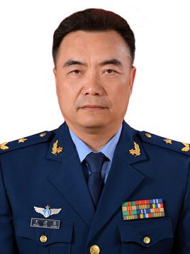 Jianchang Wang (王建昌)合作论文数中国人民解放军空军总医院2Dingyou Yang (杨丁友)合作论文数Chinese PLA General Hospital2Min Jia (贾敏)合作论文数2
 Ping Zhou (周平)合作论文数Institute of Reproduction and Genetics, Anhui Medical University2Hongwei Zhang (张宏伟)合作论文数2
 Chunwei Wang (王纯巍)合作论文数中国人民解放军空军总医院感染内科2
 XiaoJuan Li (李晓娟)合作论文数The General Hospital of The Air Force P.L.A2Ming-Lei Jin合作论文数1Du Yanqing (杜彦青)合作论文数1qi you (尤琪)合作论文数1tianyuan xiang (相田园)合作论文数1Yu Jiang (余江)合作论文数1Wen-Hui Chai合作论文数1LI Jing (李静)合作论文数1Shan XU (徐珊)合作论文数1Zhu Feiyun (朱飞云)合作论文数1Dong-li SUN (孙冬丽)合作论文数1Hao LIU (刘昊)合作论文数1Xu Guan (关旭)合作论文数1Qiu Jie (邱杰)合作论文数1RuiYan Tian (田蕊艳)合作论文数1YaJing Zhang (张亚晶)合作论文数1TieHui Xiao (肖铁卉)合作论文数1XinChun Zhou (周新春)合作论文数1HuiBin Xu (许会彬)合作论文数1XueDong Yang (杨学东)合作论文数1




