Oxidative stress after ischemia reperfusion can cause irreversible brain damage. Thus, it is vital to timely consume excessive reactive oxygen species (ROS) and conduct molecular imaging monitoring on the brain injury site. However, previous studies have focused on how to scavenge ROS while ignoring the mechanism of relieving the reperfusion injury. Herein, we reported a layered double hydroxide (LDH)-based nanozyme (denoted as ALDzyme), which was fabricated by the confinement of astaxanthin (AST) with LDH. This ALDzyme can mimic natural enzymes, which include superoxide dismutase (SOD) and catalase (CAT). Furthermore, the SOD-like activity of ALDzyme is 16.3 times higher than that of CeO2 (a typical ROS scavenger). Based on these enzyme-mimicking properties, this one -of-a-kind ALDzyme offers strong anti-oxidative properties as well as high biocompatibility. Importantly, this unique ALDzyme can establish an efficient magnetic resonance imaging platform, thus guiding the in vivo details. As a result, the infarct area can be reduced by 77% after reperfusion therapy, and the neurological impairment score can be lowered from 3-4 to 0-1. Density functional theory computations can reveal more about the mechanism of this ALDzyme's significant ROS consumption. These findings provide a method for unraveling the neuroprotection application process in ischemia reperfusion injury using an LDH-based nanozyme as a remedial nanoplatform.
In the current global crisis of antibiotic resistance, delivery systems are emerging to combat resistant bacteria in a more efficient manner. Despite the significant advances of antibiotic nanocarriers, many challenges like poor biocompatibility, premature drug release, suboptimal targeting to infection sites and short blood circulation time are still challenging. To achieve targeted drug delivery and enhance antibacterial activity, here we reported a kind of pH-responsive nanoparticles by simply self-assembly of an amphiphilic poly(ethylene glycol)-Schiff-vancomycin (PEG-Schiff-Van) prodrug and free Van in one drug delivery system. The acid-liable Schiff base furnished the PEG-Schiff-Van@Van with good storage stability in the neutral environment and susceptible disassembly in response to faintly acidic condition. Notably, on account of the combination of physical encapsulation and chemical conjugation of vancomycin, these nanocarriers with favorable biocompatibility and high drug loading capacity displayed a programmed drug release behavior, which was capable of rapidly reaching high drug concentration to effectively kill the bacteria at an early period and continuously exerting an bacteria-sensitive effect whenever needed over a long period. In addition, more Schiff-base moieties within the PEG-Schiff-Van@Van nanocarriers may also make great contributions on promoting the antimicrobial activity. Using this strategy, this system was designed to have programmable structural destabilization and sequential drug release due to changes in pH that were synonymous with bacterial infection sites, thereby presenting prominent antibacterial therapy both in vitro and in vivo. This work represents a synergistic strategy on offering important guidance to rational design of multifunctional antimicrobial vehicles, which would be a promising class of antimicrobial materials for potential clinical translation.
Phototherapy and multimodal synergistic phototherapy (including synergistic photothermal and photodynamic therapy as well as combined phototherapy and other therapies) are promising to achieve accurate diagnosis and efficient treatment for tumor, providing a novel opportunity to overcome cancer. Notably, various nanomaterials have made significant contributions to phototherapy through both improving therapeutic efficiency and reducing side effects. The most key factor affecting the performance of phototherapeutic nanomaterials is their microstructure which in principle determines their physicochemical properties and the resulting phototherapeutic efficiency. Vacancy defects ubiquitously existing in phototherapeutic nanomaterials have a great influence on their microstructure, and constructing and regulating vacancy defect in phototherapeutic nanomaterials is an essential and effective strategy for modulating their microstructure and improving their phototherapeutic efficacy. Thus, this inspires growing research interest in vacancy engineering strategies and vacancy-engineered nanomaterials for phototherapy. In this review, we summarize the understanding, construction, and application of vacancy defects in phototherapeutic nanomaterials. Starting from the perspective of defect chemistry and engineering, we also review the types, structural features, and properties of vacancy defects in phototherapeutic nanomaterials. Finally, we focus on the representative vacancy defective nanomaterials recently developed through vacancy engineering for phototherapy, and discuss the significant influence and role of vacancy defects on phototherapy and multimodal synergistic phototherapy. Therefore, we sincerely hope that this review can provide a profound understanding and inspiration for the design of advanced phototherapeutic nanomaterials, and significantly promote the development of the efficient therapies against tumor.
Bioprinting has gained immense attention and achieved the revolutionized progress for application in the multifunctional tissue regeneration. On account of the precise structural fabrication and mimicking complexity, hydrogel-based bio-inks are widely adopted for cartilage tissue engineering. Although more and more researchers have reported a number of literatures in this field, many challenges that should be addressed for the development of three-dimensional (3D) bioprinting constructs still exist. Herein, this review is mainly focused on the introduction of various natural polymers and synthetic polymers in hydrogel-based bioprinted scaffolds, which are systematically discussed via emphasizing on the fabrication condition, mechanical property, biocompatibility, biodegradability, and biological performance for cartilage tissue repair. Further, this review describes the opportunities and challenges of this 3D bioprinting technique to construct complex bio-inks with adjustable mechanical and biological integrity, and meanwhile, the current possible solutions are also conducted for providing some suggestive ideas on developing more advanced bioprinting products from the bench to the clinic.
Bioprinting has gained immense attention and achieved the revolutionized progress for application in the multifunctional tissue regeneration. On account of the precise structural fabrication and mimicking complexity, hydrogel-based bio-inks are widely adopted for cartilage tissue engineering. Although more and more researchers have reported a number of literatures in this field, many challenges that should be addressed for the development of three-dimensional (3D) bioprinting constructs still exist. Herein, this review is mainly focused on the introduction of various natural polymers and synthetic polymers in hydrogel-based bioprinted scaffolds, which are systematically discussed via emphasizing on the fabrication condition, mechanical property, biocompatibility, biodegradability, and biological performance for cartilage tissue repair. Further, this review describes the opportunities and challenges of this 3D bioprinting technique to construct complex bio-inks with adjustable mechanical and biological integrity, and meanwhile, the current possible solutions are also conducted for providing some suggestive ideas on developing more advanced bioprinting products from the bench to the clinic.</p>
Rapid hemostasis and formation of stable blood clots are very important to prevent massive blood loss from the excessive bleeding for living body, but their own clotting process cannot be completed in time for effective hemostasis without the help of hemostatic materials. In general, traditionally suturing and stapling techniques for wound closure are prone to cause the additional damages to the tissues, activated inflammatory responses, short usage periods and inevitable second operations in clinical applications. Especially for the large wounds that require the urgent closure of fluids or gases, these conventional closure methods are far from enough. To address these problems, various tissue adhesives, sealants and hemostatic materials are placed great expectation. In this review, we focused on the development of two main categories of tissue adhesive materials: synthetic polymeric adhesives and naturally derived polysaccharide adhesives. Research of the high performance of hemostatic adhesives with strong adhesion, better biocompatibility, easy usability and cheap price is highly demanded for both scientists and clinicians, and this review is also intended to provide a comprehensive summarization and inspiration for pursuit of more advanced hemostatic adhesives for biological fields.
Bone tuberculosis (TB) caused by mycobacterium tuberculosis continues to present a formidable challenge to humans. To effectively cure serious bone TB, a novel kind of composite scaffolds with long-term dual drug release behaviours were prepared to satisfy the needs of both bone regeneration and antituberculosis drug therapy. In virtue of an improved O/W emulsion technique, water-soluble isoniazid (INH)-loaded gelatin microparticles were obtained by tailoring the content of β-tricalcium phosphate (β-TCP), which played significant roles in INH entrapment efficiency and drug release behaviours. By mixing with the poly(ε-caprolactone)-block-poly (lactic-co-glycolic acid) (b-PLGC) solution containing oil-soluble rifampicin (RFP) via the particle leaching combined with phase separation technique, the dual drugs-loaded composite scaffolds were fabricated, which possessed interconnected porous structures and achieved the steady release of INH and RFP drugs for three months. Moreover, this dual drugs-loaded system could basically achieve their expectant roles of respective drugs without obvious influences with each other. This strategy on preparation of intelligent composite scaffolds with the multi-drugs loading capacity and controlled long-term release behaviour will be potential and promising substrates in clinical treatment of bone tuberculosis.
Oral cancer is an aggressive tumor that invades the local tissue and can cause metastasis and high mortality. Conventional treatment strategies, e.g., surgery, chemotherapy, and radiation therapy alone or in combinations, possess innegligible issues, and significant side and adverse effects for the clinical applications. Currently, targeting drug delivery is emerging as an effective approach for oral delivery of different therapeutics. Herein we provide a state-of-the-art review on the current progress of targeting drug delivery for oral cancer therapy. Variously oral delivery systems including polymeric/inorganic nanoparticles, liposomes, cyclodextrins, nanolipids, and hydrogels-based forms are emphasized and discussed, and biomimetic systems with respect to oral delivery like therapeutic vitamin, exosomes, proteins, and virus-like particles are also described with emphasis on the cancer treatment. A future perspective is also provided to highlight the existing challenges and possible resolution toward clinical translation of current oral cancer therapies.
Recent advances in self-assembled nanogel carriers have allowed precise design of hierarchical structures by a low-cost solution-phase approach. Typically, photochemical strategy on the tailor of morphology and dimension has emerged as a powerful tool, because light-trigger has exceptional advantages of an instant “on/off” function and spatiotemporal precision at arbitrary time. Herein, we report a tunable manipulation of sequentially morphological transition via a “living” thiol-disulfide exchange reaction from a UV-tailored hierarchical self-assembly strategy. By varying the irradiation time, the photochemical method can easily fabricate and guide a series of attractively architectural evolution in dilute aqueous solutions, by which the improving hydrophobicity and sensitive redox-responsiveness endowed these disulfide-linked nanoparticles with remarkable capacities of abundant encapsulation, effective separation, and controlled release of hydrophobic cargoes. Notably, once the exchange reaction is suspended at any point of time by removing the UV lamp, these active sites within the nanogel carriers are instantaneous deactivated and the correspondingly structural transformations are also not conducted any more. However, if the stable inert sites are reactivated as needed by turning on the UV light, the interrupting morphology evolution can continue its previous steps, which may provide a simple and novel approach to fabricating the desired self-assemblies in solutions. With regard to this advanced functionality, various nanogel carriers with customizable structures and properties have been yielded and screened for cancer therapy. Thus, this “living” controlled self-assembled method to program morphology evolution in situ is a universal strategy that will pave novel pathways for creating sequential shape-shifting and size-growing nanostructures and constructing uniform nanoscopic functional entities for advanced bio-applications.
Artificial disc replacement alone is unable to completely cure cervical degenerative diseases; thus, a stabilization device markedly improves patient recovery. In order to meet this requirement, an anterior spinal instrumentation combining a prosthetic disc nucleus with a flexible stabilization device (ASI combining PDN/FD) was developed. An artificial disc was designed and manufactured using polyvinyl alcohol hydrogel (PVA-H) with a repeated freeze-melting technique, and subsequently the dehydration and swelling properties of the PVA-H were investigated. A canine animal model was then used to compare the differences in the degeneration of cervical discs adjacent to the operative segment with PDN/FD against a plate fixation system, without the interferences of brace intensity, which was ensured by pressure monitoring the dog's intervertebral disc adjacent to operative segments. The in vivo results demonstrated a clear decrease in the degeneration of the adjacent disc with the use of PDN/FD as compared with the plate fixation system. In conclusion, PDN/FD may offer a promising method for the treatment of cervical degenerative disease.
Currently, the major issues in the treatment of osteoarticular tuberculosis (TB) after implant placement are low drug concentration at the infected focus and drug resistance resulting from the long-term chemotherapy. The application of drug-loaded polymeric multilayers on implantable devices offers a promising solution to the problems. Herein, a poly(ethylene glycol)-based hydrogel film embedded with isoniazid (INH)-loaded alginate microparticles was fixed to Ti implants via adhesive polydopamine, subsequently capped by poly(lactic-co-glycolic acid) membranes for the sustained and localized delivery of the anti-TB drug. The antibacterial efficacy of the released INH was confirmed by a 4.5 ± 0.8 cm inhibition zone formed in the fourth week after inoculation of Mycobacterium tuberculosis. The INH-loaded Ti implants showed no toxicity to the osteoblast cell and provided a consistent drug release for nearly one week in vitro. The release profile in vivo showed a high local concentration and low systemic exposure. The local INH concentration could be kept higher than its minimum inhibitory concentration over a period of 8 weeks, which proves that it is a promising strategy to improve the severe osteoarticular TB treatment.
Here we demonstrate a type of pH and reduction dual-sensitive biodegradable micelles, which were self-assembled by a cationic polymer in an aqueous solution. Due to tumor cells or tissues showing low pH and high reduction concentration, these micelles possessed specific tumor targetability and maximal drug-release controllability inside tumor cells upon changes in physical and chemical environments, but presented good stability at physiological conditions. CCK-8 assay showed that the DOX-loaded micelles had a similar cytotoxicity for MCF-7 tumor cells as free DOX, and blank micelles had a very low cytotoxicity to the cells. Fluorescent microscopy observation revealed that the drug-loaded micelles could be quickly internalized by endosomes to inhibit cancer cell growth. These results indicated these biodegradable micelles, as a novel and effective pH- and redox-responsive nanocarrier, have a potential to improve drug delivery and enhance the antitumor efficacy.
Here we demonstrate a simple and straightforward approach to prepare POSS-embedded hyperbranched (HB-POSS) polymers with customized molecular weights and sizes just by controlling the polymerization time. The polymers can be further used for building amphiphilic polymers, presenting morphological transition from micelle to vesicle in aqueous solution.
Bone tuberculosis (TB) is one of the most common extrapulmonary TB. Effective integration of chemotherapy and bone regeneration is an optimal solution for bone TB therapy. Herein, we produce a composite scaffold drug delivery system fabricated with an isoniazid conjugated star poly(lactide-co-glycolide) (PLGA-INH4) and β-TCP. The cytological assay indicated the composite system possesses good biocompatibility. The in vitro and in vivo drug release evaluations showed that the composite system can intactly release the pristine INH and maintain effective INH concentration in a controlled manner for more than 100 days, and achieve high localized drug concentration and low systemic drug concentration. The rabbit radius repair experiment testified the scaffold has good bone regeneration capacity. Our work demonstrate the composite system can simultaneously achieve localized long-term drug controlled release and bone regeneration, which provides a promising route for improved bone TB treatment.
DOI: 10.1097/BRS.0000000000000618 TO THE EDITOR: Re : He B, Yan L, Guo H, et al. The Difference in Superior Adjacent Segment Pathology After Lumbar Posterolateral Fusion by Using 2 Different Pedicle Screw Insertion Techniques in 9-Year Minimum Follow-up. Spine (Phila Pa 1976) 2014;39:1093–8. We read with interest the recent article “The difference in superior adjacent segment pathology after lumbar posterolateral fusion by using 2 different pedicle screw insertion techniques in 9-year minimum followup” by He et al . 1 The authors are to be congratulated on designing this simple randomized control trial study to provide strong evidence that a more lateral starting point results in less radiographical and clinical adjacent segment disease (ASD). However, we think that the study still has some limitations. Sagittal alignment has been shown in some studies 2–4 to infl uence adjacent level degeneration after lumbar or cervical fusion, which is one of the most important proposed surgical risk factors for ASD. However, there is no information about the sagittal parameters of each group in this article. Whether the sagittal malalignment is the causative factor for ASD or just the screw insertion technique can only be determined by also measuring the preoperative parameters for sagittal alignment and comparing the changes over time between each group. However, this comparison was not conducted by the authors of this study. The patient-selection and surgical intervention in this study might also have an enlarged and amplifi ed effects of screw insertion techniques on ASD, which could also help explain why their ASD rate (61.5%) was signifi cantly higher than others. 2–5 First, this prospective study includes 210 patients with low grade isthmic spondylolisthesis, these patients inherently, have a greater loss of disc height, and consequently, some loss of lordosis, which have more chance to be sagittal malalignment. Second, surgical decompression intervention they used is “removal of the spinous process and bilateral laminectomy, and partial bilateral foraminotomy,” which means the posterior ligament complex of the superior adjacent segment is completely removed. According to the tension band mechanism, loss of posterior ligament complex is another risk factor for adjacent segment degeneration. Third, the restoration of sagittal balance after spinal reconstruction has been identifi ed as an increasingly important risk factor for subsequent degeneration at adjacent levels. 6 Nevertheless, the authors did not mention any surgical technique for spondylolisthesis reduction procedures, nor use interbody fusion (just posterior lateral fusion instead), in these circumstances, we can only speculate there will not be any signifi cant improvement of sagittal imbalance, if it does exist.
 Xing Wang (王兴)合作论文数College of Life Science and Technology, Beijing University of Chemical Technology7
 Fei Yang (杨飞)合作论文数Institute of Chemistry, Chinese Academy of Sciences6
 Chunlin Li合作论文数5
 Decheng Wu (吴德成)合作论文数Department of Biomedical Engineering, Southern University of Science and Technology5
 Yuanzheng Ma (马远征)合作论文数The 8th Medical Center of Chinese People's Liberation Army General Hospital4
 Hong Shen (沈鸿)合作论文数中华人民共和国机械工业部4Yaping Zhuang合作论文数3
 Zhang Mingming (张明明)合作论文数辽宁师范大学脑与认知神经科学研究中心3
 Yanyu Yang (杨艳宇)合作论文数School of Materials Science and Engineering, Zhengzhou University3Jin Zhou (周晋)合作论文数3Tunan Sun合作论文数2Zhanpeng Luo合作论文数2Li Litao合作论文数2
 Guan Shan-Yue合作论文数Key Laboratory of Photochemical Conversion and Optoelectronic Materials, Technical Institute of Physics and Chemistry Chinese Academy of Sciences2Li Li-tao合作论文数2Xiaojun Cai (才晓军)合作论文数2
 Da Bao (鲍达)合作论文数The 8th Medical Center of Chinese People's Liberation Army General Hospital2
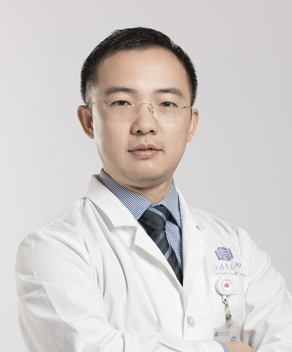 Suxi Gu (顾苏熙)合作论文数Beijing Tsinghua Changgung Hospital, Tsinghua University2
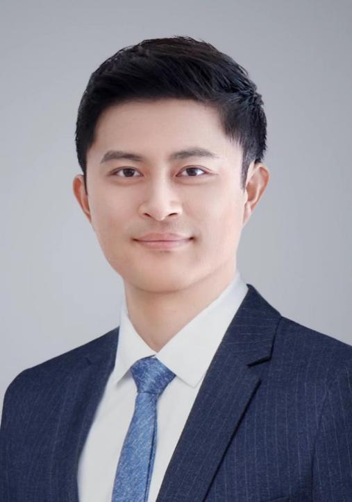 Jing Chen (陈景)合作论文数Nanjing University of Chinese Medicine1Xiangbing Bian合作论文数1Yan Miao合作论文数1Xin Liang合作论文数1Jifeng Zhang合作论文数1Hui Yao合作论文数1Dan LI (厉丹)合作论文数1
 Xiaobo Luo (罗晓波)合作论文数1Chuxuan Liu合作论文数1Wang Xing合作论文数1Guoke Tang合作论文数1Baorui Zhang合作论文数1Yunlong Ma合作论文数1Xiaotong Ma合作论文数1Shuai Chang合作论文数1Xue Han合作论文数1Chao Li合作论文数1Jianqin Liang合作论文数1TianTian Wang (王天天)合作论文数1WuRen Bao (包乌仁)合作论文数1HuiZe Liang (梁会泽)合作论文数1Xing Chen (陈兴)合作论文数1
 Da Huang (黄达)合作论文数College of Biological Science and Engineering, Fuzhou University1
 YuQi Zhu (朱瑜琪)合作论文数中国中医科学院眼科医院1
 Shenggang Wang (王胜刚)合作论文数Institute of Metal Research, Chinese Academy of Sciences1Shuyun Zhou (周树云)合作论文数1
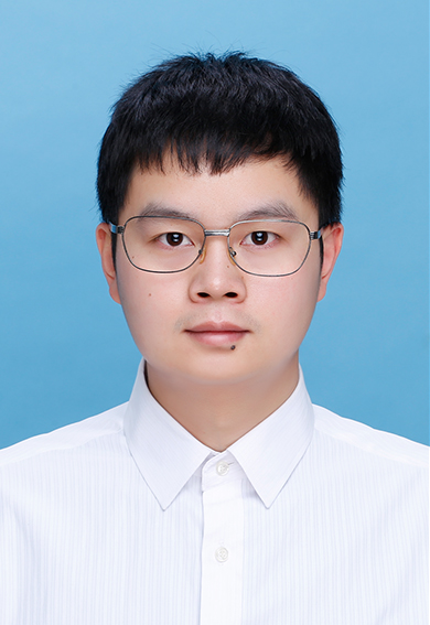 Yazhong Bu (补亚忠)合作论文数School of Basic Medical Sciences, Xi'an Jiaotong University1
 Xiaozhong Qu (屈小中)合作论文数College of Materials Science and Opto-Electronic Technology, University of Chinese Academy of Sciences1Niu Yuguang合作论文数1Liu Baoxia合作论文数1Zhao Pei合作论文数1




