Robotic-assisted total knee arthroplasty (RA-TKA) has emerged as an advanced surgical technique. However, direct comparisons between Chinese and American robotic systems are limited. Exploratory comparison of surgical accuracy and short-term clinical outcomes between Chinese HURWA and American MAKO robotic systems in total knee arthroplasty. A prospective exploratory cohort study was conducted from September 2023 to January 2024. Eighty-five patients were assigned to undergo RA-TKA with either the HURWA system (n = 43) or the MAKO system (n = 42) based on the sequential clinical introduction of the systems. The primary outcomes were radiographic accuracy, including the restoration of the mechanical axis within ± 3°. Secondary outcomes encompassed surgical efficiency metrics and patient-reported clinical scores (WOMAC, HSS, VAS) assessed preoperatively and at one-year postoperatively. All included patients have completed surgery and follow-up.The two systems achieved a comparable and high proportion of mechanical axis restoration within ± 3° (HURWA: 86.05
Objective To evaluate long-term survival and clinical outcomes of patients with knee osteo-arthritis undergoing total knee arthroplasty(TKA)through long-term follow-up.Methods This study was based on a previous cohort study that had completed follow-up.We retrospectively collected clinical data of patients with knee arthropathy(including knee osteoarthritis and knee rheumatoid arthritis)who received the first TKA opera-tion in Peking Union Medical College Hospital from 1993 to 2002 and were followed up for more than 20 years,and conducted a unified follow-up on them in November 10,2024(the last follow-up).Kaplan-Meier curve was used to evaluate the survival rate.Hospitals for special surgery(HSS)scores and joint range of motion(ROM)were compared before surgery,10 years after surgery and at the last follow-up to evaluate the clinical efficacy of TKA.Likert scale was used to evaluate patient satisfaction at the last follow-up.Results A total of 226 patients(246 knees)received their first TKA in Peking Union Medical College Hospital from 1993 to 2002 and were fol-lowed up for more than 10 years.Among them,104 patients(131 knees)were included in the study at the last follow-up,including 21 patients(24 knees)with prosthesis in place,18 patients(18 knees)who underwent reo-peration for various reasons,and 65 patients(89 knees)who died from non-TKA surgical causes.Up to the last follow-up,there were 29 patients(35 knees)with an average follow-up of more than 20 years,and 12 patients(16 knees)completed HSS score,ROM measurement and patient satisfaction evaluation.Kaplan-Meier curve showed that the 10-year,15-year,20-year,and 25-year survival rates were 93.6%,92.4%,89.8%,and 71.8%,respectively.The HSS score at the last follow-up was lower than that at 10-year postoperative follow-up[(84.69±11.03)scores vs.(95.25±13.32)scores,P<0.05],but significantly improved compared with the preoperative[(84.69±11.03)scores vs.(58.75±7.19)scores,P<0.05].The ROM of knee joint were(93.44±17.30)°,(101.88±13.33)°,and(91.56±15.98)° at preoperative,10-year of postoperative and last follow-up,respectively,and there was no statistical significance between the groups(P>0.05).In terms of pa-tient satisfaction,the 10 knees(62.50%)scored 4 points(very satisfied),the remaining 6 knees(37.50%)scored 3 points(satisfied),and the satisfaction rate was 100%.Conclusion TKA treatment for knee arthropathy has high long-term prosthesis survival rate,significant improvement of knee joint function and high patient satis-faction.
As an effective treatment for end-stage knee osteoarthritis, total knee arthroplasty (TKA) has been extensively performed globally over the past years. Despite its effectiveness, patients who undergo TKA are at risk of a series of complications, from infection to thromboembolism,[1] and substantial blood loss, which remains a concern among orthopedic surgeons, causing increased possibility for blood transfusion, prolonged hospitalization, and higher cost.[2,3] Postoperative TKA blood loss is now partially reduced due to the progress of surgical techniques and adjuvant medications. And many efforts on preoperative estimation of postoperative blood loss have been made over the past decade. However, there are still no systemic ways to assess the independent predictive value of potentially correlated risk factors; therefore, it is clinically critical to identify patients at an increased risk of blood loss and establish an effective blood management strategy.[4] This study analyzed total blood loss in patients receiving primary TKA, aiming to (i) identify influence factors for blood loss after TKA, especially among hematological parameters; (ii) develop a model for individualized prediction of blood loss following TKA using readily available clinical variables; and (iii) propose potential management strategies for adverse outcomes. This study was approved by the Institutional Review Board of the Peking Union Medical College Hospital (No. S-K2005), and because this was a retrospective study, the patients' informed consent was waived. The flowchart for experimental design is shown in Supplementary Figure 1, https://links.lww.com/CM9/C355. For all 1587 patients, the age, gender, body mass index (BMI), tobacco use (a positive status was defined as current smoker within 1 year, 1+ pack/day), alcohol use (a positive status was defined as current drinker within 1 year, 1+ drink/day) were recorded.[5] In addition, perioperative laboratory parameters were recorded. Total blood loss was calculated using Rosencher et al's[6] formula: Total red blood cell (RBC) loss (mL) = [Uncompensated RBC loss (mL)] + [Compensated RBC loss (mL)] Uncompensated RBC loss (mL) = [Initial RBC (mL)] – [Final RBC (mL)] Compensated RBC loss = [Sum of RBCs received from the various sources of transfusion] Initial RBC = [Estimated blood volume (mL)] × [Initial Hematocrit (HCT) level (%)] at Day – 1 Final RBC (mL) = [Estimated blood volume (mL)] × [Final HCT level (%)] at Day + 3 The estimated blood volume in milliliters was calculated for women and men separately with the use of the formulas[7]: Women: [Body surface area (m2)] × 2430 Men: [Body surface area (m2)] × 2530 where body surface area was calculated as: 0.0235 × [height (cm)]0.42246 × [weight (kg)]0.51456 Finally, the total blood loss was obtained at a HCT level of 35%: Total blood loss (mL) = [Total RBC loss (mL)]/0.35 Analyses were performed using SPSS software version 22.0 (SPSS Inc, Chicago, USA) and RStudio version 1.1.461 (RStudio, Boston, USA). Multivariable stepwise linear regression were established to determine potential independent influence factors for blood loss. P <0.05 was considered statistically significant. Considering that when blood loss is over 1500 mL, patients' physical condition and prognosis will be greatly reduced, so we converted the dependent variables into binary categorical variables at the threshold of 1500. The association between the pre-specified predictors and the primary outcome was assessed using LASSO Cox regression model analysis. And we used the Cox regression coefficients to generate nomograms. The model was internally validated using 1000 bootstrap samples. At the same time, the model was externally validated using another cohort of patients from January 2021 to December 2023. Agreement between predicted and observed outcomes was also evaluated graphically using receiver operating characteristic (ROC) analysis and the area under curve (AUC). The calibration curve was plotted to evaluate the agreement between nomogram-derived probability and actual observations of the model. A decision curve analysis (DCA) was also performed to evaluate the clinical benefit of our model. In this study, the total blood loss on the third day after the TKA surgery reached 961.29 ± 489.40 mL. Male patients lost an average of 1218.33 ± 549.67 mL of blood, whereas female patients lost an average of 898.89 ± 452.53 mL. We conducted a multivariate analysis of demographics, clinical, surgical variables, peroperative hematological parameters. Results were summarized in Supplementary Table 1, https://links.lww.com/CM9/C355. For these variables showing a correlation with postoperative blood loss, they were used for LASSO Cox regression model analysis, and the results are presented in Figure 1 and Supplementary Table 2, https://links.lww.com/CM9/C355.Figure 1: Development and performance of the nomogram. (A) Tuning parameter (λ) selection in the LASSO model used 10-fold cross-validation via minimum criteria. The AUC curve was plotted versus log (λ). Dotted vertical lines were drawn at the optimal values by using the minimum criteria and the 1 standard error of the minimum criteria (the 1-SE criteria). A λ value of 0.01623, with log (λ), 10 was chosen (1-SE criteria) according to 10-fold cross-validation. (B) LASSO coefficient profiles of the 13 features. A coefficient profile plot was produced against the log (λ) sequence. Vertical line was drawn at the value selected using 10-fold cross-validation, where optimal λ resulted in 10 non-zero coefficients. (C) A nomogram was created to evaluate the possibility of postoperative hemorrhage (total blood loss >1500 mL). To use the nomogram, find the position of each variable on the corresponding axis, draw a line to the points axis, add the points for the ten variables to create a total point value, and draw a line to the possibility axis to determine the probability on the lower line of the nomogram. ALB: Albumin; APTT: Activated partial thromboplastin time; BMI: Body mass index; HCT: Hematocrit; MONO: Monocyte; PLT: Blood platelet; TBIL: Total bilirubin.These independently associated risk factors were used to form a blood loss risk estimation nomogram [Figure 1C]. The optimal cutoff value of the total nomogram scores was determined to be 200. The sensitivity, specificity, positive predictive value, and negative predictive value when used in predicting risk of postoperative hemorrhage were 81%, 65%, 24%, and 96%, respectively. The resulting model was internally validated using the bootstrap validation method. The nomogram demonstrated good accuracy in estimating the risk of blood loss, with an unadjusted C index of 0.78 and a bootstrap-corrected C index of 0.77. ROC analysis and AUC graphically showed good agreement on the presence between the risk estimation by the nomogram (the mean AUC of the nomogram was 0.784), with high sensitivity and specificity of 79% and 64%, respectively, which indicated good discrimination. The calibration curve suggested that the nomogram had good calibration and fit in the internal validation set. The DCA results demonstrated that the nomogram added more net benefit than the "treat all" strategy or the "treat none" strategy in the training cohort. Overall, 500 patients after TKA from January 2021 to December 2023 from another center were finally included for external validation of the nomogram. The area under the ROC curve remained stable in the external validation (0.774). The calibration plots of the nomogram showed high consistency between the ideal line and the observed survival probability in the external validation cohorts. In the external validation, the DCA also revealed that the nomogram had an overall net benefit. The results of intetnal and external validation are shown in Supplementary Figure 2, https://links.lww.com/CM9/C355. In this study, we developed a prediction model based on demographics and hematological characteristics to improve the prediction of risk of hemorrhage after surgery for patients with TKA. Our results showed that this model can predict patients with high risk of postoperative blood loss (>1500 mL). Few studies investigated the relationship between smoking and postoperative blood loss. Nordestgaard et al[8] reported two potential mechanisms of smoking's influence on surgical bleeding. The chemicals inside the tobacco increased the recruitment of inflammatory cells to the vessel wall, causing injury to endothelial cells and vessel walls to be more fragile, resulting in increased perioperative bleeding. The other mechanism is reduced oxygenation and altered function of inflammatory cells during the initial healing process, which would increase short-term bleeding risk (within 72 hours). McCunniff et al[9] found platelet membrane change and impaired function caused by tobacco use. Tobacco use was also found to alter the membrane of red blood cells to be more fragile and hemolytic, causing postoperative blood loss. Multivariate analysis showed a positive correlation between preoperative HCT and postoperative blood loss (P <0.001). In a postoperative bleeding study in cervical cancer, Zhao et al[10] reported a significant positive correlation between preoperative HCT and insidious postoperative blood loss (P = 0.003), because high HCT level could increase blood accumulation in interstitial space by participating in postoperative hyperfibrolysis, thus increasing total blood loss by adding to insidious loss. In addition, for the same volume of blood loss, a higher HCT level means more lost visible components in the blood, resulting in more relative blood loss. We also found a positive relationship between preoperative ALB and postoperative blood loss. Possible explanations include compromised coagulation and inhibited platelet aggregation by albumin, as reported in a previous study.[11] Second, a relatively higher level of albumin could maintain colloid osmotic pressure, which would help replenish circulation volume, resulting in a higher relative volume of blood loss. Moreover, elevated preoperative albumin would cause volume expansion secondary to increased colloid osmotic pressure and diluting clotting factors, resulting in more bleeding intraoperative and postoperatively bleeding. Herein, we have explored many demographic and hematological parameters for blood loss after TKA, and developed a prediction model to improve the prediction of risk of hemorrhage after surgery for patients with TKA. The use of the nomogram in estimating the risk of a patient harboring postoperative hemorrhage to direct clinical treatment is a new concept. For clinical use of the model, we summarized the sensitivity, specificity, negative predictive value, and positive predictive value in estimating the risk of postoperative hemorrhage using 200 as the cutoff value. Patients with a score of 200 or more are a high-risk subgroup of blood loss. Based on these preoperative and postoperative predictions, the nomogram might serve as a tool to select patients at an increased risk of blood loss and establish an effective blood management strategy. This study still had some limitations. There is a geographical limitation as the sample was sourced from a single center. And the study adopted a retrospective design and relied on past medical records. The completeness and accuracy of the data may be constrained by the standard of medical record keeping. A further prospective study is needed to verify some hypotheses we proposed. Conflicts of interest None. Funding This work was supported by CAMS Innovation Fund for Medical Sciences (CIFMS) (No. 2023-I2M-C&T-B-044).
Introduction Preoperative patients with knee osteoarthritis have a significantly increased risk of venous thromboembolism (VTE). While the Caprini risk assessment model offers some clinical guidance in predicting deep vein thrombosis (DVT), it has a relatively low predictive accuracy. Enhancing the model by integrating biomarkers, such as D-dimers, can potentially improve its accuracy. In this study, we explored the effectiveness of combining the Caprini risk model with D-dimer levels for individualized DVT risk assessment in patients with knee osteoarthritis. Materials and Methods This retrospective cohort study included 1605 knee osteoarthritis patients scheduled for total knee arthroplasty from Peking Union Medical College Hospital, screened between January 2015 and December 2018. A revised Caprini risk stratification model was developed, and a predictive DVT model was developed based on this revised system. The sensitivity, specificity, and the area under the curve (AUC) were used to determine predictive effectiveness of the model. Results In the revised Caprini risk stratification, the incidence of DVT increased with higher risk levels: 2.52% in the low-risk group (scores 0-2), 2.88% in the moderate-risk group (score 3), 6.47% in the high-risk group (score 4), and 9.09% in the highest-risk group (score ≥ 5). The incidence of DVT was 3.869-fold higher in the highest-risk group and 2.676-fold higher in the high-risk group compared to the low-risk group (p = 0.013 and p = 0.014, respectively). Combining the revised Caprini risk stratification with D-dimer level demonstrated an improved AUC of 0.792, compared to D-dimer level alone (AUC 0.774) and the revised Caprini model alone (AUC 0.598). Furthermore, applying specific D-dimer thresholds across the four Caprini risk stratifications outperformed the combination of the revised Caprini model and D-dimer level in terms of AUC, specificity, and reduction in unnecessary ultrasonography. Using the Youden index, the AUC for the threshold-based method was slightly higher (0.775 vs 0.754, p = 0.310), with significantly better specificity (76.8% vs 63.6%, p < 0.001) and a greater reduction in ultrasound use (74.1% vs 61.4%). At a sensitivity of 85.5%, the differences were modest but still favored the threshold-based approach. At a sensitivity of 100%, the specificity (36.0% vs 24.7%, p < 0.001) and ultrasound reduction (34.8% vs 23.9%) were significantly better. Conclusion The revised Caprini risk stratification improves preoperative DVT prediction in patients with knee osteoarthritis. Incorporating specific D-dimer thresholds into the four-level Caprini risk model enhances specificity and reduces unnecessary ultrasonography, outperforming both the use of individual indicators and the combination of the revised Caprini model with D-dimer level.
BACKGROUND:Three-dimensional facial stereophotogrammetry, a convenient, noninvasive and highly reliable evaluation tool, has in recent years shown great potential in plastic surgery for preoperative planning and evaluating treatment efficacy. However, it requires manual identification of facial landmarks by trained evaluators to obtain anthropometric data, which takes much time and effort. Automatic 3D facial landmark localization has the potential to facilitate fast data acquisition and eliminate evaluator error. OBJECTIVES:The aim of this work was to describe a novel deep-learning method based on dimension transformation and key-point detection for automated 3D perioral landmark annotation. METHODS:After transforming a 3D facial model into 2D images, High-Resolution Network is implemented for key-point detection. The 2D coordinates of key points are then mapped back to the 3D model using mathematical methods to obtain the 3D landmark coordinates. This program was trained with 120 facial models and validated in 50 facial models. RESULTS:Our approach achieved a satisfactory mean [standard deviation] accuracy of 1.30 [0.68] mm error in landmark detection with a mean processing time of 5.2 [0.21] seconds per model. Subsequent analysis based on these landmarks showed mean errors of 0.87 [1.02] mm for linear measurements and 5.62° [6.61°] for angular measurements. CONCLUSIONS:This automated 3D perioral landmarking method could serve as an effective tool that enables fast and accurate anthropometric analysis of lip morphology for plastic surgery and aesthetic procedures.
Deep vein thrombosis (DVT) is a common and serious risk in elderly patients with knee osteoarthritis (OA), making preoperative detection crucial. Despite this, identifying OA patients at high risk for preoperative DVT and appropriately targeting them for venous ultrasound screening remains a challenge. There is limited research-based evidence on the risk factors for preoperative DVT in elderly patients with end-stage OA. We examined the incidence of and risk factors for preoperative DVT in elderly patients with end-stage OA scheduled for total knee arthroplasty. We retrospectively analyzed the demographic data (age, sex, body mass index, current smoking, alcohol consumption, walking status, and Barthel index score), medical history, and laboratory test indices of 1411 patients with end-stage OA aged ≥ 60 years scheduled for total knee arthroplasty from January 2015 to December 2018. Risk factors for preoperative DVT were evaluated by univariate and multivariate logistic analyses. Receiver operating characteristic analysis was performed to determine optimal cut-off values. The incidence of preoperative DVT was 4.5
Objective To investigate the perioperative rate of allogeneic red blood cell (RBC) transfusion in patients who underwent total knee arthroplasty (TKA) and its risk factors, and to identify its cross-match to transfusion ratio (C∶T ratio). Methods Anesthetic data of patients who underwent TKA from January 2014 to October 2019 in Peking Union Medical College Hospital were collected and analyzed retrospectively. Perioperative allogeneic RBC transfusion rate was calculated, and binary Logistic regression analysis was performed to identify its risk factors in these patients. The overall C∶T ratio was calculated and divided into subgroups based on surgery type and age group. Results The study enrolled 2 903 patients. The perioperative rate of allogeneic RBC transfusion in TKA patients was 10.9% (95% CI 9.8%~12.0%) and overall C∶T ratio was 5.6∶1. The independent risk factors leading to perioperative allogeneic RBC transfusion included advanced age(OR=1.025, 95% CI 1.009~1.042, P<0.01), preoperative hemoglobin level(OR=0.966, 95% CI 0.954~0.978, P<0.001), preoperative anemia(OR=3.543, 95% CI 2.052~6.119, P<0.001), hematological diseases(OR=6.462, 95% CI 2.479~16.841, P<0.001), bilateral surgery(OR=7.681, 95% CI 5.759~10.245, P<0.01) and revision surgery(OR=9.584, 95% CI 4.360~21.065, P<0.001). Conclusion The risk factors for perioperative allogeneic RBC transfusion in TKA patients included advanced age, preoperative low hemoglobin level, preoperative anemia, hematological diseases, bilateral surgery and revision surgery. Only type and screen tests are recommended if patients receiving unilateral primary TKA surgery are less than 75 years old without anemia and hematological diseases, while at least one to four units of blood should be cross-matched if patients are with preoperative anemia and hematological diseases or will receive bilateral and revision arthroplasty.
The American College of Rheumatology (ACR) and the American Association of Hip and Knee Surgeons (AAHKS) convened a writing group to develop a consensus report on the management of anti-rheumatic medication in patients with rheumatic diseases undergoing elective total hip or total knee arthroplasty in June 2022. In particular, the consensus provides significant updates on target synthetic disease modifying anti-rheumatic drugs and perioperative medication management in patients with systemic lupus erythematosus, as well as the addition of newly approved antirheumatic medications for administration. This article will interpret the consensus and provide a reference for the perioperative management of antirheumatic medications for hip and knee arthroplasty in patients with rheumatic diseases in China.
Abstract Background: Total knee arthroplasty (TKA) can reduce severe joint pain and improve functional disability in hemophilia. However, the long-term outcomes have rarely been reported in China. Therefore, this study aimed to evaluate the long-term outcomes and complications of TKA in Chinese patients with hemophilic arthropathy. Methods: We retrospectively reviewed patients with hemophilia who underwent TKA between 2003 and 2020, with at least 10 years of follow-up. The clinical results, patellar scores, patients' overall satisfaction ratings, and radiological findings were evaluated. Revision surgery for implants during the follow-up period was recorded. Results: Twenty-six patients with 36 TKAs were successfully followed up for an average of 12.4 years. Their Hospital for Special Surgery Knee Score improved from an average of 45.8 to 85.9. The average flexion contracture statistically significantly decreased from 18.1° to 4.2°. The range of motion (ROM) improved from 60.6° to 84.8°. All the patients accepted patelloplasty, and the patients' patellar score improved from 7.8 preoperatively to 24.9 at the last follow-up. There was no statistically significant difference in clinical outcomes between the unilateral and bilateral procedures, except for a better ROM at follow-up in the unilateral group. Mild and enduring anterior knee pain was reported in seven knees (19%). The annual bleeding event was 2.7 times/year at the last follow-up. A total of 25 patients with 35 TKAs were satisfied with the procedure (97%). Revision surgery was performed in seven knees, with 10- and 15-year prosthesis survival rates of 85.8% and 75.7%, respectively. Conclusions: TKA is an effective procedure for patients with end-stage hemophilic arthropathy, which relieves pain, improves knee functions, decreases flexion contracture, and provides a high rate of satisfaction after more than ten years of follow-up.
Background: The number of patients with primary Sjögren’s syndrome (pSS) who require total knee arthroplasty (TKA) is expected to increase, and there are few studies describing their outcomes. This research was focused on the evaluation of a TKA cohort in pSS patients and to compare outcomes with those of matched individuals from the general population. Methods: From 2004 to 2020, we found 36 TKAs in 30 patients with pSS from the single-institution arthroplasty registry, and they were matched for age, gender, bilateral or unilateral surgery, American Society of Anesthesiologists (ASA) score, and year of surgery with 72 TKAs in 60 osteoarthritis patients without rheumatic diseases (1:2 ratio). Perioperative outcomes were obtained, and clinical evaluations were performed at the last follow-up. Results: After a mean six-year follow-up, both cohorts had similar knee function and health-related quality of life outcomes. The pSS group had more patients with post-operative anemia and hypoalbuminemia and more patients needing platelet transfusion. There were no significant differences in other complications, the rates of 90-day readmission, or overall revision. By multivariate analysis, the influencing factor for anemia in pSS patients was lower preoperative hemoglobin (OR = 0.334, 95% CI (0.125–0.889), p < 0.05). Conclusions: Our study demonstrated that pSS patients who received TKA could achieve comparable clinical outcomes to the general population. However, more attention should be paid to the perioperative hematological management of pSS patients who underwent TKA.
目的:比较采用增强(Enhanced)模式MAKO机器人辅助后外侧入路全髋关节置换术(RTHA)与常规THA手术的早期临床疗效.方法:回顾性分析2021年5—12月采用增强模式MAKO RTHA治疗的77例患者的临床资料,并与同期采用传统后外侧入路全髋关节置换术(THA)治疗的77例患者进行对比.比较RTHA组与常规THA组患者的手术时间、总失血量.术后测量髋臼外展角、前倾角、双下肢长度差(LLD)、两侧联合偏心距差异(ΔCO).比较RTHA组术中与术后LLD及ΔCO的一致性,同时比较两组髋臼外展角、前倾角,术后LLD及ΔCO的差异.术后随访收集两组Harris髋关节评分.结果:RTHA组术中测量的LLD和ΔCO与术后影像学测量LLD和ΔCO比较,差异无统计学意义.RTHA组术中机器人测量股骨前倾角14.9°±7.8°、联合前倾角37.4°±9.1°.RTHA组术后LLD(0.65±3.40)mm,常规THA组术后LLD(2.54±4.46)mm,差异有统计学意义(P=0.013).RTHA组术后ΔCO(-0.15±6.27)mm,常规THA组(2.65±6.40)mm,差异有统计学意义(P=0.020).RTHA组髋臼前倾角平均23.1°±4.3°,常规THA组26.6°±4.1°,差异有统计学意义(P<0.001).RTHA组髋臼外展角平均42.1°±2.8°,常规THA组40.9°±5.8°,差异无统计学意义.两组患者随访6~12个月,RTHA组末次随访Harris髋关节评分平均(90.5±6.8)分,常规THA组(90.1±6.5)分,差异无统计学意义.结论:增强模式下MAKO机器人辅助THA能提高髋臼杯植入的准确性,可更精确恢复下肢长度及联合偏心距,对股骨侧前倾的判断有助于个体化调整髋臼前倾.RTHA和传统THA手术术后短期疗效无统计学差异.
Background: The objective of this study was to compare the radiologic and clinical outcomes of HURWA robotic-assisted total knee arthroplasty (TKA) to those of conventional TKA. Methods: A total of 150 patients were randomized into two groups - 73 and 77 patients underwent robotic-assisted TKA and conventional TKA, respectively. Preoperative and postoperative Western Ontario McMaster University Osteoarthritis Index (WOMAC) score, Hospital for Special Surgery (HSS) score, 36-item Short Form Health Survey (SF-36) score, Knee Society Score (KSS) and range of motion (ROM) were obtained and compared between these two groups. The preoperative and postoperative hip-knee-ankle (HKA) angle and the rate of HKA <= 3 degrees in the two groups were also compared. Results: The postoperative mean HKA angle was 1.801 degrees +/- 1.608 degrees of varus for the robotic-assisted TKA group and 3.017 degrees +/- 2.735 degrees of varus for the conventional TKA group; these values were significantly different. The alignment rate for mechanical axis lower than 3 degrees in the robotic-assisted TKA group and the conventional TKA group were 81.2% and 63.5%, respectively. Patients undergone robotic-assisted TKA or conventional TKA had similarly improved knee flexion and functional recovery reflected by WOMAC score, HSS score, SF-36 score and KSS. Conclusion: HURWA robotic-assisted TKA is a safe and effective, resulting in better alignment for mechanical axis than conventional TKA. The improvement in knee flexion and functional recovery after HURWA robotic-assisted TKA were similar to those after conventional TKA. However, longer follow-up is needed to determine whether the improved alignment of mechanical axis will produce better long-term clinical outcomes. The translational potential of this article: Recently, the robotic-assisted TKA system has been introduced to clinical practice for TKA. Several robotic-assisted TKA systems, including CASPAR, Tsolution, ROSA, ROBODOC and Mako, have been implemented into clinical application.However, the clinical application of these robotic systems was limited due to their technical complexity, insufficient versatility and increased operative time. Until now, there are still no robotic-assisted TKA systems approved by the National Medical Products Administration of China. Therefore, more robotic-assisted TKA systems need to be designed and improved, particularly in China. Through our randomized, multicenter, single blind and parallel controlled trial, we showed that HURWA robotassisted TKA system is a safe and effective system for TKA, which had improved knee flexion.
As compulsory content for orthopedic graduate students, total knee arthroplasty (TKA) is characterized by requirements of surgical accuracy and so is difficult to train since there is limited opportunity for young surgical trainees especially design of surgical operation and long term training to get experience. Traditional teaching of TKA for graduates is embarrassed by these barriers. Recent years, new techniques such as robotic-arm assisted surgery and 3D-printing technology have been extensively used in clinical training. For example the hybrid model with combined surgical design in virtue of robotic system and vivid simulation produced by 3D-printing for training. This paper reviewed is current situation in this field and evaluated the feasibility in clinical application
Objective Total knee arthroplasty is one of the most common orthopedic surgeries. Readmission due to severe complications after total knee arthroplasty is a grave concern to surgeons. In this study, we evaluated the risk factors for severe complications after primary total knee arthroplasty. Methods We retrospectively collected clinical data of 2,974 patients who underwent primary total knee arthroplasty from July 2013 to June 2019 in our hospital. Postoperative complication ≥ grade Ⅲ was defined as severe complication according to Clavien-Dindo classification system. Binary logistic regression was used to identify the predictive risk factors for severe complications. Results The complication rate after primary total knee arthroplasty was 6.8% and severe complication rate was 2.5%. Male (OR = 2.178, 95%CI: 1.324-3.585, P= 0.002), individuals above 75 years old (OR = 1.936, 95%CI: 1.155-3.244, P= 0.012), arrhythmia (OR = 2.913, 95%CI: 1.350-6.285, P= 0.006) and cerebrovascular disease (OR = 2.804, 95%CI: 1.432-5.489, P= 0.003) were predictive risk factors for severe complications after primary total knee arthroplasty. Conclusion Advanced age, male, arrhythmia, and cerebrovascular disease might be patients-related risk factors for postoperative severe complications after primary total knee arthroplasty. Special attention should be paid to patients with risk factors.
Background Neuromyelitis Optica Spectrum Disorders (NMOSD) are rare inflammatory diseases of the central nervous system that cause transverse myelitis and optic neuritis. Steroids are commonly administered in NMOSD patients. The use of steroids may lead to osteonecrosis, which makes some of the NMOSD patients candidate for total hip arthroplasty (THA). To date, the clinical outcome of THA in NMOSD patients have not been investigated. Aim Investigate the patient reported outcome measures (PROM), radiographic outcome and complication in NMOSD patients after THA, compared with that of non-NMOSD patients. Methods Patients from Jan. 2016 to October. 2020 were identified in our database. 12 NMOSD cases which met the inclusion criteria were matched to non-NMOSD cases in a ratio of 1:2 based on age, sex, Charlson Comorbidity Index (CCI) and surgical date. Relevant outcome were analyzed and compared between the two groups. Results There was a significantly increased risk of dislocation in NMOSD patients. Post-operative HOOS score was similar between the two groups even though the pre-operative HOOS score is significantly higher in the non-NMOSD group. NMOSD patients had poor performance in EQ-5D and EQ-VAS. The cups were placed more anteverted in NMOSD cases ( P = 0.01). Conclusion There is a significantly increased risk of dislocation after THA in NMOSD patients. However, satisfactory improvement in functional outcome of the hip was achieved. Due to the natural process of NMOSD, rehabilitation and hip precaution should be patient-specific and time-specific.
Multimodal cocktail periarticular injections comprising corticosteroids are the most suggested therapy for postoperative discomfort and swelling following total knee arthroplasty (TKA). Nevertheless, previous findings cannot be applied to instances of unilateral total knee arthroplasty on bilateral knees. This randomized, prospective, double-blind, controlled clinical study examines the efficacy as well as safety of periarticular multimodal cocktail injection along or sans corticosteroids in certain situations. The 60 patients (120 knees) that experienced concurrent bilateral total knee arthroplasty were provided periarticular injections along additional betamethasone (7 mg) in the randomized knee, as well as the other knee, where corticosteroid was not administered. Key results were "pain scores at rest as well as in action" on a visual analogue scale of 11 pt. Other results included motion range, swelling of the thigh, Hospital for Special Surgery score (HSS score), and adverse effects were measured between the two sides. No statistically promising variations were found in the visual analogue scale ranking, motion range, girth of the thigh, and HSS score, as well as complications between the two sides. The impact on treatment outcomes was maintained between the knees on postoperative day 3 or at 3 months of follow-up. Multimodal periarticular injection without corticosteroid will alleviate postoperative swelling and pain. More studies are needed for the use of betamethasone as a corticosteroid in periarticular multimodal cocktail injections. This Chinese Clinical Trial Registry is registered with ChiCTR-OPC-17013503, dated 2017-11-23, available from http://www.chictr.org.cn/showproj.aspxproj=23146.
BACKGROUND:We investigated the accuracy and safety of a new HURWA robotic-assisted total knee arthroplasty (TKA) system in a sheep model.METHODS:Ten male small-tailed Han sheep were used in this study. Sheep were imaged by computed tomography scan before and after bone resection and the cutting errors between actual bone preparation and preoperative planning of the femur and tibia in three dimensions were measured.RESULTS:The overall accuracies after surgery compared with that from preoperative surgical planning of the left and right femurs were 1.93 ± 1.02° and 1.93 ± 1.23°, respectively. Additionally, similarly high overall accuracies for the left and right tibia of 1.26 ± 1.04 and 1.68 ± 0.92°, respectively, were obtained. The gap distances of the distal cut, anterior chamfer, anterior cut, posterior chamfer and posterior cut on the medial side were 0.47 ± 0.35 mm, 0.41 ± 0.37 mm, 0.12 ± 0.26 mm, 0.41 ± 0.44 mm and 0.12 ± 0.23 mm, respectively. No intraoperative complications, such as intraoperative fracture, massive bleeding or death, occurred.CONCLUSION:This new HURWA robotic-assisted TKA system is an accurate and safe tool for TKA surgery based on the sheep model.
BACKGROUND:The purpose of this study was to compare the accuracy of this new HURWA robotic-assisted total knee arthroplasty (TKA) technique to the accuracy of the conventional technique in a sawbone model.METHODS:The HURWA robotic-assisted TKA system was applied in the robotic group. After bone resection, all of these sawbones were scanned by the use of a structured light scanning system. Measurements of bone resections, femoral coronal and sagittal measurements, and tibial coronal and sagittal measurements were recorded.RESULTS:Compared to the conventional technique, the HURWA robotic-assisted system significantly improved the accuracy of the bone resection levels and angles. In the robotic group, the accuracy of all of the bone resection levels was below 0.6 mm (with standard deviation [SD] below 0.6 mm), and all of the bone resection angles were below 0.6° (with SD below 0.4°).CONCLUSION:Our data suggest that this novel HURWA robotic-assisted system can significantly improve the accuracy of bone resection levels and angles.
The study aims to evaluate the clinical outcomes and surgery survivorship for over 10 years following patellar resurfacing or nonresurfacing in total knee arthroplasty (TKA) in a cohort of Chinese patients. From 1998 to 2003, 355 patients underwent primary TKA in our institute. The survivorship of TKA between the patellar resurfacing and nonresurfacing groups and the clinical outcome of Hospital for Special Surgery knee score, Western Ontario and McMaster Universities index score, patellar score, patellar related complications, and radiological results were studied at latest follow-up. There was no statistically significant difference for the Hospital for Special Surgery score, Western Ontario and McMaster Universities score, and the patellar score between the 2 groups after an average 12.4-year follow-up. Nonresurfacing group had higher anterior knee pain than the resurfacing group (13.2% vs 5.6%). The patients with rheumatoid arthritis had a 2.9-fold higher rate of patellar-related complications than did the patients with osteoarthritis. The 10-year survival rate was not significantly different between the 2 groups both for revision surgery (P = .505) and for patellar-related complication (P = .194). There was no significant difference in the long-term clinical outcome and survivorship between patellar resurfacing and nonresurfacing. Patellar nonresurfacing could be advisable during primary TKA for osteoarthritis patients. Selective patellar resurfacing for RA patients could achieve lower patellar-related complications.
Osteoarthritis (OA) is a degenerative disease of middle-aged and elderly people, contributed a higher burden of disease in China and the world. In 2017, under the support of the Rheumatology and Immunology Expert Committee of the Cross-Strait Medical and Health Exchange Association. The objective was to develop an evidence-based diagnosis and treatment guideline for OA in China based on emerging new evidence. The guideline was registered at International Practice Guidelines Registry Platform (IPGRP-2018CN028). The grading of recommendations assessment, development and evaluation (GRADE) approach was used to rate the quality of evidence and the strength of recommendations, and the RIGHT (Reporting Items for Practice Guidelines in Healthcare) checklist was followed to report the guideline. The guideline provides recommendations for the OA diagnosis, disease risks monitoring and evaluate, treatment purpose and physical, medical and surgical interventions. This guideline is intended to serve as a tool for Chinese clinicians for the best decisions-making on diagnosis and treatment of OA.
 Xisheng Weng (翁习生)合作论文数Peking Union Medical College Hospital109
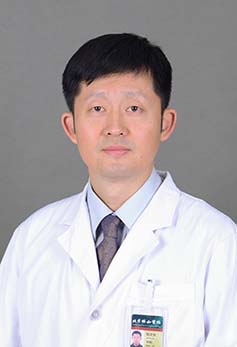 Wenwei Qian (钱文伟)合作论文数Peking Union Medical College Hospital63
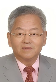 Guixing Qiu (邱贵兴)合作论文数Peking Union Medical College Hospital57
 Jin Jin (金今)合作论文数Department of Orthopaedics,Peking Union Medical College Hospital,Beijing ,China44
 Yipeng Wang (王以朋)合作论文数Peking Union Medical College Hospital18
 Huiming Peng合作论文数16
 Jianguo Zhang (仉建国)合作论文数Peking Union Medical College Hospital15
 Hong Zhao (赵宏)合作论文数Department of Orthopaedic,PUMC Hospital,CAMS/PUMC,Beijing ,China14
 Bin Feng (冯宾)合作论文数Peking Union Medical College Hospital13
 Bian Yanyan合作论文数13JiLiang Di (翟吉良)合作论文数12
 Jianxiong Shen (沈建雄)合作论文数Beijing Union Medical College Hospital12Wei WANG (王炜)合作论文数11Wei Wang合作论文数10XinYu Yang (杨新宇)合作论文数10
 Shugang Li (李书纲)合作论文数Peking Union Medical College Hospital10LiJuan Zhao (赵丽娟)合作论文数9
 Yu Fan (范彧)合作论文数北京协和医学院北京协和医院9
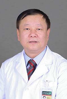 Baozhong Zhang (张保中)合作论文数Peking Union Medical College Hospital9
 Zhihong Wu (吴志宏)合作论文数Peking Union Medical College Hospital7
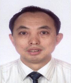 Ye Tian (田野)合作论文数Peking Union Medical College Hospital7
 Jiliang Zhai (翟吉良)合作论文数Peking Union Medical College Hospital7
 Qibin Ye (叶启彬)合作论文数Peking Union Medical College Hospital7Qing Zhao (赵庆)合作论文数6Xiao Chang (常晓)合作论文数5
 Zengxin Gao (高增鑫)合作论文数Department of Spine Surgery, Zhongda Hospital, Southeast University5Jia Zhang (张嘉)合作论文数4Na Gao (高娜)合作论文数4ZhiFeng Huang (黄志峰)合作论文数4Peng Gao (高鹏)合作论文数4
 Yixin Zhou (周一新)合作论文数Beijing Jishuitan Hospital;Peking University Health Science Center4Zhao Hong合作论文数3Long-Chao Wang合作论文数3Lin Sheng合作论文数3Xiaofeng Zhang合作论文数3Zheng Li合作论文数3Jun Yan合作论文数3QiBin Ye (叶启彬)合作论文数3Ye Li (李晔)合作论文数3
 Jun Qian (钱军)合作论文数北京协和医学院北京协和医院3
 Qianyu Zhuang (庄乾宇)合作论文数Peking Union Medical College Hospital3
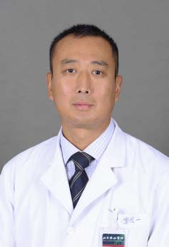 Qiyi Li (李其一)合作论文数Peking Union Medical College Hospital3
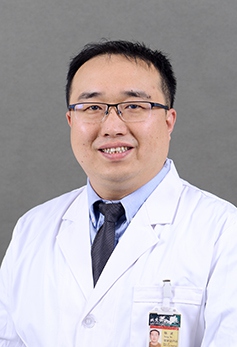 Bo Yang (杨波)合作论文数Peking Union Medical College Hospital3
 Yu Zhao (赵宇)合作论文数Peking Union Medical College Hospital;Peking Union Medical College, Chinese Academy of Medical Sciences3
 Yuguang Huang (黄宇光)合作论文数Peking Union Medical College Hospital;Peking Union Medical College3Xi Chen合作论文数2Lianhua Li合作论文数2




