Excessive accumulation of reactive oxygen species (ROS) impairs bone regeneration and angiogenesis in steroid-induced osteonecrosis of the femoral head (SONFH), yet current antioxidant therapies remain limited by low catalytic efficiency and short duration. Single-atom nanozymes (SANs), with their well-defined structures and maximal atomic efficiency, show great potential for treating ROS-induced diseases by mimicking natural enzymes. However, the strong binding between transition metal sites and electron-donating intermediates (e.g., O*, OH*, OOH*) creates high energy barriers, limiting their catalytic activities. Herein, single-atomic platinum is successfully embedded into CeO2-x to form CeO2-x/Pt SANI, which enhanced catalytic activity via an "island-sea" synergistic effect. Leveraging the unique charge-transfer structures and confinement effect of nanoislands, CeO2-x/Pt SANI exhibits superior enzymatic activities than CeO2, attribute to the island-sea synergistic effect that facilitates strong electron transfer, as proved by density functional theory (DFT) calculations. DFT calculations further demonstrate that Pt incorporation increases oxygen vacancies and tunes the d-band center toward the Fermi level, facilitating ROS adsorption and accelerating redox reactions. Single-cell sequencing and experimental results confirm that CeO2-x/Pt SANI reprograms the oxidative microenvironment, leading to significant therapeutic effects in SONFH. This study provides insights into the rational design of an advanced "island-sea" structured single-atom nanozyme to optimize the catalytic activity.
Abstract Nanozymes, which mimic the catalytic activities of natural enzymes, have been investigated as therapeutic agents for treating reactive oxygen species (ROS)-related diseases. Despite progress having been made in nanozyme engineering, their diverse nanostructures and poor atom utilization efficiency severely limit further applications. Symmetric Fe–N4 coordination in iron-based single-atom nanozymes (SANs) confers intrinsic ROS scavenging activity; however, excessive adsorption of oxygen intermediates restricts catalytic turnover. Here, we describe a spin-state engineering strategy to address this limitation through asymmetric coordination design. Phosphorus incorporation transforms the symmetric Fe–N4 motif into an Fe–PN3 configuration, inducing a transition of Fe 3d orbitals from a low-spin to a high-spin state. Density functional theory calculations indicate that this spin modulation raises the count of unpaired electrons and moves the d-band center nearer the Fermi level, which in turn tunes the binding energies of oxygen intermediates and speeds up the catalytic cycle. Consequently, the Fe–PN3 nanozyme exhibits substantially enhanced ROS detoxification efficiency compared with its Fe–N4 counterpart. In a glucocorticoid-induced osteonecrosis model, the spin-engineered nanozyme modulates the pathological redox microenvironment, restores impaired autophagic flux, and preserves tissue integrity. Single-cell RNA sequencing further indicates coordinated modulation of oxidative stress-responsive and autophagy-related pathways. Collectively, this work supports asymmetric coordination-induced spin-state regulation as a strategy for designing high-performance iron-based single-atom nanozymes with translational potential in ROS-driven degenerative diseases.
Steroid-induced osteonecrosis of the femoral head (SONFH) is a debilitating condition resulting from the use of glucocorticoids, commonly prescribed for immune-related and inflammatory diseases. Understanding the mechanisms driving SONFH remains a significant challenge, complicating efforts to prevent and treat the condition. While genetic predispositions, impaired blood supply, and metabolic changes are recognized contributors, the complex interplay between these factors is not yet fully understood. Recent research has shed light on the pathogenesis of SONFH, exploring it from multiple perspectives, including tissue-level damage, cellular dysfunction, and molecular pathways. This review summarizes these recent advancements, providing an integrated understanding of the onset and progression of the condition. Additionally, it highlights emerging therapeutic strategies that potentially pave the way for more effective treatments in the future.
Layered double hydroxide (LDH)-based materials have been explored as efficient photosensitizers for photodynamic therapy (PDT) or functional agents for bone regeneration, holding great promise in the synergistic PDT-mediated osteosarcoma treatment and bone reconstruction. Herein, we report the functionalization of 5-iodoisophthalic acid (I-IPA)-intercalated MgZnAl-LDH onto bioactive glass scaffold (BGS) to construct a bifunctional composite scaffold (BGS/I-LDH) for integrated osteosarcoma treatment and bone regeneration. The surface functionalization of I-LDH onto BGS confers BGS/I-LDH high-efficiency reactive oxygen species generation performance under 1270 nm laser irradiation, resulting in a singlet oxygen quantum yield up to 1.53, which is significantly superior to that of previously reported NIR-excited PSs. Moreover, thanks to the sustained release of Mg2+ and Zn2+ ions from I-LDH, BGS/I-LDH exhibits exceptional anti-osteosarcoma and osteogenic properties with 3.8-fold bone volume, 3.0-fold bone mineral density, and 11.4-fold new bone mass increments compared with the pristine BGS, which is remarkably more preponderant than other previously reported modified BGS.
OBJECTIVE:Postoperative inflammation and pain remain key barriers to recovery after total knee arthroplasty (TKA). Metformin has anti-inflammatory properties, but its perioperative impact in TKA is unclear. This study evaluated whether regular metformin use reduces postoperative inflammation and pain in T2DM patients undergoing TKA. METHODS:In this single-center retrospective cohort study, patients with Type 2 diabetes mellitus (T2DM) who underwent primary TKA between December 2024 and May 2025 were included. Patients were grouped based on whether they had regularly taken metformin. 1:1 propensity score matching was performed to reduce baseline differences between groups, yielding 60 matched pairs. Inflammatory markers including C-reactive protein (CRP), interleukin-6 (IL-6), erythrocyte sedimentation rate (ESR), and D-dimer were measured preoperatively and on postoperative days 1 and 3. Pain was assessed using the Visual Analogue Scale (VAS) on postoperative days 1 and 3. Linear mixed-effects models were used to compare outcomes between groups after adjusting for residual confounders. RESULTS:In the matched cohort, the metformin group showed significantly lower CRP levels on postoperative day 3 (107.04 ± 49.19 vs. 130.91 ± 59.62 mg/L, p = 0.02), lower IL-6 levels on postoperative day 1 (62.22 ± 47.17 vs. 91.07 ± 41.55 pg/mL, p < 0.01), and postoperative day 3 (31.33 ± 20.84 vs. 57.74 ± 59.98 pg/mL, p < 0.01), and lower ESR levels on postoperative day 3 (45.40 ± 17.85 vs. 47.05 ± 20.19 mm/h, p = 0.03) than the non-metformin group. VAS pain scores on postoperative day 3 were also significantly lower in the metformin group (3.03 ± 1.03 vs. 4.15 ± 1.25, p < 0.01). These differences remained significant after adjusting for confounders. CONCLUSION:Long-term regular use of metformin is associated with reduced acute inflammation and postoperative pain in T2DM patients undergoing TKA. As a widely used and well-tolerated medication, metformin holds promise as a low-cost, adjunctive therapeutic strategy to enhance early recovery and improve perioperative outcomes in diabetic patients undergoing TKA.
Osteoarthritis (OA) is a progressive and disabling joint disease driven by oxidative stress, chondrocyte senescence and extracellular matrix (ECM) degradation, yet lacks effective disease-modifying treatments. In this study, we identified miR-197-3p as a previously unrecognized, cartilage-protective miRNA significantly downregulated in both aged and osteoarthritic cartilage. Functional studies revealed that miR-197-3p restores ECM anabolism, suppresses senescence and directly targets G3BP1, a stress granule protein linked to redox imbalance and inflammatory signaling. To enable effective intra-articular delivery, we engineered a multifunctional microsphere platform (miR/PBNP@Gel) by co-encapsulating miR-197-3p and ultrasmall Prussian blue nanozymes (PBNPs) into GelMA hydrogel microspheres. This composite design synergistically enhances miRNA stability, facilitates cellular internalization and provides continuous reactive oxygen species (ROS) scavenging to protect mitochondrial function. miR/PBNP@Gel reversed mitochondrial dysfunction and senescence in OA chondrocytes, while promoting cartilage repair and joint function in vivo. Metabolomic profiling further revealed reprogramming of TCA cycle and antioxidant pathways. This work established miR-197-3p as a novel therapeutic regulator in OA and introduced a bioinstructive, injectable, and cell-free strategy that integrates miRNA therapy and redox modulation for disease modification and cartilage regeneration.
BACKGROUND:Osteoarthritis (OA) in working-age individuals (aged 30-64 years) adversely affects health and reduces productivity. In this study, the disease burden and economic impact of OA on this demographic are examined, and the trends from 1990 to 2021 are analyzed. METHODS:Using data from the 2021 Global Burden of Disease Study, incident cases, prevalent cases, years lived with disability (YLDs), and the corresponding age-standardized rates of OA in the working-age population from 1990 to 2021 were examined. To evaluate the changes in trends, the average annual percentage change (AAPC) of these age-standardized rates was calculated. Subgroup analyses based on sex, age, sociodemographic index (SDI) level, and joint site were conducted. The economic burden was assessed by integrating data from the World Health Organization (WHO), the World Bank, and the International Labour Organization (ILO). RESULTS:By 2021, 329 million working-age individuals had OA, a 123% increase over 1990. Age-standardized rates of incidence, prevalence, and YLDs increased globally by 116%, 123%, and 125%, respectively, with the most rapid growth occurring in low-middle-SDI regions. The total economic burden in 2021 was $350 billion, with $165 billion in direct medical costs and $185 billion in productivity losses, representing 0.32% of the global GDP. High-SDI regions bore a greater economic burden, representing approximately 50% of the total. CONCLUSION:The increasing prevalence of OA and its significant economic impact on the working-age population highlight the need for targeted policies and preventive strategies. The growing burden, especially in low-middle-SDI countries, underscores the importance of sustainable health development.
BACKGROUND:The long-term benefits of prophylactic factor replacement following total knee arthroplasty in patients with hemophilia remain insufficiently evaluated. METHODS:This retrospective cohort study included 70 patients (108 knees) with hemophilia A or B who underwent primary total knee arthroplasty (TKA) between January 2003 and October 2020. Based on their long-term postoperative pattern of factor replacement therapy, patients were classified into the prophylactic group and the on-demand group. Patient-reported outcomes (WOMAC, VAS, FJS-12, and satisfaction scores) were compared between the two groups. Kaplan-Meier survival analysis was conducted to compare prosthesis survival between groups. RESULTS:Over a mean follow-up period of 9.7 years (range, 5 to 22 years), the prophylaxis group demonstrated superior clinical outcomes compared to the on-demand group, with lower WOMAC total scores (14.9 vs. 20.1, p = 0.03) and higher satisfaction scores (17.7 vs. 15.2, p < 0.01), greater ROM (93.5° vs. 60.3°, p < 0.01), and less flexion contracture (5.3° vs. 13.0°, p < 0.01). Kaplan-Meier survival analysis indicated superior prosthesis survival in the prophylaxis group (HR = 0.19, 95% CI: 0.06-0.56, p = 0.01). Restricted cubic spline (RCS) demonstrated that when the annual joint bleeding rate (AJBR) exceeded 4 episodes per year, the odds ratio for adverse clinical outcomes was greater than 1. CONCLUSION:Long-term prophylactic factor replacement after primary TKA seemed to optimize surgical outcomes and improve prosthesis survival. However, due to the limitations of this study, its conclusions should be confirmed by future, better-designed studies (prospective, double-blind, randomized).
BACKGROUND:A structured classification describing knee alignment in flexion would be valuable in total knee arthroplasty (TKA). This study introduces the axial plane alignment of the knee (APAK) classification, which integrates distal femoral rotation and tibial coronal alignment at 90° of flexion, and evaluates its association with intraoperative flexion-gap patterns. METHODS:Radiological analysis of 500 healthy and 500 osteoarthritic knees defined APAK using the arithmetic hip-knee-ankle angle in flexion (varus/neutral/valgus) and joint-line obliquity in flexion (apex distal/neutral/apex proximal). A cohort of 180 robot-assisted TKAs was used to examine the association between APAK phenotypes with intraoperative flexion-gap patterns and compare medial-lateral differences at 90° between mechanical alignment and functional alignment across APAK phenotypes. RESULTS:Both healthy and osteoarthritic cohorts showed mild varus alignment at 90° of flexion, with comparable arithmetic hip-knee-ankle angle in flexion values (-1.7 ± 4.2° and -1.8 ± 3.9°, respectively). The APAK phenotype distributions were similar between healthy and osteoarthritic populations, with types I and II most common (healthy: 35.6 and 41.4%; osteoarthritic: 45.0 and 37.0%), whereas types VII to IX were rare. The APAK and the coronal plane alignment of the knee phenotypes were moderately correlated, but not predictive. There was a significant association between APAK phenotype and intraoperative flexion laxity patterns (Pearson Chi-square = 83.321, P < 0.001), with a moderate-to-strong effect size (Cramer's V = 0.481). The functional alignment significantly reduced medial-lateral gap differences at 90° compared with mechanical alignment across major APAK types (type I: 3.12 ± 1.7 to 0.49 ± 1.1 mm; type II: 1.31 ± 1.6 to -0.17 ± 1.0 mm). Postoperative phenotypes converged primarily to APAK types IV (62.2%) and V (33.3%). CONCLUSIONS:The APAK provides a structured framework for describing knee alignment at 90° of flexion and may serve as a complementary tool for flexion-plane phenotyping and research standardization in TKA.
ABSTRACT Glucocorticoid (GC)‐induced osteonecrosis of the femoral head (ONFH) involves stem cell senescence, mitochondrial dysfunction, and impaired bone regeneration. However, the molecular basis linking GC stress to bone marrow stromal cell (BMSC) dysfunction remains unclear. Here, we identify miR‐146a‐5p as a key regulator of BMSC fate under GC exposure, through comprehensive transcriptomic analysis of clinical bone marrow samples from GC‐induced ONFH patients. Exosomes engineered to deliver miR‐146a‐5p restored mitochondrial membrane potential, suppressed oxidative stress, and reactivated mitophagy by targeting the TRAF6‐NF‐κB axis. These exosomes reversed GC‐induced senescence and enhanced osteogenic and angiogenic capacity in vitro and in vivo. In a rat ONFH model, intraosseous delivery of miR‐146a‐5p exosomes improved trabecular structure and vascularization. Single‐cell RNA‐seq revealed a shift toward osteogenic and immunomodulatory BMSC subtypes. Our findings demonstrate that miR‐146a‐5p‐engineered exosomes rejuvenate skeletal regeneration by restoring mitochondrial homeostasis and inflammatory balance, offering a promising cell‐free therapy for GC‐associated ONFH.
The repair of bone defects remains a major clinical challenge, as autologous grafts are constrained by donor limitations and bioinert implants often necessitate secondary surgeries. Absorbable materials, including polymers, ceramics, and metals, are rapidly transforming orthopedic care by offering temporary support and synchronized degradation to match tissue regeneration. This review highlights cutting-edge advances in composite scaffolds that enhance mechanical integrity, degradation kinetics, and osteoinductivity. We further explore innovative systems integrating stimuli-responsive smart materials (e.g., photothermal, piezoelectric), 4D printing smart materials, regenerative therapies, and bone organoids. Emerging clinical data are discussed in the context of diseases such as osteonecrosis, osteomyelitis, and osteoporosis. Looking forward, we outline transformative directions, including AI-driven scaffold design, gradient architectures, and immunomodulatory strategies to resolve the spatiotemporal mismatch between degradation and regeneration. This review offers a forward-looking framework for next-generation multifunctional, bioresponsive orthopedic implants.
The purpose of this study is to preliminarily evaluate the performance of our AI-assisted decision-making system compared to the final intraoperative parameters obtained under the supervision of the operating surgeon. This prospective study included 52 patients (54 knees) undergoing primary robotic-assisted TKA from November 2024 to February 2025. The AI system, developed from retrospective data, incorporated 17 parameters including resection depths, angles and gap measurements. We compared initial osteotomy parameters, AI predictions, and final implemented parameters. Accuracy was categorized as high (0–1 mm/°), medium (1–2 mm/°), or low precision (> 2 mm/°). The AI system achieved high precision across all parameters. For femoral varus/valgus angle, 100.00% of predictions fell with high precision, with 59.26% exactly matching final resection. Medial distal femoral resection showed 87.04% high precision. Similar high precision rates were observed for lateral distal femoral resection (83.33%), femoral rotation (88.89%), posterior condylar resections (98.15% medial, 92.59% lateral), and tibial resections (83.33% medial, 94.44% lateral and 88.89% for tibia varus/valgus angle). The AI decision support system demonstrated remarkable accuracy in predicting bone resection parameters for robotic-assisted TKA. These findings support AI integration in preoperative planning to enhance surgical precision and efficiency.
Objective To evaluate long-term survival and clinical outcomes of patients with knee osteo-arthritis undergoing total knee arthroplasty(TKA)through long-term follow-up.Methods This study was based on a previous cohort study that had completed follow-up.We retrospectively collected clinical data of patients with knee arthropathy(including knee osteoarthritis and knee rheumatoid arthritis)who received the first TKA opera-tion in Peking Union Medical College Hospital from 1993 to 2002 and were followed up for more than 20 years,and conducted a unified follow-up on them in November 10,2024(the last follow-up).Kaplan-Meier curve was used to evaluate the survival rate.Hospitals for special surgery(HSS)scores and joint range of motion(ROM)were compared before surgery,10 years after surgery and at the last follow-up to evaluate the clinical efficacy of TKA.Likert scale was used to evaluate patient satisfaction at the last follow-up.Results A total of 226 patients(246 knees)received their first TKA in Peking Union Medical College Hospital from 1993 to 2002 and were fol-lowed up for more than 10 years.Among them,104 patients(131 knees)were included in the study at the last follow-up,including 21 patients(24 knees)with prosthesis in place,18 patients(18 knees)who underwent reo-peration for various reasons,and 65 patients(89 knees)who died from non-TKA surgical causes.Up to the last follow-up,there were 29 patients(35 knees)with an average follow-up of more than 20 years,and 12 patients(16 knees)completed HSS score,ROM measurement and patient satisfaction evaluation.Kaplan-Meier curve showed that the 10-year,15-year,20-year,and 25-year survival rates were 93.6%,92.4%,89.8%,and 71.8%,respectively.The HSS score at the last follow-up was lower than that at 10-year postoperative follow-up[(84.69±11.03)scores vs.(95.25±13.32)scores,P<0.05],but significantly improved compared with the preoperative[(84.69±11.03)scores vs.(58.75±7.19)scores,P<0.05].The ROM of knee joint were(93.44±17.30)°,(101.88±13.33)°,and(91.56±15.98)° at preoperative,10-year of postoperative and last follow-up,respectively,and there was no statistical significance between the groups(P>0.05).In terms of pa-tient satisfaction,the 10 knees(62.50%)scored 4 points(very satisfied),the remaining 6 knees(37.50%)scored 3 points(satisfied),and the satisfaction rate was 100%.Conclusion TKA treatment for knee arthropathy has high long-term prosthesis survival rate,significant improvement of knee joint function and high patient satis-faction.
ObjectiveThis trial aims to evaluate the effectiveness and safety of a Tibetan medicine pain relieving plaster (PRP) in knee osteoarthritis (KOA) management, generating evidence-based support for clinical application.MethodThis multi-center, randomized, positive drug-controlled, non-inferiority trial will evaluate the effectiveness and safety of the PRP in symptomatic participants with KOA. The trial will enroll 440 participants, allocated in a 1:1 ratio to the PRP group and flurbiprofen cataplasms group. All participants will undergo a 7-day treatment period followed by 14-day post-treatment monitoring. We will assess pain severity by the pain dimension score of the Western Ontario and McMaster Universities Osteoarthritis Index (WOMAC) scale, and evaluate knee joint function using the stiffness and joint function dimension scores of the WOMAC scale and knee joint range of motion. We will assess joint swelling by measuring knee circumference. The quality of life will be evaluated using EQ-5D-5L scale, with utility scores calculated based on utility values and changes in the frequency of each level across dimensions. For safety assessment, we will perform blood laboratory tests, detailed recording of adverse events, and assessment of the severity of local skin reactions to the plaster based on a four-point Likert scale. In addition, we will record information related to plaster use, compliance, and the use of rescue therapy.DiscussionThis methodologically robust randomized controlled trial will comprehensively characterize PRP’s therapeutic potential in KOA management, specifically examining its impacts on core clinical manifestations (pain, mobility restriction, joint swelling) and patient-reported outcomes. The findings will inform evidence-based utilization of this approved Tibetan medicinal formulation in real-world clinical practice.
The aim of this study is to compare mid-term clinical outcome of direct superior approach (DSA) and posterolateral approach (PLA) in THA. Seventy-eight patients who underwent THA from January 2021 to June 2021 were prospectively enrolled and randomly allocated into DSA group and PLA group. The surgical duration, incision length, total blood loss, ambulation time, visual analogue scale (VAS), patient satisfaction, Harris hip score (HHS), Western Ontario and McMaster Universities Osteoarthritis (WOMAC) score, postoperative leg length discrepancy (LLD), abduction angle, anteversion angle and complications were compared between the two groups. Thirty three patients were randomized to the PLA group and 32 patients allocated to the DSA group. The average follow-up period was 39.8 months in DSA group and 40.1 months in PLA group. Compared with PLA group, DSA group had longer surgical duration ([124.1 ± 11.2] min vs. [103.3 ± 20.7] min), less total blood loss ([482.4 ± 236.1] ml vs. [680.2 ± 299.4] ml), shorter incision ([8.7 ± 1.9] cm vs. [15.6 ± 1.7] cm), and earlier ambulation time ([15.8 ± 4.0] h vs. [20.5 ± 3.5] h), with statistically significant differences (P < 0.0001). Furthermore, the DSA group had significantly lower WOMAC Scores compared to the PLA group at 1-year postoperatively (P = 0.011), and had significantly lower WOMAC Function Scores and WOMAC Stiffness Scores both at 1-year postoperatively and at the last follow-up. The anteversion angle of the cup in the DSA Group was significantly smaller than the PLA Group at postoperative day (POD) 3 and the last follow-up (P = 0.006 and P = 0.010, respectively). However, there was no difference in postoperative patient satisfaction, complication, LLD or HHS between the two groups. DSA demonstrated benefits in terms of reduced blood loss, improved pain scores, shorter incisions, and earlier ambulation. These advantages support the use of DSA for promoting early recovery and better mid-term functional outcomes. The study protocol was registered on Chinese Clinical Trial Register (ChiCTR) with the register number ChiCTR2000041186 (registration date: 2020/12/21).
Meniscus is vital for maintaining the anatomical and functional integrity of knee. Injuries to meniscus, commonly caused by trauma or degenerative processes, can result in knee joint dysfunction and secondary osteoarthritis, while current conservative and surgical interventions for meniscus injuries bear suboptimal outcomes. In the past decade, there has been a significant focus on advancing meniscus tissue engineering, encompassing isolated scaffold strategies, biological augmentation, physical stimulus, and meniscus organoids, to improve the prognosis of meniscus injuries. Despite noteworthy promising preclinical results, translational gaps and inconsistencies in the therapeutic efficiency between preclinical and clinical studies exist. This review comprehensively outlines the developments in meniscus tissue engineering over the past decade (Scheme 1). Reasons for the discordant results between preclinical and clinical trials, as well as potential strategies to expedite the translation of bench-to-bedside approaches are analyzed and discussed.
Osteoarthritis (OA) is a common degenerative disorder characterized by joint inflammation and progressive loss of cartilage. Anti-OA therapies are still lacking. The hallmark of OA pathoetiology is an imbalance in metabolic activities that causes persistent inflammation and an intrinsic loss of regenerative capacity. In the present study, we discovered that lysyl oxidases (LOXs) were downregulated in clinical samples of patients with OA. LOX supplementation remodeled the metabolic microenvironment via an anti-inflammatory effect. We constructed a positively charged LOX-based (p-LOX) nanodrug delivery system guided by a hydrogel (HG), which consisted of photo-crosslinked hyaluronic acid and dopamine. The HG-p-LOX system delivered sustained release of the p-LOX nanodrug for up to 20 days following a one-time intra-articular administration, which effectively repaired cartilage defects and inhibited the pathoetiology of OA. Mechanistically, we discovered that HG-p-LOX delivery altered the OA microenvironment by targeting glycolysis-mediated lactate production in chondrocytes. p-LOX was observed to exert a hitherto unrecognized role in modulating histone lactylation, which is essential for the transcription of target genes linked to inflammatory conditions. We also used SRY-Box Transcription Factor 9-Cre recombinase mutated estrogen receptor 2/lactate dehydrogenase Afloxed/floxed transgenic mice to validate the critical role of lactate in LOX-mediated inflammation. Thus, the HG-p-LOX nanodrug delivery system may represent a novel drug target and a potential clinical treatment for OA repair.
Spondylometaphyseal Dysplasia, Kozlowski Type (SMDK) is an autosomal dominant skeletal disorder characterized by marked scoliosis, platyspondyly, overfaced pedicles, and mild metaphyseal changes. Pathogenic variants in TRPV4, which encodes a calcium-permeable nonselective cation channel, are known to underlie SMDK. In this study, we identified a previously unreported missense variant in NM_021625.5(TRPV4): c.2354G > C (p.Trp785Ser), in a patient clinically diagnosed with SMDK. This variant affects a highly conserved residue and is predicted to alter protein conformation. Functional validation through cellular experiments revealed that the p.W785S substitution markedly reduces agonist-induced calcium influx and membrane currents, indicating a loss-of-function effect on TRPV4 channel activity. This deviates from the typical gain-of-function paradigm observed in most TRPV4-related skeletal dysplasias and may explain the relatively milder phenotype in our case. Our findings establish p.W785S as a novel pathogenic variant and highlight loss of TRPV4 activity as an alternative mechanism contributing to disease pathogenesis in SMDK.
Background This study evaluated the restoration of trochlear orientation following primary functional alignment (FA) total knee arthroplasty (TKA) using an image-based robotic-assisted system and investigated the relationship between trochlear orientation and clinical outcomes. Methods This retrospective study included 110 robotic-assisted TKAs performed between September 2021 and October 2023. The average age of this cohort was 67 ± 8.1 years, and the average follow-up duration was 24 ± 6.5 months. Radiographic and clinical analyses were conducted, including visual analog scale (VAS), range of motion (ROM), the Knee Society Score (KSS), and the Feller’s patellar score (FPS). We measured the angles between the transepicondylar axis and posterior condylar axis (PCA), between the anterior trochlear line (ATL) and PCA, and between the lateral trochlear inclination and PCA, between the preoperative and postoperative Whiteside line, and sulcus angle (SA) to analyze the restoration of the trochlear orientation. Results The VAS, ROM, KSS, and FPS all showed significant improvement after FA-TKA (P < 0.001). Postoperatively, the proportion of abnormal SA was 88.7%, abnormal ATL was 56.4%, abnormal lateral trochlear inclination was 77.2%, and abnormal Whiteside line was 31.8%. Subgroup analysis showed that there was no significant correlation between the restoration of trochlear orientation and early- to mid-term knee function scores (VAS, ROM, KSS, and FPS; P > 0.05). Furthermore, multivariable logistic regression identified abnormal SA (odds ratio = 1.15; 95% confidence interval: 1.03 to 1.27; P = 0.009) and abnormal ATL (odds ratio = 1.57; 95% confidence interval: 1.26 to 1.97; P < 0.001) as independent risk factors for the severity of anterior knee pain at 30 days postoperatively. Conclusions In robotic-assisted FA-TKA, most cases failed to restore natural trochlear orientation and shape. This discrepancy did not significantly affect patellar scores or knee function at 30-day and 2-year follow-up. But abnormal SA and ATL may be related to anterior knee pain at 30 days postoperatively.
 Guixing Qiu (邱贵兴)合作论文数Peking Union Medical College Hospital242
 Bin Feng (冯宾)合作论文数Peking Union Medical College Hospital111
 Wenwei Qian (钱文伟)合作论文数Peking Union Medical College Hospital110
 Jin Lin (林进)合作论文数Peking Union Medical College Hospital109
 Bian Yanyan合作论文数84
 Jianguo Zhang (仉建国)合作论文数Peking Union Medical College Hospital83
 Yipeng Wang (王以朋)合作论文数Peking Union Medical College Hospital82
 Huiming Peng合作论文数65
 Jin Jin (金今)合作论文数Department of Orthopaedics,Peking Union Medical College Hospital,Beijing ,China65
 Jianxiong Shen (沈建雄)合作论文数Beijing Union Medical College Hospital56
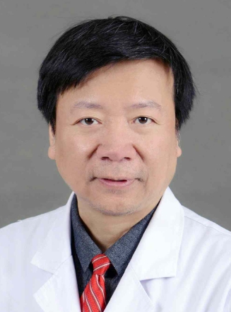 Zhihong Wu (吴志宏)合作论文数Peking Union Medical College Hospital55Yingjie Wang合作论文数State Key Lab Complex Severe & Rare Dis, Peking Union Med Coll Hosp53
 Shugang Li (李书纲)合作论文数Peking Union Medical College Hospital38
 Yu Zhao (赵宇)合作论文数Peking Union Medical College Hospital;Peking Union Medical College, Chinese Academy of Medical Sciences34JiLiang Di (翟吉良)合作论文数32wei zhu合作论文数31XinYu Yang (杨新宇)合作论文数31
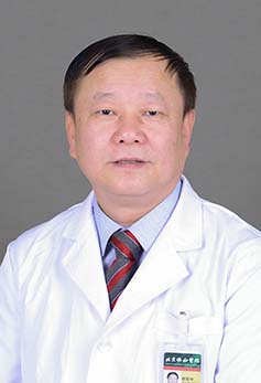 Baozhong Zhang (张保中)合作论文数Peking Union Medical College Hospital31
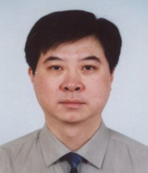 Hong Zhao (赵宏)合作论文数Department of Orthopaedic,PUMC Hospital,CAMS/PUMC,Beijing ,China30
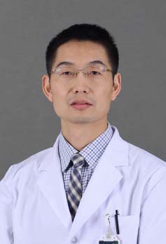 Jiliang Zhai (翟吉良)合作论文数Peking Union Medical College Hospital30Yixin Bian合作论文数29Li Zeng合作论文数25Bin Yu (于斌)合作论文数24LiJuan Zhao (赵丽娟)合作论文数23Yulei Dong (董玉雷)合作论文数19
 Ye Tian (田野)合作论文数Peking Union Medical College Hospital19Wei Zhu合作论文数18Wei Wang合作论文数18Wei WANG (王炜)合作论文数18
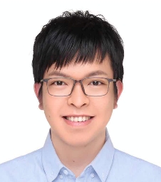 Ruizheng Liang (梁瑞政)合作论文数College of Chemistry, Beijing University of Chemical Technology18
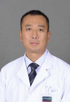 Qiyi Li (李其一)合作论文数Peking Union Medical College Hospital17Zehui Lv合作论文数16Xiao Chang (常晓)合作论文数16
 Qianyu Zhuang (庄乾宇)合作论文数Peking Union Medical College Hospital16Zhanqi Wei合作论文数15Shengru Wang合作论文数15Ke Xiao合作论文数15Zheng Li合作论文数15
 Yu Fan (范彧)合作论文数北京协和医学院北京协和医院15
 Fuxing Pei (裴福兴)合作论文数West China Hospital, Sichuan University15
 Tao Li (李涛)合作论文数Emergency General Hospital15Yong Liu (刘勇)合作论文数Cancer Hospital, Chinese Academy of Medical Sciences15
 Han Wang合作论文数14
 Zengxin Gao (高增鑫)合作论文数Department of Spine Surgery, Zhongda Hospital, Southeast University14Tingting Hu合作论文数13JianHua Hu (胡建华)合作论文数13JunWei Li (李军伟)合作论文数12
 Zongke Zhou (周宗科)合作论文数West China Hospital of Sichuan University12




